
Remember me


In this study, we aimed to assess the morbidity and mortality associated with BTTS, in comparison to PDA stenting, as well as the effect on pulmonary artery growth over the course of time. Most of the patient demographics and preoperative conditions did not differ. There were significantly more patients diagnosed with pulmonary atresia/intact ventricular septum or critical pulmonary stenosis in the PDA stent group, probably due to center preference, as seen in other studies such as Glantz et al. and McMullan et al. [7, 10]. Although the PDA morphology in patients with pulmonary atresia tends to be more favorable, making the ductal stenting technically simpler [11], the two overall groups did not differ in terms of PDA tortuosity. Furthermore, there was no difference between the numbers of patients with univentricular physiology or antegrade flow in the two groups, so we do not believe that the percent of pulmonary atresia/stenosis patients accounted for the overall improved outcomes of the PDA stent group.
The need for mechanical ventilation before the procedure was significantly more common in the BTTS group. Many of the patients who were ventilated before the procedure were transferred from other medical centers due to a lack of appropriate diagnostic and treatment equipment. It was decided not to extubate these patients before the procedure. In contrast, those born at our medical center often had a prenatal diagnosis of the specific cardiac defect and were monitored and treated with Prostaglandin E, eliminating the need for mechanical ventilation. If the initial ventilation status were the only factor influencing outcomes, we would expect post-procedure ventilation days to be roughly twice as high in the BTTS group. However, we observed that post-procedure ventilation days were actually ten times higher in the BTTS group compared to the PDA stent group.
The prevalence of previous surgeries before the initial palliation was more common among the PDA stent group. The patients in PDA stent group who underwent previous procedures were mostly patients diagnosed with pulmonary atresia or critical pulmonary stenosis. At our center, these patients are initially treated with pulmonary valve perforation and dilation percutaneously, then a wait-and-see period to assess if they can be weaned from oxygen support and prostaglandin E treatment. If not, they are taken to another catheterization for PDA stenting.
When we first performed PDA stenting our center, only babies with high risk for surgery and two sources of pulmonary blood flow were referred for this procedure. Over the years of the study, PDA stenting has become the go-to intervention for babies with duct dependent pulmonary circulation. Therefore, the surgical vs. stenting groups assessed did not differ in terms of antegrade blood flow or complicated pre-interventional findings, and we believe that this era difference did not have significant effect. There was no significant difference in terms of pulmonary artery measurements prior to intervention. If anything, the ductal stenting group exhibited qualitatively more pre-intervention pulmonary artery stenosis.
Our study demonstrates that PDA stenting was associated with significantly shorter ICU length of stay, shorter hospitalization, fewer ventilation days, and less need for inotropic support after the procedure. A large meta-analysis carried by Alsagheir et al. exhibited similar results with a median of 4.6 days in ICU after PDA stent and 5.8 days of hospital length of stay [8], and a similar trend was demonstrated by Ratnayaka et al. [12]. Bentham et al. also showed the length of ventilation was significantly shorter after PDA stenting with a median of 1 day after the procedure, compared to 4 after BTTS. Longer hospitalizations as well as the use of mechanical ventilation increase the risk for serious hospital-acquired infections and other complications [13]. Findings of less need for inotropic support are also of great importance, reflecting the improved hemodynamic condition of the PDA stent patients after intervention. A high VIS score, associated with morbidity, mortality, and poor clinical outcome in infants undergoing cardiac surgery [9], is reflected in our findings that the BTTS group had higher ICU mortality.
All patients in the BTTS group received a 4 mm shunt, regardless of age and weight, with a median weight of 3.2 kg in the study group. This uniform shunt size may have contributed to post-procedure over-circulation and related complications, such as major hemodynamic instability and congestive heart failure, which were observed more frequently in the BTTS group. In contrast, PDA stenting conforms to the patient’s anatomy, resulting in fewer over-circulation complications, and stent diameter was adjusted according to patient weight: 2.5 kg babies received a 3.5 mm diameter stent, whereas babies above 3 kg received a 4 mm stent.
The high cardiopulmonary bypass rate in patients who underwent BTTS, was primarily due to surgical preference aimed at improving the anastomosis site of the BTTS, rather than concomitant surgery. However, as demonstrated in our study, the use of cardiopulmonary bypass did not improve surgical outcomes and instead led to higher morbidity and mortality, suggesting that ductal stenting, a less invasive procedure has significant survival advantages. Bentham et al. concluded that since PDA stenting eliminates the need for thoracic surgical dissection of the neonate, it may be related with better post-procedural stability and survival [14]. What is more, babies with branch pulmonary artery stenosis referred for initial shunt with PA plasty, actually demonstrated more pulmonary artery stenosis during follow-up. In comparison, babies with neonatal branch PA stenosis who had initial ductal stenting followed by pulmonary artery repair at the second stage procedure, exhibited significantly better pulmonary artery growth. Although the number of patients compared in this respect are small, the results are very suggestive, and may be due to the improved ease of performing surgical arterial repair in larger babies (Figs. 4, 5) [15].
Li et al. assumed that preforming BTTS with concomitant pulmonary arterioplasty may have favorable results on pulmonary blood flow. However, it was discovered that when these two procedures were combined, there was a higher rate of hospital mortality [16]. In our study we examined the mid-term effects of combining two procedures, BTTS and PA plasty as initial surgery, on mid-term PA stenosis, in comparison to patients who underwent BTTS or PDA stenting at first, then pulmonary artery repair during their second surgery. Our findings indicate that patients who underwent BTTS along with PA plasty as part of an initial surgical procedure had a higher rate of RPA stenosis and shunt site stenosis compared to those who underwent PDA stenting followed by pulmonary arterioplasty at the second procedure. It therefore appears that a child with duct dependent pulmonary blood flow presenting with pulmonary artery stenosis at birth is better palliated with an initial PDA stent until pulmonary artery plasty is safely performed surgically on larger, more developed pulmonary arteries later in infancy (Fig. 8).
Fig. 8
Surgical images showing A the PDA stent in situ as encountered by the surgical team at the definitive operation nine months after implantation, and B following removal of the stent, prior to arterioplasty. Surgical video available in Online Appendix
Santoro et al. found that both BTTS and PDA stent groups experienced significant PA growth as determined by the Nakata index and McGoon ratio, with no difference in the overall growth of the pulmonary arteries. However, the group treated with PDA stents had more symmetrical growth of the pulmonary arteries as measured by the ratio of left pulmonary artery to right pulmonary artery diameter [17]. We also found that the BTTS group experienced a significantly narrower proximal RPA diameter over time, which may have been caused by the shunt implantation distorting the PA anatomy. This suggests that the BTTS group may have less favorable mid-term effects such as PA distortion and stenosis at the shunt site [11]. In addition, although the PDA stent group had a higher prevalence of pulmonary artery stenosis before initial intervention, they experienced significantly better PA growth compared to the BTTS group as expressed in a higher McGoon ratio.
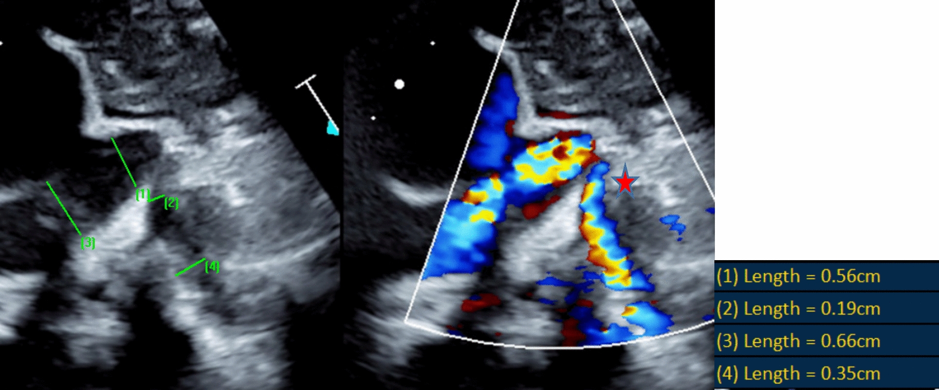
Given the small size of the pulmonary arteries in these infants, the small study group, and the difficulty achieving statistically significant quantitative differences, we decided to include a qualitative assessment of the pulmonary arteries. This assessment was defined as a 50% reduction in PA diameter. We believe the categorical assessment holds clinical importance since clinical decisions are often based on qualitative evaluations rather than specific numerical measurements.
Comments (0)