Oncocytic Mucoepidermoid Carcinoma of the Parotid Gland
Case presentation:
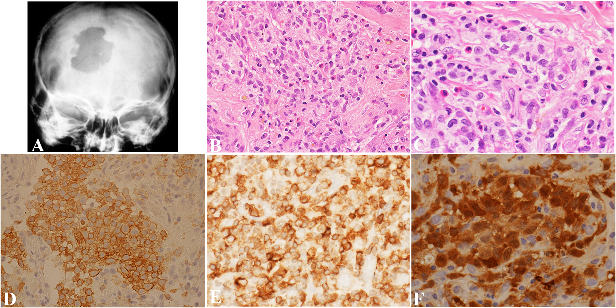
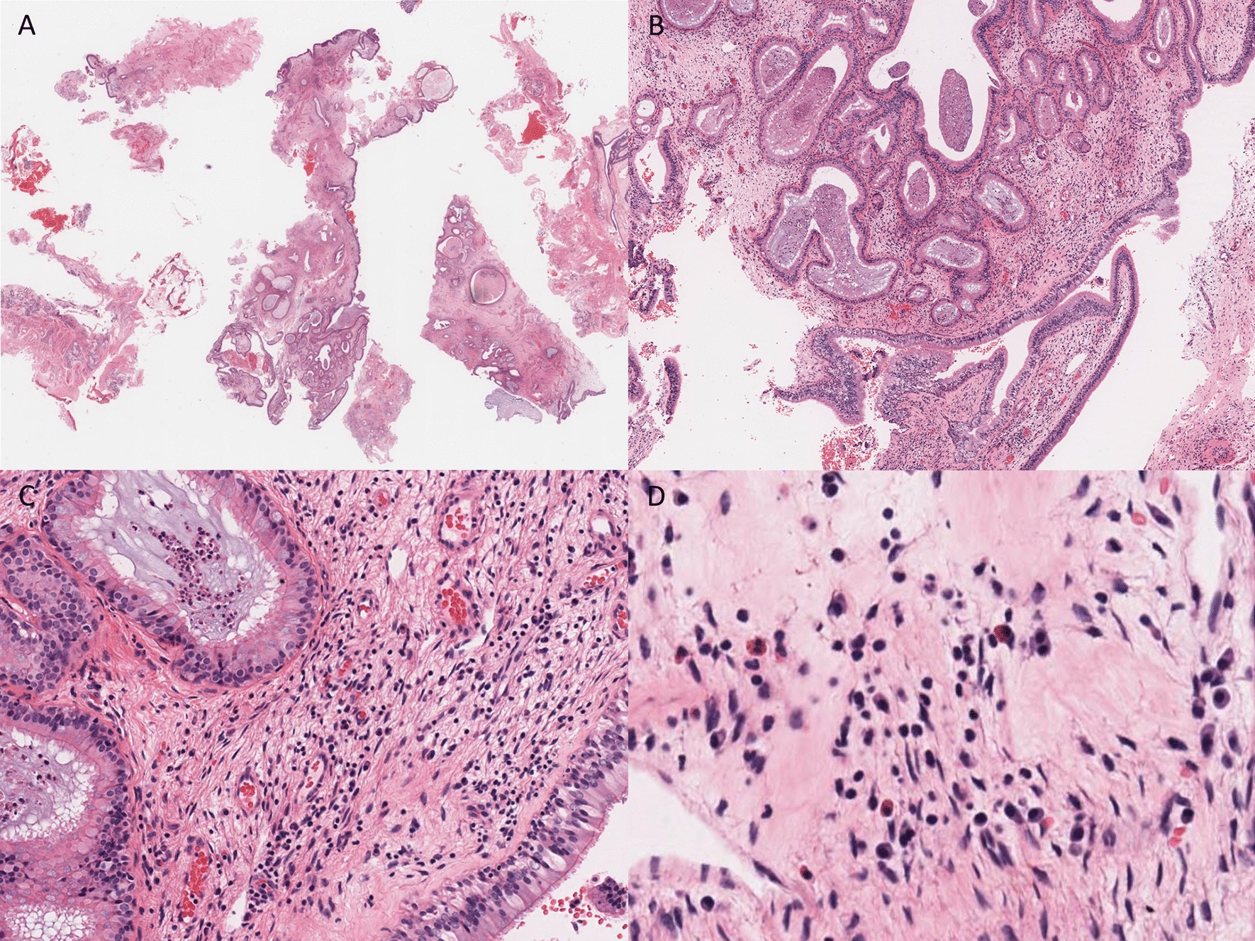
A 30-year-old man presented with a multilobulated left parotid mass measuring 5.2 × 4.1 × 2.4 cm by imaging, and numerous enlarged left cervical lymph nodes, suspicious for metastasis. FNA cytopathology of the mass showed loose clusters of large cells displaying increased N/C ratios and ample granular oncocytic cytoplasm. A superficial left parotidectomy with radical resection of the cheek and cervical lymphadenectomy was performed. Histopathologic examination disclosed a circumscribed, unencapsulated neoplasm exhibiting a solid growth pattern composed of infiltrative islands and nests of cohesive, polygonal, oncocytoid cells in a densely fibrous stroma. Lesional cells exhibited enlarged, oval, open-face nuclei with coarse chromatin and a single acidophilic macronucleolus, voluminous eosinophilic granular cytoplasm and distinct cell membrane borders. Mitotic activity and necrosis were absent. Microcystic architecture was noted solely in a single tumor nest at the periphery. These spaces contained mucinous secretions and were lined by cuboidal oncocytic and intermediate cells with interspersed mucocytes, highlighted by mucicarmine stain. Immunohistochemically, oncocytic cells were strongly and diffusely positive for cytokeratin AE1/AE3, p63 and p40, and uniformly negative for androgen receptor, GATA3, S100, SOX10 and Her-2. A MAML2 rearrangement was identified by FISH, thus confirming the diagnosis of oncocytic variant of mucoepidermoid carcinoma.
Conclusion:
In this illustrative example, we present the clinicoradiologic, cytologic, histopathologic, and immunophenotypic characteristics of this rare variant of mucoepidermoid carcinoma, together with molecular documentation.


Comments (0)