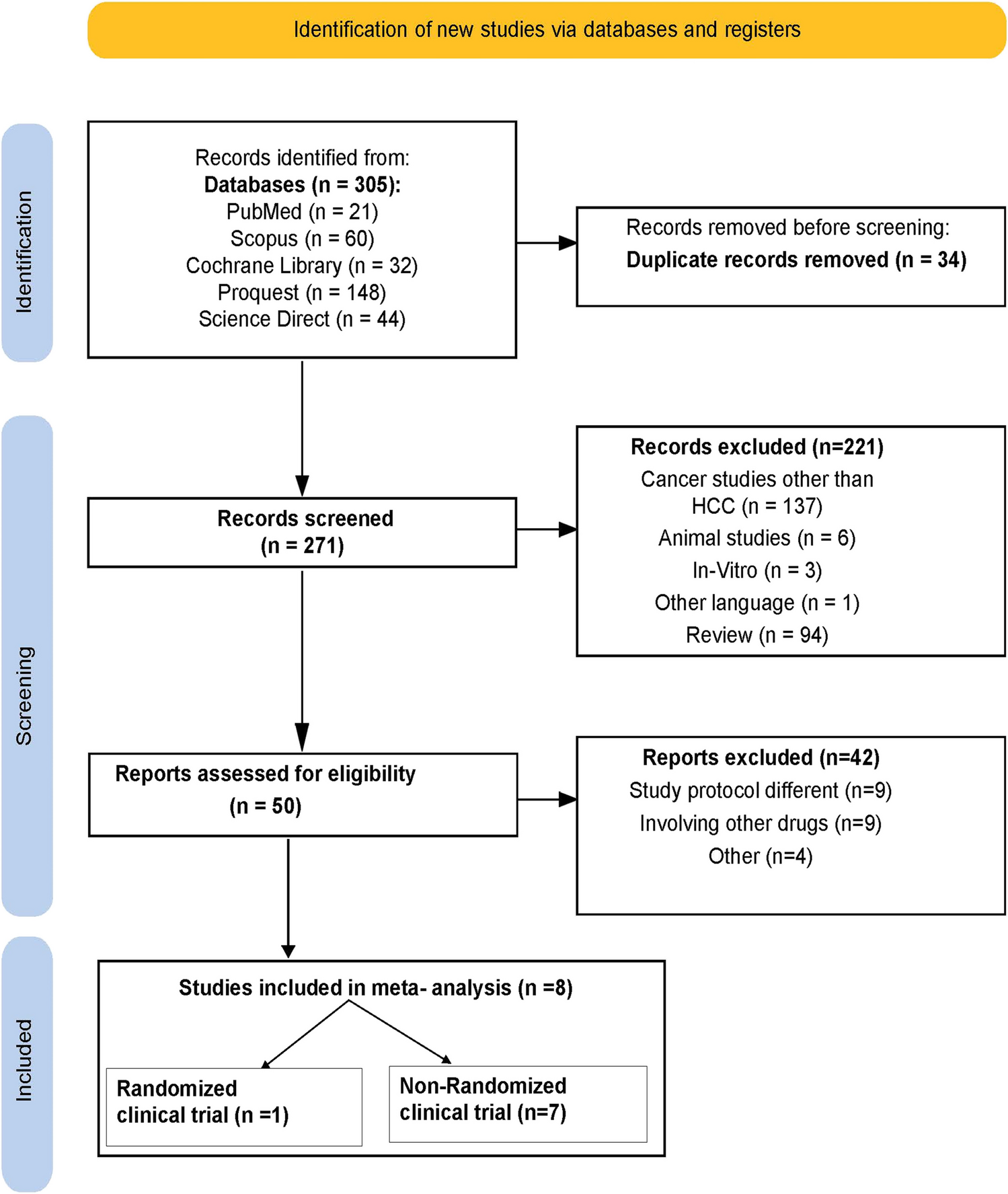
Hematological toxicity, particularly CIT, is a major dose-related adverse effect of many cytotoxic chemotherapeutics. CIT often increases bleeding, delays chemotherapy, and limits the dose of chemotherapeutics, consequently reducing the efficacy of chemotherapy, affecting the quality of life of patients, and increasing mortality and medical costs. Therefore, assessing the risk of CIT in patients with gastrointestinal malignancies undergoing chemotherapy is of great clinical significance.
Studies have shown that women are less likely to suffer from low platelets due to their high basal platelet count [9,10,11,12]. However, in a pooled analysis of five randomized trials, Abdel-Rahman demonstrated that not all grades of thrombocytopenia significantly differed between women and men [13]. In our study, thrombocytopenia incidence did not significantly differ between men and women. We speculate that it may be because male and female patients were not stratified for analysis. In subsequent studies, we will stratify male and female patients to separately analyze its relationship with CIT.
Using multivariate analysis, Yamada [11] found thrombocytopenia to be more frequent in patients aged > 70 years after CS treatment. Similarly, studies have reported that with increasing age, the hematopoietic function of bone marrow slows down and is more vulnerable to myelosuppression during chemotherapy [14,15,16]. Conversely, Professor Yo Han showed that thrombocytopenia incidence did not significantly differ between different age groups (6.4% over 65 years vs. 3.9% under 65 years) [17]. Our study only found that age is an influencing factor of CIT in univariate analysis; we can combine multiple indicators to further analyze their relationship with CIT in future.
Some studies have pointed out that patients with a lower body mass index (BMI), moderate/severe malnutrition, and an Eastern Cooperative Oncology Group (ECOG) score of 1 are more likely to suffer from thrombocytopenia [18,19,20]. Our study included nutrition-related indicators such as serum albumin, phase angle, and SMI. The results showed that serum albumin is an independent risk factor for CIT, while phase angle and SMI were only found to affect platelets in univariate analysis. But this provides us with ideas for subsequent research, whether it is possible to include more body composition analysis indicators, conduct combined analysis of different indicators, or calculate unit area to further study the relationship with CIT.
Woo et al. [21] reported the incidence of thrombocytopenia due to myelosuppression as 45–77%. Platelet count reduces below 75 × 109/L usually after 10 days of oxaliplatin administration, with few symptoms of thrombocytopenia, such as congestion, purpura, or mucosal bleeding. Studies have suggested that oxaliplatin causes CIT mainly through the following three mechanisms [22]: (1) oxaliplatin has a myelosuppressive effect, and thus, it reduces the activity and function of hematopoietic stem cells in the bone marrow, resulting in decreased platelet production; (2) repeated oxaliplatin treatment induces and maintains immune responses, leading to immune thrombocytopenia [23]; and (3) oxaliplatin causes liver sinusoidal injury, leading to portal hypertension, splenomegaly, and hypersplenism, which reduce the platelet count. The main mechanism of oxaliplatin-induced thrombocytopenia is myelosuppression, whereas the incidence of splenomegaly and immune response mechanisms is relatively low in clinical practice. Studies have shown that the incidence of CIT increases significantly with the increase in chemotherapy cycles. An observational study of 320 patients with CIT published in 2011 showed that the incidence of CIT was 0.16%, 17.8%, 37.8%, and 43.8% in the first, second, third, and fourth cycles of chemotherapy, respectively [24]. Some studies have shown that the number of chemotherapy cycles [median (25%, 75%)] at the first occurrence of CIT in patients receiving a platinum-containing regimen were 3 and the cumulative dose of oxaliplatin received at the time of the first occurrence of CIT was 569.7 mg ± 273.4 mg [25]. We found that total oxaliplatin dose was an independent risk factor for CIT, consistent with the above studies. In the future, we will include indicators such as body surface area to further analyze the relationship between unit oxaliplatin dose and CIT.
Other studies have shown that different races have different adverse reactions to capecitabine. For example, the incidence of III–IV CIT was significantly higher in Chinese patients at a dose intensity of 900 mg/m2 (9%; overall CIT incidence, 84%) than in the Western population at a dose intensity of 1000 mg/m2 (2.1%; overall CIT incidence, 49%), suggesting that capecitabine has a greater effect in Asian populations [26]. In our study, patients with gastric cancer were treated with the SOX regimen, and patients with colorectal cancer were treated with the XELOX or FOLFOX regimen. By stratifying different types of gastrointestinal cancers and different chemotherapy regimens, the total doses of S-1, capecitabine, and 5-FU were calculated, and then their correlation with CIT was analyzed; our study did not conclude that total fluorouracil dose was an independent risk factor for CIT. In combination with clinical practice, it is considered that it may be due to the different metabolic pathways and mechanisms of action of fluorouracil and oxaliplatin, fluorouracil belongs to the antipyrimidine antimetabolite, which inhibits deoxythymididate synthase by converting to 5-fluorouracil deoxynucleotide in vivo, blocking the conversion of deoxyuriglycate to deoxythymidylate and inhibiting DNA synthesis. It may cause a heavier myelosuppressive response, mainly manifested by varying degrees of granulocytopenia, which occurs earlier but relatively rarely [27].
The rate of depletion of hematopoietic cells in the bone marrow depends on the half-life of the cells. The half-lives of white blood cells, platelets, and red blood cells were 6 h, 5–7 days, and 120 days, respectively, indicating that platelets were more easily affected and decreased. The low platelet level before treatment sensitively reflects compromised hematopoietic function of bone marrow granulosa system. Clinically, such patients are more prone to myelosuppression after chemotherapy. Our current findings reveal that the baseline platelet count is an independent risk factor for CIT, emphasizing the significance of a low baseline platelet count before treatment in predicting CIT occurrence. Combined with the situation of some studies, the occurrence of thrombocytopenia before treatment can be attributed to the following: (1) bone metastases occur in the late stage of the tumor, and tumor cells have spread to the marrow hematopoietic system, thus directly inhibiting the normal hematopoietic function of the bone marrow [28]; (2) poor eating and malnutrition in patients with gastrointestinal tumors affect bone marrow hematopoiesis. Therefore, pretreatment of patients with low baseline platelets via prophylactic use of thrombopoietin and improved nutrition nutritional status is highly significant for clinical treatment.
Thrombopoietin (TPO) is an essential factor for megakaryocyte growth and is the only important regulator of platelet production. Endogenous TPO is mainly produced by the liver (70%) and kidney. In patients with liver metastasis, the liver structure is affected, and there are notable pathological changes in the normal liver tissue around the lesion; this further decreases chemotherapy tolerance of the liver [29]. The oxaliplatin-containing chemotherapy regimen leads to liver function damage and causes spleen enlargement, which is a potential cause of persistent thrombocytopenia after oxaliplatin chemotherapy [30]. Studies have shown that the cumulative incidence of thrombocytopenia as an adverse event was 22% (41/184), 24% (181/752), and 23% (32/142) in patients with only liver metastases, liver metastases combined with other metastases, and without liver metastases, respectively, and the difference in these incidence rates was not statistically significant [31]. Our study found that lung metastases distant metastases to other sites besides the liver and lung seemed to affect platelet counts after chemotherapy only in univariate analysis. We herein could not identify whether liver metastases can affect the generation of endogenous TPO and consequently the platelet count and thus may need to be further explored in clinical practice in the future.
Our study identified NK cell as an independent risk factor for CIT; however, the predictive value of NK cell for inducing CIT remains unknown. Clinically, albumin indicators can roughly reflect the patient’s nutritional status, and NK cells can also reflect the patient’s immune function to a certain extent. The nutritional status of patients may affect their immune function, so NK cells may be related to CIT, but this conclusion may require further clinical research and analysis. Unfortunately, the area under the ROC curve for either factor was small, and the predictive value was low. Given the high reliability of joint prediction, we can carry out joint factor analysis for patients with gastrointestinal tumors in the clinic to intervene and treat patients who are prone to CIT and improve the outcomes of these patients.
Thus far, few studies have established CIT risk prediction models based on clinical and laboratory indicators. In our study, the best cutoff value for the area under the curve of the predicted probability was 0.3579613 (sensitivity, 78.9%; specificity, 81.8%). These differences may be attributed to a number of different factors, such as the number of patients, the definition of CIT, and differences in the chemotherapy agent administered.
The total dose of oxaliplatin, M stage, albumin, baseline platelet count, and NK cell was independent risk factors for CIT. The sequentially constructed histogram model had a good predictive effect on the risk of thrombocytopenia caused by oxaliplatin chemotherapy in patients with gastrointestinal malignancies.
The present study had several limitations. The selection of subjects was from a single center, and the variety of potential predictors collected was limited by clinical practice. Furthermore, this study was a retrospective study with limited collection of clinical data. The conclusions obtained in this study need to be further verified in multicenter studies and clinical practice. In a future study, we hope to increase the sample size and include other variables in clinical practice to identify the patients at risk of CIT with greater accuracy, guide clinical practice, and improve the treatment effect.


Comments (0)