Clinical studyClinical samples
The blood samples were collected from the peripheral veins of healthy volunteers aged between 20 and 30 years (young group) and between 55 and 70 years (elderly group). There are at least 6 subjects in each group. Subjects with diabetes mellitus, significant systemic diseases, who had received major operations in the past 6 months were excluded. Demographic and clinical data were obtained at enrollment. The human study was approved by the institute research committee (IRB No. 2021-02-010AC in Taipei Veterans General Hospital) and was conducted in accordance with the guidelines detailed in the Declaration of Helsinki.
In vitro studyCell culture
After blood was collected, the total mononuclear cells were separated by using a Histopaque-1077 (Sigma-Aldrich, 10,771, Darmstadt, Germany) and centrifuged at 500 × g at room temperature for 30 min. The mononuclear cells were cultured in an endothelial cell basal medium (EBM-2; Lonza, CC-3156, Basel, Switzerland) with supplements, including hydrocortisone, human fibroblast growth factors, vascular endothelial growth factor (VEGF), R3-insulin-like growth factor-1, ascorbic acid, human epidermal growth factor, gentamicin sulfate-amphotericin, and 20% fetal bovine serum on fibronectin-coated 6-well plates. After 2–4 weeks culture, attached EPCs emerged. The EPCs were in the shape of cobblestones; this kind of shape is the typical monolayer growth pattern of mature endothelial cells.
Primary HAECs (ScienCell, Catalog #6100, Carlsbad, CA, USA) were cultured in fibronectin-coated plates with endothelium cell medium containing 5% fetal bovine serum and 1% of endothelial cell growth supplement in an atmosphere of 95% air and 5% CO2 at 37 °C. Different dosages of CCL4 (0.1, 1 ng/mL; R&D Systems, Minnesota, USA) were added to the cells as the cells grown to a certain circumstance. The cells were then cultured at 37 °C and 5% CO2 for 3 days after CCL4 stimulation. Edaravone (MCI186) were purchased from Cayman Chemical (Ann Arbor, MI, USA) and was used as a free radical scavenger.
Transfection of siRNA
Cells were transfected with ccl4 siRNA and p65 siRNA (Santa Cruz Biotechnology, sc-43,932 and sc29410, Dallas, TX, USA) using Lipofectamine 2000 (Invitrogen, Carlsbad, CA, USA) in culture medium.
Cell proliferation assay
Cell Counting Kit-8 (CCK-8; Dojindo Molecular Technologies, Inc., Rockville, MD, USA) was used to evaluate cell viability according to the manufacturer’s instructions. In brief, reagents were added and incubated with cells at 37 °C for 2 h and the absorbance at 450 nm was determined.
β-Galactosidase staining
The senescence phenotype was detected using the β-galactosidase staining kit (Merck, Darmstadt, Germany). The number of positive senescence-associated β-galactosidase cells were observed by light microscopy in 10 randomly chosen low-power fields.
ROS generation assay
Hydrogen peroxide production from cells was measured using the Amplex Red Hydrogen Peroxide/Peroxidase Assay Kit (Invitrogen, Carlsbad, CA, USA). Briefly, horseradish peroxidase catalyzed the stoichiometric reaction of Amplex Red with hydrogen peroxide (H2O2), composing of water and resorufin. Then, cells were scraped into phosphate buffer (pH 7.4). After spin-down of cell debris, 50 µL of supernatant was loaded with 50 µL of working solution. Following a 30-minute incubation, fluorescence was measured using a microplate reader with excitation at 540 nm and emission at 590 nm.
Migration and tube formation assay
The migration was evaluated by a chamber assay. Cells (1 × 10⁴ cells) were resuspended in the culture medium with 5% FBS. The cells were added to the upper chamber of a 24-well transwell plate with a polycarbonate membrane. Culture medium supplemented with fetal bovine serum was added to the lower chamber, and the chambers were incubated for 18 h. After incubation, the membrane was fixed with 4% paraformaldehyde and stained using hematoxylin solution. The numbers of migrated cells were counted in random high-power (×100) microscopic fields.
The in vitro tube formation assay was performed with an angiogenesis assay kit (Invitrogen, Carlsbad, CA, USA). ECMatrix gel solution was mixed with ECMatrix diluent buffer and placed in a 96-well plate. Then, cells (1 × 10⁴ cells) were placed on a matrix solution with 10% FBS culture medium and incubated for 16 h. Tube formation was inspected under an inverted light microscope (× 40). The average of the total area of complete tubes formed by cells was compared using Image-Pro Plus (Media Cybernetics, Inc. Rockville, MD, USA).
Western blot analysis
Total cell or tissue lysates were extracted using a lysis buffer, and proteins were separated in 8–12% (v/v) SDS-PAGE gels. After electrophoresis (Bio-Rad Laboratories, Hercules, CA, USA), the proteins were transferred onto nitrocellulose membranes (Millipore, Darmstadt, Germany). The membranes were incubated with antibodies against CCL4 (Santa Cruz Biotechnology, sc-393,441, Dallas, TX, USA); p-AKT (BD Biosciences, 550,747; NJ, USA); AKT (BD Biosciences, 610,868; NJ, USA); VEGF (Santa Cruz Biotechnology, sc-152; Dallas, TX, USA); SDF-1 (Cell Signaling, 3530 S; Boston, MA, USA); IL-1β (Santa Cruz Biotechnology, sc-7884; Dallas, TX, USA); IL-6 (Cell Signaling, 12,153 S; Boston, MA, USA); TNF-α (Cell Signaling, 3707 S; Boston, MA, USA); p-p65 (Cell Signaling, 3031 S; Boston, MA, USA); p65 (BD Biosciences, 610,868; NJ, USA); SIRT1 (Cell Signaling, 8469 S; Boston, MA, USA); p53 (Cell Signaling, 2524 S; Boston, MA, USA); p16 (Cell Signaling, 80,772 S and 29,271 S ; Boston, MA, USA); xanthine oxidase (Santa Cruz, sc-398,548, Dallas, TX, USA); NADPH oxidase subunit p47 (Santa Cruz, sc-17,845, Dallas, TX, USA); p-eNOS (Cell Signaling, 9571 S; Boston, MA, USA); eNOS (Cell Signaling, 32,027 S; Boston, MA, USA) and β-actin (Merck, MAB1501, Darmstadt, Germany) [14] at 4 °C overnight. The above protein expressions were normalized to that of actin expression.
In vivo studyAnimal preparation
Six-week-old male C57BL/6JNarl-Ccl4em1 knock out (CCL4KO) mice were design and purchased from the National Laboratory Animal Center (Taipei, Taiwan). CCL4KO mice were generated in a C57BL/6JNarl genetic background by using the CRISPR/Cas9 system. All mice were genotyped using PCR with specific primers (forward, 5′-TCTCCCTCCTTTCTCTTCCGTG-3′, and reverse, 5′-TCTACTCCCAATGATGGCTGACC-3′). C57BL/6JNarl mice were used as the wild-type (WT) control. Six-month-old mice were defined as the young group and eighteen-month-old mice were defined as the elderly group. Some animals were randomized into the CCL4 neutralizing antibody-injected group (100 µg, intraperitoneal injection; R&D Systems, MAB451, Minneapolis, MN, USA). The monoclonal neutralizing antibody was injected 3 times a week for 2 weeks. The animals were raised under specific pathogen-free conditions and all mice were kept in microisolator cages on a 12-hour day/night cycle in the animal center of National Yang Ming Chiao Tung University (Taipei, Taiwan) according to the regulations of the Animal Care Committee of National Yang Ming Chiao Tung University. The animal study was approved by the Animal Care Committee of National Yang Ming Chiao Tung University (IACUC No. 1101114).
Wound healing assay and evaluations of morphological changes in the wound area
The circular full-thickness excisional wounds of 3 mm of diameter were generated with biopsy punch without injuring the muscle. The wounds were recorded using a digital camera (Nikon, Tokyo, Japan) after they were generated. Transverse slices of the wound areas were fixed in 10% formaldehyde, embedded in paraffin, and mounted onto slides for staining. Hematoxylin/Eosin (H&E) stains were used to evaluate the morphological changes in the wound area.
Capillary densities, cell proliferation, and collagen deposition levels in the wound area
Sections were de-paraffinized and incubated with a rat-monoclonal antibody against murine CD31 (Abcam, 124432, Waltham, MA, USA) and a rabbit-polyclonal antibody against the murine marker of proliferation Ki67 (Novus, NB500-170, Minneapolis, MN, USA). Antibody distribution was visualized with the avidin-biotin-complex technique and Vector Red chromogenic substrate, followed by counterstaining with hematoxylin. Sections were allowed to dry overnight and stained with H&E and Masson’s trichrome-stained for histological analysis.
Matrigel plug neovascularization assay
Mice were injected subcutaneously with growth factor reduced basement membrane matrix (Corning® Matrigel, Glendale, AZ, USA) containing 30 ng/mL VEGF (Peprotech, Rocky Hill, CT, USA) and 50 U heparin (Sigma-Aldrich, Darmstadt, Germany). Plugs were collected after 14 days and homogenized in 500 µL of cell lysis buffer and centrifuged at 6000 g at 4 °C for 60 min. Hemoglobin was detected at 400 nm wavelength by using a colorimetric assay (Sigma-Aldrich, MAK115, Darmstadt, Germany). Also, plugs were harvested for histological and immunohistochemistry analysis.
Aortic ring assay
The detailed method and timing of tissue culture have been well indicated in our recent publication [15]. In brief, the aortic rings were cut a 0.5 mm and embedded 1 mg/mL type 1 rat tail collagen matrix (Millipore, Darmstadt, Germany) and incubated for 1 h at 37 °C. Aortic rings were cultured in EBM-2 (Lonza, Basel, Switzerland) containing 2.5% bovine serum (Gibco, Carlsbad, CA, USA), 50 U/mL penicillin and 0.5 mg/mL streptomycin (Sigma-Aldrich, Darmstadt, Germany) and 30 ng/mL VEGF (Peprotech, Rocky Hill, CT, USA) in 24-wells for 7 days. Images were captured using a microscope (× 100).
ELISA
The concentrations of CCL4 were analyzed using a CCL4 ELISA kits (R&D, Minneapolis, MN, USA) according to the manufacturer’s instructions.
Statistical analysis
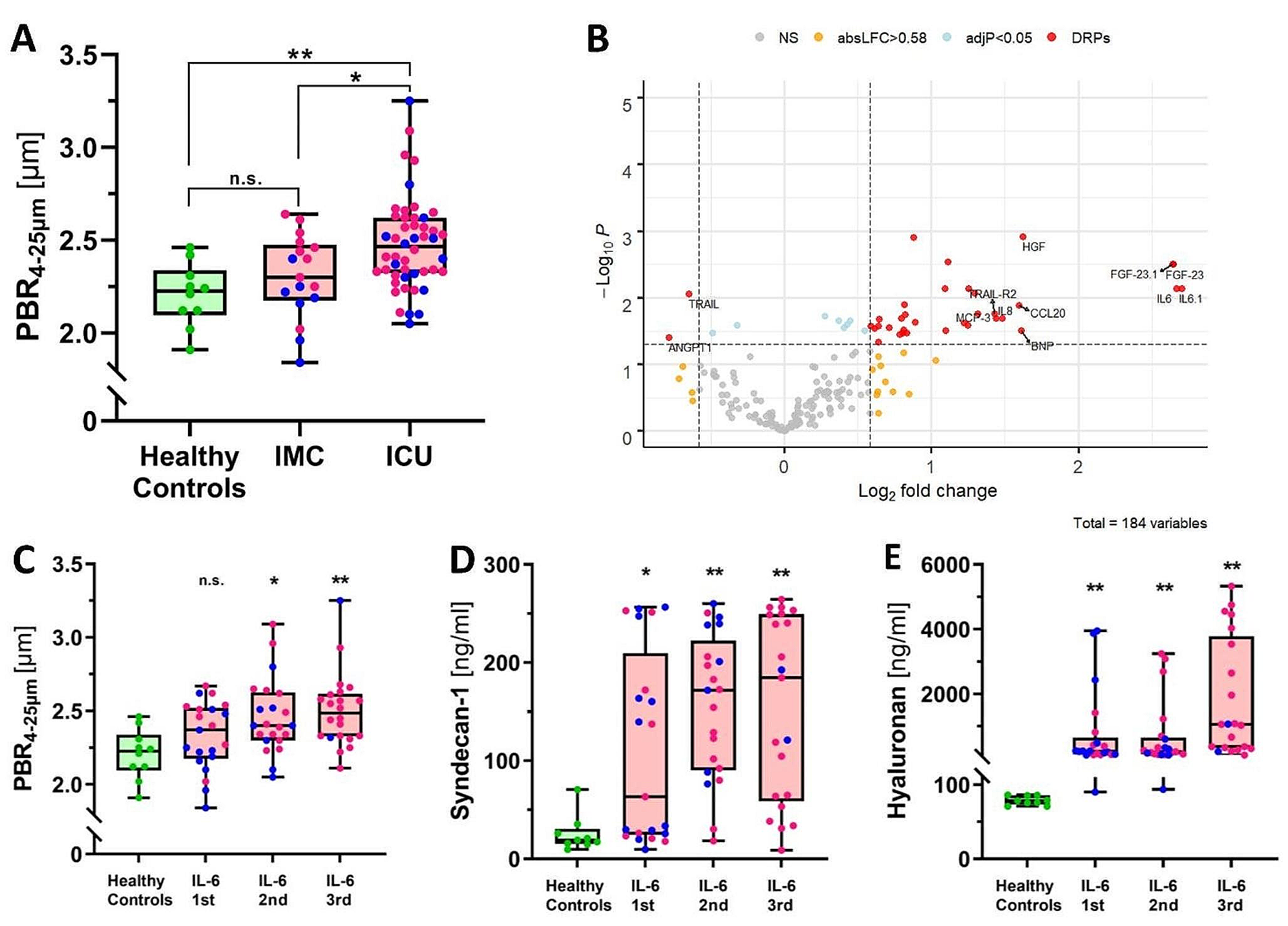
Given our data’s non-parametric nature, we utilized the median and interquartile for descriptive statistics and the Mann-Whitney U test to identify group differences. The statistical analysis software GraphPad Prism vision 6 was used for the statistical analysis. A p-value of less than 0.05 was deemed statistically significant.


留言 (0)