Saeidnia S, Manayi A, Abdollahi M. From in vitro experiments to in vivo and clinical studies; pros and cons. Curr Drug Discov Technol. 2015;12(4):218–24.
Article
CAS
PubMed
Google Scholar
Abdinian M, Moshkforoush S, Hemati H, Soltani P, Moshkforoushan M, Spagnuolo G. Comparison of cone beam computed tomography and digital radiography in detecting separated endodontic files and strip perforation. Appl Sci. 2020;10(23):8726.
Article
CAS
Google Scholar
Miri S, Mehralizadeh S, Sadri D, Motamedi MRK, Soltani P. The efficacy of the reverse contrast mode in digital radiography for the detection of proximal dentinal caries. Imag Sci Dentist. 2015;45(3):141–5.
Article
Google Scholar
Sirin Y, Yildirimturk S, Horasan S, Guven K. Diagnostic potential of panoramic radiography and CBCT in detecting implant-related ex vivo injuries of the inferior alveolar canal border. J Oral Implantol. 2020;46(3):206–13.
Article
PubMed
Google Scholar
Molteni R. Prospects and challenges of rendering tissue density in Hounsfield units for cone beam computed tomography. Oral Surg Oral Med Oral Pathol Oral Radiol. 2013;116(1):105–19.
Article
PubMed
Google Scholar
Souza PH, da Costa NP, Veeck EB. Influence of soft tissues on mandibular gray scale levels. Braz Oral Res. 2004;18(1):40–4.
Article
PubMed
Google Scholar
Blake GM, McKeeney DB, Chhaya SC, Ryan PJ, Fogelman I. Dual energy x-ray absorptiometry: the effects of beam hardening on bone density measurements. Med Phys. 1992;19(2):459–65.
Article
CAS
PubMed
Google Scholar
Borg E, Källqvist A, Gröndahl K, Gröndahl HG. Film and digital radiography for detection of simulated root resorption cavities. Oral Surg Oral Med Oral Pathol Oral Radiol Endod. 1998;86(1):110–4.
Article
CAS
PubMed
Google Scholar
Caldas Mde P, Ramos-Perez FM, de Almeida SM, Haiter-Neto F. Comparative evaluation among different materials to replace soft tissue in oral radiology studies. J Appl Oral Sci. 2010;18(3):264–7.
Article
PubMed
Google Scholar
Cook JE, Cunningham JL. The assessment of fracture healing using dual x-ray absorptiometry: a feasibility study using phantoms. Phys Med Biol. 1995;40(1):119–36.
Article
CAS
PubMed
Google Scholar
Hildebolt CF, Rupich RC, Vannier MW, Zerbolio DJ, Shrout MK, Cohen S, et al. Inter-relationships between bone mineral content measures. Dual energy radiography (DER) and bitewing radiographs (BWX). J Clin Periodontol. 1993;20(10):739–45.
Article
CAS
PubMed
Google Scholar
Richards AG, Webber RL. Constructing phantom heads for radiation research. Oral Surg Oral Med Oral Pathol. 1963;16:683–90.
Article
CAS
PubMed
Google Scholar
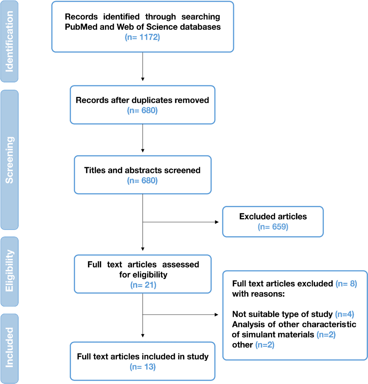
Page MJ, McKenzie JE, Bossuyt PM, Boutron I, Hoffmann TC, Mulrow CD, et al. The PRISMA 2020 statement: an updated guideline for reporting systematic reviews. BMJ. 2021;372:n71.
Article
PubMed
PubMed Central
Google Scholar
Sheth VH, Shah NP, Jain R, Bhanushali N, Bhatnagar V. Development and validation of a risk-of-bias tool for assessing in vitro studies conducted in dentistry: the QUIN. J Prosthet Dent. 2022. https://doi.org/10.1016/j.prosdent.2022.05.019.
Article
PubMed
Google Scholar
Ariji Y, Ariji E, Araki K, Kanda S. CT values of dental materials and preparation of phantoms with CT values equivalent to those of some human soft tissues. Radiat Prot Dosimetry. 1993;49(1–3):129–31.
Article
Google Scholar
Oenning AC, Salmon B, Vasconcelos KdF, Pinheiro Nicolielo LF, Lambrichts I, Sanderink G, et al. DIMITRA paediatric skull phantoms: development of age-specific paediatric models for dentomaxillofacial radiology research. Dentomaxillofac Radiol. 2018;47(2):20170285.
Article
PubMed
PubMed Central
Google Scholar
Lopes PA, Santaella GM, Lima CAS, Vasconcelos KdF, Groppo FC. Evaluation of soft tissues simulant materials in cone beam computed tomography. Dentomaxillofac Radiol. 2019;48(1):20180072.
Article
PubMed
Google Scholar
Santaella GM, Visconti MAPG, Devito KL, Groppo FC, Haiter-Neto F, Asprino L. Evaluation of different soft tissue–simulating materials in pixel intensity values in cone beam computed tomography. Oral Surg Oral Med Oral Pathol Oral Radiol. 2019;127(4):e102–7.
Article
PubMed
Google Scholar
Grehn M, Stille M, Ziemann C, Cremers F, Rades D, Buzug TM. A new phantom for individual verification of the dose distribution in precision radiotherapy for head-and-neck cancer. Anticancer Res. 2019;39(12):6931–8.
Article
CAS
PubMed
Google Scholar
Lee DI, Yang Z, Kim JH, eds. A 3D printing-based realistic anthropomorphic dental phantom and its imaging evaluation. In: Medical Imaging 2019: Imaging Informatics for Healthcare, Research, and Applications; 2019: SPIE
Nascimento EHL, Fontenele RC, Lopes PA, Santaella GM, Vasconcelos KF, de Freitas DQ, et al. Development of a model of soft tissue simulation using ballistic gelatin for CBCT acquisitions related to dentomaxillofacial radiology research. Dentomaxillofac Radiol. 2021;50(3):20200191.
Article
PubMed
Google Scholar
Badiuk SR, Sasaki DK, Rickey DW. An anthropomorphic maxillofacial phantom using 3-dimensional printing, polyurethane rubber and epoxy resin for dental imaging and dosimetry. Dentomaxillofac Radiol. 2022;51(1):20200323.
Article
PubMed
PubMed Central
Google Scholar
Brand JW, Kuba RK, Braunreiter TC. An improved head-and-neck phantom for radiation dosimetry. Oral Surg Oral Med Oral Pathol. 1989;67(3):338–46.
Article
CAS
PubMed
Google Scholar
Schropp L, Alyass NS, Wenzel A, Stavropoulos A. Validity of wax and acrylic as soft-tissue simulation materials used in in vitro radiographic studies. Dentomaxillofac Radiol. 2012;41(8):686–90.
Article
CAS
PubMed
PubMed Central
Google Scholar
De Molon R, Batitucci R, Spin-Neto R, Paquier G, Sakakura C, Tosoni GM, et al. Comparison of changes in dental and bone radiographic densities in the presence of different soft-tissue simulators using pixel intensity and digital subtraction analyses. Dentomaxillofac Radiol. 2013;42(9):20130235.
Article
PubMed
PubMed Central
Google Scholar
Shin JM, Lee C, Kim JE, Huh KH, Yi WJ, Heo MS, et al. Contrast reference values in panoramic radiographic images using an arch-form phantom stand. Imaging Sci Dent. 2016;46(3):203–10.
Article
PubMed
PubMed Central
Google Scholar
Payne T, Mitchell S, Bibb R, Waters M. The evaluation of new multi-material human soft tissue simulants for sports impact surrogates. J Mech Behav Biomed Mater. 2015;41:336–56.
Article
CAS
PubMed
Google Scholar
Cha BK, Jeon S, Seo C-W, Back J, Choi S, Lee C, et al. Optimization of X-ray image acquisition and reconstruction for a C-arm CBCT system with a flat-panel detector. Nucl Instrum Methods Phys Res, Sect A. 2019;924:343–9.
Article
ADS
CAS
Google Scholar
Wu P, Sisniega A, Stayman J, Zbijewski W, Foos D, Wang X et al., eds. Clinical study of soft-tissue contrast resolution in cone-beam CT of the head using multi-resolution PWLS with multi-motion correction and an electronic noise model. In: 15th international meeting on fully three-dimensional image reconstruction in radiology and nuclear medicine; 2019: SPIE
Santaella GM, Visconti M, Devito KL, Groppo FC, Haiter-Neto F, Asprino L. Evaluation of different soft tissue-simulating materials in pixel intensity values in cone beam computed tomography. Oral Surg Oral Med Oral Pathol Oral Radiol. 2019;127(4):e102–7.
Article
PubMed
Google Scholar
Oenning AC, Salmon B, Vasconcelos KF, Pinheiro Nicolielo LF, Lambrichts I, Sanderink G, et al. DIMITRA paediatric skull phantoms: development of age-specific paediatric models for dentomaxillofacial radiology research. Dentomaxillofac Radiol. 2018;47(3):20170285.
Article
PubMed
PubMed Central
Google Scholar

留言 (0)