Cell culture
The MCF7, MDA-MB-231 (MB231) and HEK293T cell lines were procured from the National Centre for Cell Science (NCCS), Pune, India. MCF7 and HEK293T cell lines were cultured in DMEM (Thermo Scientific, USA), while L15 medium (Thermo Scientific) was used to culture MB231 cells. The culture medium was supplemented with 10% FBS (Thermo Scientific) and antibiotics (100 IU/ml penicillin, 100 µg/ml streptomycin, and 0.25 µg/ml amphotericin B; MP Biomedicals, USA). The cells cultured in DMEM were incubated at 37 °C in a humid incubator with 5% CO2 whereas the cells cultured in L15 medium were incubated without CO2. A 5 mM stock of XCT790 in dimethyl sulfoxide (DMSO) was used to treat the cells. The corresponding solvent (DMSO) has been used as vehicle control.
Overexpression and knockdown of ERRα
The human ERRα gene with an N-terminal FLAG was subcloned from pCMV-flag-ERRα (Addgene plasmid 10975) into the pCDH-CMV-MCS-EF1-Puro (pCDH) lentiviral vector (System Biosciences, USA). Additionally, lentiviral human shRNA plasmids (shERRα) and control shRNA plasmid-A (shControl) were purchased from Santa Cruz Biotechnology, USA, (sc-44706; sc-108060) for the knockdown of ERRα. Recombinant lentivirus was generated in HEK293T cells using ViraPower Lentiviral Packaging Mix (Invitrogen, USA) following the manufacturer’s instructions. Lentiviruses produced using pCDH control and pCDH-ERRα were transduced into MCF7 cells for 12 h to produce the control and ERRα overexpressing cells. Similarly, lentiviruses produced using shControl and shERRα plasmids were transduced into MB231 cells to knock down the ERRα. Subsequently, MCF7-pCDH, MCF7-pCDH-ERRα, MB231-shControl and MB231-shERRα cells were selected for puromycin resistance (2.5 µg/ml) for 10 days and maintained in puromycin (0.5 µg/ml) containing medium.
Mammosphere culture and mammosphere forming efficiency (MFE)
The complete mammosphere media, composed of DMEM/F12 with 2 mM L-glutamine, 10 ng/ml bFGF (Peprotech, Israel), 20 ng/ml EGF (MP Biomedicals, USA), 1x B27 (Thermo Scientific, USA), 100 IU/ml penicillin, 100 µg/ml streptomycin and 0.25 µg/ml amphotericin B, was used for mammosphere culture. Briefly, breast cancer cells with ERRα overexpression and knockdown were seeded (20,000 cells per well) in mammosphere media into 6-well ultra-low attachment plates (Corning, USA). For the XCT790 treatment group, the cells were seeded in media containing 2.5 and 5 µM XCT790. The plates were incubated for 7 days to facilitate the formation of mammospheres. Subsequently, the mammospheres were collected and the MFE was calculated by counting the mammospheres with a diameter greater than 40 μm under a microscope. The MFE was expressed as the percentage of mammospheres formed out of the initially seeded cells. For subsequent assays, the collected mammospheres were trypsinized and passed through a 23-gauge needle to dissociate into single cells.
Transwell migration and invasion assays
The mammospheres were generated with XCT790 treatment (2.5 and 5 µM) as well as from the cells with ERRα overexpression and knockdown in mammosphere medium for 7 days. The mammospheres were collected by centrifugation, trypsinized, and then passed through a 23-gauge needle to dissociate into single cells. For the transwell migration assay, 40,000 cells from the mammospheres suspended in serum-free DMEM/F12 were seeded into the upper chamber of 8 μm transwell inserts placed in 24 well plates. The lower chambers of the transwells were filled with DMEM/F12 containing 10% FBS to induce cell migration. After 24 h incubation, the cells in the upper chamber were removed using a cotton swab, and the transwell membrane was fixed with 70% ethanol for 15 min. Subsequently, the membrane was washed with PBS and stained with Giemsa for 30 min. Images of the cells that had migrated to the lower surface of the membrane were captured and then counted.
For the invasion assay, 50 µl Matrigel (Corning, USA) was added to the upper chamber of the 8 μm transwell and allowed to solidify for 30 min. Then, the single-cell suspension of the mammospheres (40,000 cells/well) in 200 µl of serum-free DMEM/F12 medium was plated on top of the Matrigel layer. The lower chambers of the transwells were filled with DMEM/F12 containing 10% FBS and incubated for 24 h. Then, the cells from the upper chamber, along with the matrigel, were carefully removed. Subsequently, the membrane was stained with Giemsa, and the cells that had invaded the lower surface of the membrane were imaged using a microscope.
Chorioallantoic membrane (CAM) assay
The CAM assay is commonly utilized for investigating the angiogenic potential. The assay was conducted following the protocol outlined by Naik et al. [19] with some modifications. Experimentation on chick embryos up to embryonic day 15 does not require approval from an animal ethics committee as the embryos younger than embryonic day 16 are assumed to be unable to experience pain [20, 21]. In brief, the mammospheres were cultured from both MCF7 and MB231 cells with XCT790 treatment as well as from the cells with ERRα overexpression and knockdown. After 7 days of mammosphere culture, the respective conditioned media was collected and concentrated using a concentrator. For CAM assay,100 µg of protein was used after measuring the protein content using BCA assay (Thermo Scientific, USA). The 48 h fertilized eggs were acquired from the Central Poultry Development Organisation (CPDO), Bhubaneswar, India. The eggs were cleaned with 70% ethanol, and incubated for an additional 24 h at 37 °C. At 72 h post-fertilization, a sterile opening was created in the eggshell. The autoclaved filter paper disc (5 mm in diameter) soaked with the respective concentrated conditioned media (100 µg protein) was gently positioned over the CAM. The steady development in vascularity was observed over 48 h and the images were captured. Subsequently, the photographs were analyzed by using the AngioTool software, and the results were plotted in the graph.
Flow cytometric analysis of CD44 expression
Mammospheres were cultured from both MCF7 and MB231 cells with XCT790 (2.5 and 5 µM) treatment as well as with ERRα overexpression and knockdown. The mammospheres were trypsinized and dissociated into single cells. After a 30-min incubation with CD44-FITC antibody (Biolegend, USA) in the binding buffer, the cells were washed and the cell surface expression of CD44 was assessed using a flow cytometer (BD FACSCanto II).
Western blotting
The mammospheres were collected by centrifugation and lysed using lysis buffer containing NaCl (150 mM), NP-40 (1%), Tris (25 mM, pH 7.5), sodium deoxycholate (1%), EDTA (1 mM), SDS (0.1%), phosphatase inhibitors and protease inhibitor cocktail. The protein content of the lysates was quantified using the BCA method. The proteins (20 µg) were separated on SDS PAGE, electrotransferred to a PVDF membrane and blocked. The membranes were incubated overnight at 4 °C with primary antibodies, followed by 1 h incubation with appropriate HRP-conjugated secondary antibodies and washing with TBST. The enhanced chemiluminescence (ECL) method was used to develop the membranes. The primary antibodies and their respective dilutions are itemized in Table S1.
Statistical analysis
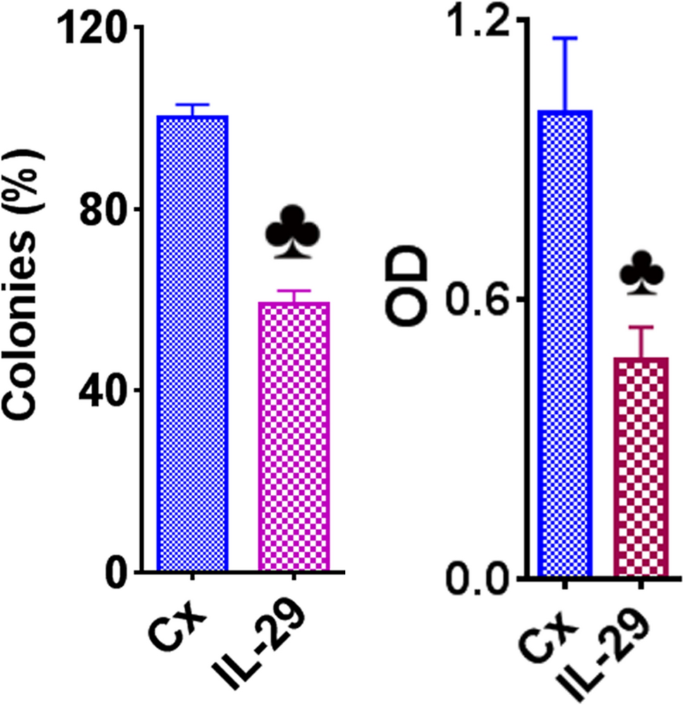
The experiments were performed three times, and the findings were presented as mean ± SD. Statistical differences were assessed using the student’s t-test, and those with p-values of < 0.05, < 0.01 and < 0.001 were considered significant differences and were given corresponding symbols in the graphs.

留言 (0)