
記住我
In our study, we acquired 48 seven-week-old male Sprague–Dawley (SD) rats, each weighing between 220 and 270 g, from the Experimental Animal Centre of Chong Qing Medical University. These rats were maintained in a specific pathogen-free (SPF) laboratory environment. Our experimental procedures were thoroughly reviewed and approved by the Ethics Committee for Animal Research at the School and Hospital of Stomatology, Chong Qing Medical University, China, with the given approval number: Ethics Review 2022 (119).
Establishment of a CFA-Induced TMJ OA ModelAfter a one-week isolation period, we randomly divided the rats into 8 groups, with each group consisting of 6 SD rats. The first group received saline injections in the temporomandibular joint (TMJ). Groups two to five had CFA injected into the TMJ, with subsequent observations at 1, 3, 7, and 14 days, respectively. The sixth group underwent a CFA injection in the TMJ and a simultaneous saline injection in the trigeminal ganglion. The seventh group received CFA in the TMJ and CGRP in the ganglion. The eighth group was administered CFA in the TMJ and CGRP 8-37 in the ganglion. For the administration of drugs via ganglion injection, after establishing an arthritis model in SD rats through CFA injection into the temporomandibular joint, drugs were administered into the trigeminal ganglion of the rats. Anesthesia was administered using 1.5% isoflurane in oxygen, sourced from Abbott Laboratories, North Chicago, IL, USA. Following the established protocol (Kameoka et al. 2010), a 27-gauge 0.5-inch needle was precisely inserted at the anterosuperior portion of the zygomatic arch root of the rat. We then administered a bilateral slow injection of 50 µL of either saline or CFA (product number F5881 from Sigma-Aldrich, USA) into the upper compartment of the TMJ.
Intra-Ganglionic Drug AdministrationIn the CFA-injected group, rats were categorized into three subgroups for trigeminal ganglion injection: CGRP, CGRP 8-37, and normal saline, with 6 rats in each subgroup. We prepared the solutions of rat CGRP (10−5 M, MCE) and CGRP 8-37 (a CGRP antagonist, 10−5 M, MCE), both at a concentration of 10–5 M, in normal saline(Afroz et al. 2019). These were aliquoted and stored at − 20 °C in accordance with the manufacturer's guidelines. For TG intra-ganglionic (IG) drug delivery, we followed the previously published protocols (Neubert et al. 2005). A 26-gauge Hamilton syringe with a 10 µL capacity was used for IG administration. The entry point for the needle was the infra-orbital foramen, positioned 1 mm medial to the zygomatic process of the maxilla. We inserted the needle approximately 22 mm, angling it toward the midline by roughly 10 degrees and 15 degrees downward from the plane of the parietal bone (Villa et al. 2010). This procedure allowed the needle to pass through the infraorbital canal and reach the ipsilateral trigeminal ganglion located in Meckel’s cave. To verify the accuracy of our injections, methylene violet was injected to visualize the trigeminal ganglion (Fig. 1E).
Fig. 1
Temporomandibular Arthritis Pain Response in Rats. A Overview of the experimental setup. B Rat pain response post-CFA induction. C Behavioral outcomes following CGRP injection in the trigeminal ganglion. D Effects of CGRP 8-37 antagonist on pain behavior. E Illustration of drug administration targeting the trigeminal area. Data are presented as mean ± SD. The t-test was used for analysis; n = 6. Number of technical replicates (NTR) = 3. Significance: *p < 0.05, **p < 0.01, ***p < 0.001
Behavioral AssessmentTo assess mechanical allodynia in inflamed animals, we implemented a previously established protocol (Ren 1999), incorporating minor modifications for our specific needs. The study began by acclimatizing the rats to the test environment for one week. This was followed by a seven-day behavioral training period prior to establishing the temporomandibular arthritis model. During this phase, SD rats were placed unrestrained in a glass box situated in a quiet room, allowing them to adapt for 30 min. The next step involved measuring the pain threshold in the preauricular temporomandibular joint area of the SD rats using Von Frey filaments (Biological Instruments, North Coast Medical, America). Notably, while the rats’ heads were not restrained during the measurement, their overall movements were still limited. This procedure was focused on testing the orofacial skin regions adjacent to the center of the temporomandibular joint on both sides. Von Frey filaments were specifically employed to assess the TMJ mechanical sensitivity, with measurements conducted at 1, 3, 7, and 14 days post-injection. For mechanical sensitivity testing, Von Frey filaments were applied in ascending order, starting with a 2 g filament for control animals and a 0.16 g filament for inflamed animals. Each filament was applied five times at intervals of a few seconds, and the response threshold was determined by the minimal force that induced three or more head withdrawal reactions during the five applications. We maintained a consistent 2-min interval between subsequent filament applications. All testing procedures were conducted in a tranquil setting to preclude the influence of diurnal variations.
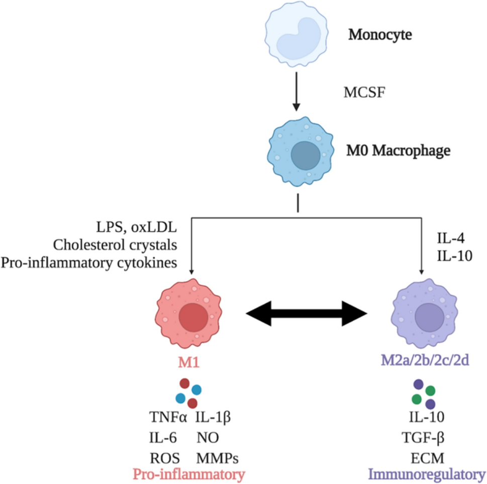
Cell Culture and α-CGRP TreatmentThe THP-1 human monocytic leukemia cell line, authenticated by short tandem repeat spectrometry, was acquired from Procell Life (Wuhan, China). It was cultured in RPMI 1640 medium (Procell Life, Wuhan) supplemented with 10% heat-inactivated fetal calf serum (FCS), and the cells were maintained at 37 °C in a humidified atmosphere containing 5% CO2. After cultivating, the THP-1 cells underwent centrifugation at 800 RPM/min for 5 min. Post centrifugation, the supernatant was removed, and the cell density was adjusted to 1 × 10⁶/mL using fresh culture medium. Subsequently, Phorbol ester (PMA) was introduced to the cell suspension until it reached a concentration of 100 ng/ml. Following gentle mixing, the cell suspension was allocated into 6-well plates, with each well containing 2 ml. These plates were then incubated under controlled light exposure. After a 48-h treatment with PMA, a notable change was observed in the cells: they transitioned from being in suspension to displaying protruding pseudopods as adherent cells. This transformation was confirmed under a microscope. Eventually, the THP-1 cells differentiated into M0 macrophages, which were then cleansed twice with sterile PBS. To understand the impact of CGRP on the inflammatory response of these macrophages, cells were pre-treated with CGRP (10−6 M, MCE) for 30 min. Thereafter, they were either cultured in a medium containing 50 ng/mL LPS (MCE) + 20 ng/mL IFN-γ(MCE) for 24 h or in another medium with 20 ng/mL IL-4(MCE) + 20 ng/mL IL-13(MCE), specifically for a 72-h period targeting M0 macrophages.
Tissue PreparationRats were euthanized using an overdose of isoflurane for a humane endpoint. We meticulously extracted the temporomandibular joint tissues, including the mandibular condyle disc, retro-discal area, fossa, and the trigeminal ganglion, from each rat across all groups. These tissues were then fixed in 4% paraformaldehyde for a 24-h period. For histopathological evaluation, the TMJ specimens were subjected to demineralization in a 19% solution of EDTA, with the solution being refreshed every three days. Meanwhile, TG tissues were prepared for dehydration by immersion in 30% sucrose solution overnight.
Quantitative Real-Time Polymerase Chain Reaction (q RT-PCR)Total RNA was isolated from cells utilizing the RNAiso Plus reagent (TaKaRa), which was then reverse-transcribed into cDNA with TaKaRa’s reverse transcription kit. The quantitative real-time PCR (qRT-PCR) was performed using SYBR Green (TaKaRa) as the DNA-binding dye, on the 7500 Fast Real-Time PCR System (Applied Biosystems). For normalization purposes, the expression of β-actin mRNA was used as a reference against the expression of the target mRNA. For each cell sample, three replicate wells were set up to eliminate technical errors in the experiment. The specific sequences for the PCR primers utilized in this investigation were shown below: CD86 F_5ʹ-GTTTCATTCCCTGATGTTACGAG-3ʹ R_5ʹ-GAGAAAGGTGAAGATAAAAGCCG-3ʹ, CD206 F_5ʹ-GCTGAAAGGT. GACCCTACTATGT-3ʹ R_5ʹ-GCTCAGGTTTTGGTGTTTGTC-3ʹ.
HistologyDemineralized TMJs from two groups (CFA-treated, n = 6 joints; Saline-treated, n = 6 joints) were embedded in paraffin and cut into 5 µm thick mid-sagittal sections. These sections were then dewaxed, rehydrated, and triple-washed with PBS to prepare for antigen retrieval. For this process, the slides were immersed in sodium citrate (pH 6.0) and heated to 95 ℃ for 20 min. Following antigen retrieval, the sections were washed in PBS for 10 min and blocked with 10% bovine serum albumin for one hour at room temperature. Subsequently, these tissues were exposed to primary antibodies: CD86 (Huabio, ER1906, 1:400) and CD206 (CST, 24595, 1:500) and incubated overnight at 4 ℃. Upon completion, after three PBS washes, the sections were introduced to a horseradish enzyme-labeled streptavidin solution (Abcam; dilution, 1:500) for an hour at room temperature. The final step involved incubation in diaminobenzidine (DAB, Sigma-Aldrich) for coloration and a counterstain using hematoxylin. In this experiment, the immunohistochemistry assay was performed in triplicate on joint tissue from each identical biological sample, with the results being averaged across these technical replicates.
Immunofluorescence Staining (IF)TG tissues were prepared by embedding them in an optimal cutting temperature compound (OCT) (Sakura, USA) and then sectioning them serially at a thickness of 10 μm. First, the specimens underwent a 10-min wash using phosphate-buffered saline (PBS). This was followed by antigen retrieval, which entailed heating the specimens in citrate using a microwave oven for 25 min. To prevent nonspecific binding, we treated the tissues with 5% bovine serum albumin (BSA) for 1 h at 37 ℃. Subsequently, the tissues were incubated with primary antibodies: CGRP (CST, ab81887, 1:1000), CD206 (CST, 24595, 1:500), and CD86 (ABclonal, A1199, 1:100) overnight at 4 ℃. As a next step, the tissues were treated with goat anti-rabbit Alexa Fluor 488 (CST, 4412s, 1:1000) secondary antibodies for 1 h at 37 ℃. For cell nuclei visualization, DAPI (Beyotime, C1006, 1:100) was added, with a 5-min incubation period. Images were captured using a fluorescence microscope (Carl Zeiss, Baden-Wurttemberg, Germany). Within the TG, enumerations of both CGRP-positive neurons and total neurons, which were labeled with DAPI, were conducted across five fields of each section. Two independent evaluators performed these counts, using a 20 × objective lens for CGRP-labeled immunofluorescence, and a 40 × objective lens for CD86 and CD206 labeled immunofluorescence. The data was assessed based on the count of positive cells and the intensity of immunofluorescence. In the technical replicates for both immunofluorescence and immunohistochemistry experiments, this study employed a consistent procedure, where the results for each biological sample were derived from the average of three identical assays. Image J software was employed for all immunofluorescence data analyses.
Statistical AnalysesStatistical analyses were conducted utilizing GraphPad Prism version 9.5 and SPSS version 25. All results are expressed as mean ± standard deviation (SD). We employed one-way analysis of variance (ANOVA) followed by Dunnett’s t-test to compare means, assuming normality and homogeneity of variances across samples. For all identical experiments, the number of technical replicates (NTR = 3) is three to exclude technical errors. Levels of statistical significance were denoted as follows: *p < 0.05, **p < 0.01, ***p < 0.001, and ****p < 0.0001.
留言 (0)