Animals
Heterozygous App+/NL−G−F mice (C57BL/6-App < tm3(NL-G-F)Tcs >), carrying the App gene with humanized Aβ sequence (G676R, F681Y, and R684H), Swedish (KM670/671NL), Beyreuther/Iberian (I716F), and Arctic (E693G) mutations, were previously established by a knock-in strategy [27]. Homozygous AppNL−G−F/NL−G−F mice were obtained by crossbreeding. Genotyping of mice was performed as previously described Age-matched wild‐type control mice (C57BL/6J) were purchased from CLEA Japan (Tokyo, Japan). Nrf2 knockout mice (B6.129P2-Nfe2l2 < tm1Mym > /MymRbrc, RBRC01390) were obtained from the RIKEN BioResource Research Center (Tsukuba, Japan) with the kind permission of Dr. Masayuki Yamamoto (Tohoku University) [28] All mice were maintained under a standard specific pathogen-free environment (12 h light–dark cycle; 23 ± 1 ºC; 50 ± 5% humidity) with free access to food and water throughout the experiments. The animals were treated in compliance with the guidelines established by the Institutional Animal Care and Use Committee of Nagoya University.
Isolation of microglia and astrocytes from the mouse brain
Microglia and astrocytes were isolated from the cerebral cortices of mice using magnetic-activated cell sorting (MACS) as described previously [29]. In brief, after mice were transcardially perfused with phosphate-buffered saline (PBS) under deep anesthesia, the cerebral cortex was dissociated at 37 °C for 15 min using the Neural Tissue Dissociation Kit-Postnatal Neurons (Miltenyi Biotec, Bergisch-Gladbach, Germany) with a gentle MACS Dissociator (Miltenyi Biotec). To isolate microglia, we removed myelin debris using Myelin Removal Beads II (Miltenyi Biotec) and incubated purified cells with anti-CD16/CD32 antibodies (Thermo-Fisher Scientific, Waltham, MA, USA) for blocking Fc receptors, followed by incubation with anti-CD11b MicroBeads (Miltenyi Biotec). By MACS, CD11b-positive microglia were isolated through an LS column (Miltenyi Biotec). To isolate astrocytes, astrocyte-containing, CD11b-negative flow-through cells were incubated with anti-ACSA2 MicroBeads (Miltenyi Biotec), and then subjected to MACS through the LS column.
Chronic oral administration of dimethyl fumarate
DMF (Sigma, CA, USA) was dissolved in corn oil and administered by oral gavage (300 mg/kg, p.o) [30]. We administered DMF orally three times per week for 1 month (short-term administration) and 5 months (long-term administration). To minimize bias because of possible undetected changes in environmental conditions, Veh- or DMF-administered wild-type (WT)/App mice were always examined in pairs; both recordings were performed on the same day. Therefore, the experimenter was not blinded to the genotype/treatment throughout the experimental procedures. No exclusion criteria were predetermined, and no animals were excluded. The sample size for each experiment was determined based on previous studies with the relevant type of experiment [29, 31].
Behavioral experiments
The novel object recognition test was performed as described previously [31] with minor modifications. The mice were habituated to an open box (30 × 30 × 35 cm) individually for 3 days. During the training, two novel objects were placed in an open field. Under moderately lit conditions (12 lx), mice were allowed to explore for 10 min and the time spent exploring each object was recorded. During the test sessions, one of the familiar objects used in the training session was replaced by a novel object. The mice were placed back into the same box 24 h after the training session and allowed to explore freely for 5 min. The preference index in the test session, which is the ratio of the duration of time spent exploring the novel object to the total time spent exploring both objects, was used to evaluate cognitive function.
An open field test was performed for measuring exploratory behavior and anxiety in a novel environment according to the manufacturer’s protocol (O’Hara & Co., Ltd., Tokyo, Japan). The mice were placed in the center of an empty open field (40 × 40 × 30 cm) and allowed to explore the environment for 20 min without any external disturbances. TimeOFCR1 software (O’Hara & Co., Ltd.) was used to measure the movement patterns, time spent in the center and margin, velocity, and distance traveled; these measurements were used as indicators of anxiety, agility, and exploration.
Quantification of mRNA levels using real-time PCR
According to the manufacturer’s instructions, we used the RNeasy Micro Kit (Qiagen) to extract total RNA from cultured astrocytes or MACS-isolated microglia and astrocytes. Complementary DNA (cDNA) from MACS-isolated cells was generated by reverse transcription of total RNA (2.5 or 5 ng) using the PrimeScript™ RT reagent Kit (Perfect Real Time) (TaKaRa Bio, Kusatsu, Japan), and 1/50 of the yield was amplified using the SYBR Premix Ex Taq II (Tli RNaseH Plus) (TaKaRa Bio) and the Thermal Cycler Dice Real Time System II or III (TaKaRa Bio). The PCR protocol was as follows: 1 cycle at 95 °C for 30 s; 40 cycles at 95 °C for 5 s and 60 °C for 30 s; and a dissociation stage at 95 °C for 15 s, 60 °C for 30 s, and 95 °C for 15 s. Actb was used for normalization. The primers used for real-time RT-PCR are summarized in Additional file 1: Table S1.
Immunofluorescence analysis
Immunofluorescence analysis was performed as described previously [29]. In brief, mice were deeply anesthetized and perfused intracardially with PBS and 4% paraformaldehyde in PBS. The brains were dissected, postfixed with the same fixative, and cryoprotected with 30% sucrose containing PBS. Twenty-micrometer-thick coronal brain sections were fixed with 4% paraformaldehyde in PBS for 5 min, and then we performed antigen retrieval with HistoVT One (Nakarai, Kyoto, JAPAN) 70 °C for 20 min. After incubation in blocking solution (5% goat or donkey serum/PBS) for 1 h, sections were incubated with a combination of the following antibodies: rabbit anti-Iba-1 (#019–19741, 1:500; FUJIFILM Wako Osaka, Japan), goat anti-AIF-1/Iba1 (#NB100-1028, 1:250; Novus Biologicals, CO, USA), mouse anti-Aβ (#10326, 1:200; IBL, Nagoya, Japan), mouse anti-GFAP (#G3893, 1:250, Sigma), rat anti-C3 (#sc58926, 1:200; Santa Cruz Biotechnology, Inc., CA, USA), rabbit anti-BACE1 (#5606, 1:100, Cell Signaling, Inc., CA, USA), and rabbit anti-p-STAT3 (Tyr705) (#9145, 1:2000, Cell Signaling Technology, Inc.) at 4 °C overnight. After washing with PBS, the sections were incubated with fluorescent-conjugated anti-rabbit, anti-mouse, anti-goat, or anti-rat IgG (1:1000; Thermo Fisher Scientific) and 4ʹ,6ʹ-diamidino-2-phenylindole (DAPI) (1:2000) at room temperature for 1 h. After washing, the sections were mounted on slides with Fluoromount/Plus™ (Diagnostic BioSystems, Pleasanton, CA, USA) and analyzed using a confocal microscope (LSM700, Carl Zeiss, Oberkochen, Germany). Microscopic images were quantitatively analyzed using ImageJ (National Institutes of Health).
Protein extraction and immunoblotting
The half brain hemisphere of mice was homogenized in RIPA buffer (tris-buffered saline (TBS) with 1% NP-40, 1% sodium deoxycholic acid, 0.1% sodium dodecyl sulfate (SDS), complete protease inhibitor cocktails (Roche), and PhosSTOP phosphatase inhibitor cocktails (Roche)) and centrifuged at 20,000 × g for 30 min. The resulting supernatant was used as the RIPA–soluble fraction. The remaining brain hemisphere was used for Aβ extraction as described elsewhere [32]. In brief, mouse brains were homogenized in 5 × volumes of TBS (50 mM Tris–HCl pH7.6, 150 mM NaCl, complete protease inhibitor cocktail, and PhosSTOP phosphatase inhibitor cocktail) with 25 strokes using a potter homogenizer and centrifuged at 200,000 × g at 4 °C for 20 min. The resulting supernatant was collected as a TBS-soluble fraction. After the addition of the same amount of 2% Triton X-100/TBS, the pellet was homogenized on ice and centrifuged at 200,000 × g at 4 °C for 20 min. The resulting supernatant was collected as the Triton X fraction. Then, the same amount of 2% SDS containing TBS was added to the pellet. After homogenization at room temperature, the pellet was incubated at 37 °C for 2 h and centrifuged at 200,000 × g at 20 °C for 20 min. The resulting supernatant was collected as the SDS fraction. Finally, the pellet was sonicated with 500 μL of 70% formic acid (WAKO). The samples were centrifuged at 200,000 × g at 4 °C for 20 min, and the resulting supernatant was evaporated for 2 h. The pellet was dissolved in the same volume of dimethyl sulfoxide (DMSO) as the brain weight and stored at − 80 °C until use. To determine the protein concentration, the BCA assay was used. Equal amounts of total protein were separated using SDS–polyacrylamide gel electrophoresis (SDS-PAGE) and transferred to a polyvinylidene difluoride membrane (Immobilon-P; Merck Millipore, Billerica, MA, USA). The membrane was incubated with a blocking buffer (50 mM Tris–HCl (pH7.4), 150 mM NaCl, 0.05% (v/v) Tween-20, and 2% (w/v) bovine serum albumin (FUJIFILM Wako), followed by incubation with mouse anti-Aβ (#10323, 1:200; IBL, Nagoya, Japan), mouse anti-ACTB (#a5441, 1:5000; Sigma), rabbit anti-human APP (C) (#18961, 1:5000; IBL, Nagoya, Japan), mouse anti-GFAP (#G3893, 1:250, Sigma), rabbit anti-p-STAT3(Tyr705) (#9145, 1:2000, Cell Signaling Technology Inc., CA, USA), rabbit anti-Nrf2 (#137550, 1:1000, abcam, Cambridge, UK), rabbit anti-SOD1 (#ab_10616253, Enzo Life Science, Farmingdale, NY, USA), or rabbit anti-fibrillarin (C13C3) (#2639, 1:5000, Cell Signaling, Inc, CA, USA) antibodies diluted in the blocking buffer at 4 °C for at least 6 h. The membrane was further incubated with the corresponding horseradish peroxidase-conjugated secondary antibodies and visualized using Immobilon Crescendo Western HRP substrate (Merck Millipore). Images were obtained using LAS-4000 mini (Cytiva, Shinjuku, Tokyo, Japan) with the equipped software (Multi-Gauge; Cytiva). Silver staining of SDS–polyacrylamide gel was performed according to the manufacturer’s instructions (FUJIFILM Wako).
Cell culture and drug treatment
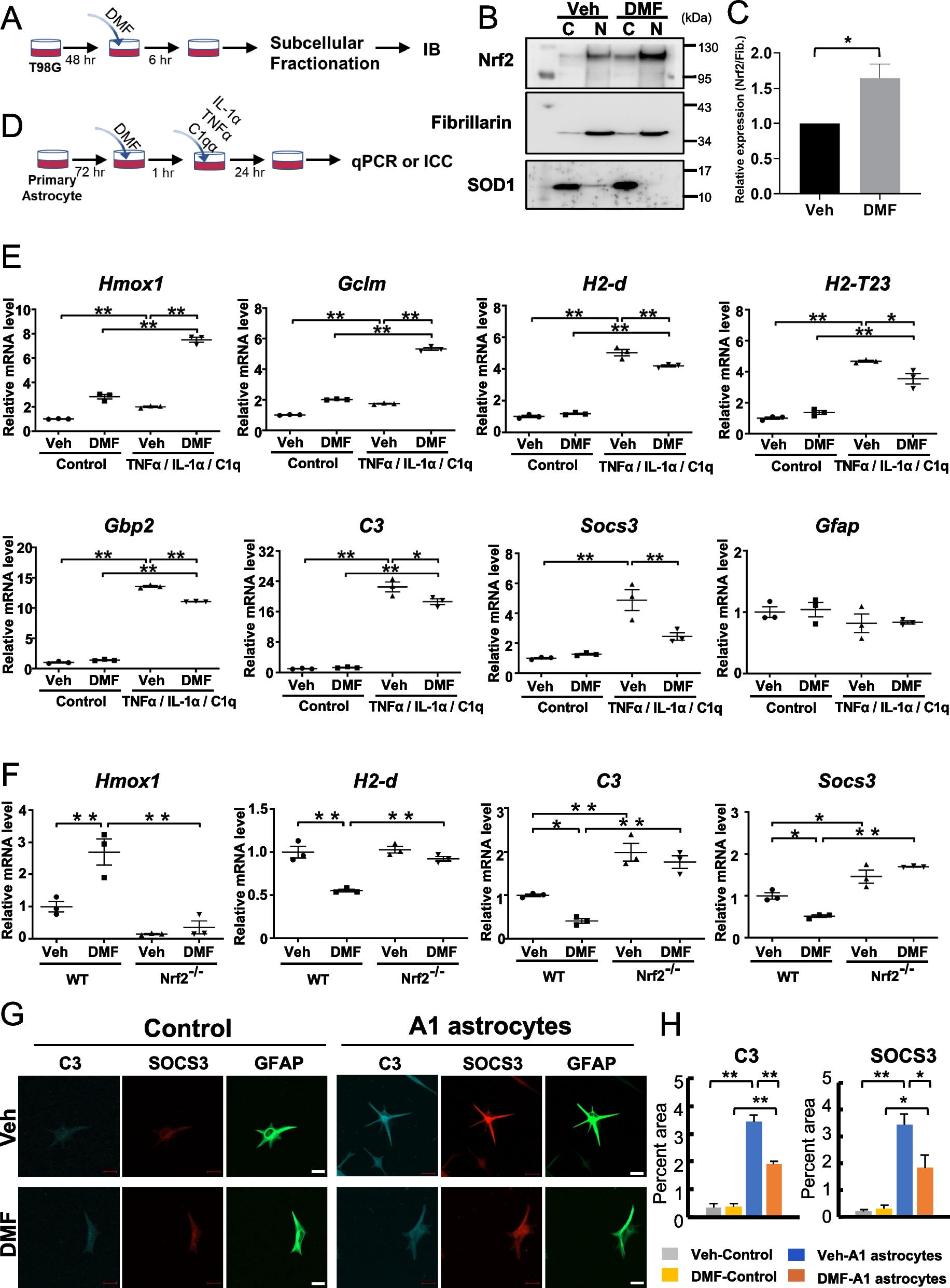
Mouse primary astrocytes were prepared as described previously with minor modifications [33]. Briefly, primary astrocytes were prepared from the brains of newborn (postnatal days 0–3) C57/BL6J or Nrf2 knockout mice. The brain was isolated and carefully stripped off the meninges in PBS. The tissue was minced into small pieces, digested by incubation with 0.25% trypsin and 0.01% DNase at 37 ºC for 10 min, and mechanically dissociated by gentle pipetting. Debris was removed by passing the samples through a 70 μm pore cell strainer. The resulting cells were plated onto a poly-L-lysine (Sigma)-coated flask (25 cm2), and cultured in glial medium (Dulbecco’s Modified Eagle’s Medium (DMEM; Sigma) containing 10% (v/v) fetal bovine serum (Gibco, Gaithersburg, MD, USA). The culture medium was replenished after 24 h and incubated for 7 days. The astrocyte layer was detached using 0.25% trypsin EDTA (FUJIFILM Wako) and replated on culture plates. After replating twice, the primary astrocytes were used in all experiments. We confirmed that more than 95% of the cells were ALDH1L1-positive and Iba1-negative by immunostaining (ALDH1L1 (+): 98%, IBA1 (+): 0% and ALDH1L1 (−) / IBA1 (−): 2%). Primary astrocytes and the human glioma cell line T98G (RRID: CVCL_0556) were cultured in glial medium at 37 °C, 5% CO2 in air, and 95% humidity. Primary astrocytes were pretreated with 35 μM DMF or DMSO (vehicle; Veh) for 1 h followed by treatment with TNFα (30 ng/mL; Peprotech, Hartford, CT, USA), IL-1α (3 ng/mL; Peprotech, Hartford, CT, USA), and C1q (400 ng/mL; Hycult Biotech, PB Uden, The Netherlands) for 24 h as indicated [34]. The T98G cell line was treated with DMF (0 and 35 μM) for 6 h [35]. Treated cells were analyzed using real-time PCR or immunoblotting.
Immunocytochemistry
Immunostaining was performed as previously described [31] with minor modifications. Astrocytes were seeded at a density of 7.0 × 10⁴ cells/well on LabTek II 4-well chamber slides (Thermo Fisher Scientific) coated with poly-D-Lysine (Sigma). The cells were fixed with 4% (w/v) paraformaldehyde for 20 min at room temperature, permeabilized with PBS containing 0.5% (v/v) Triton X-100 for 30 min., incubated in a blocking buffer (PBS containing 0.3% (v/v) Triton X-100 and 5% goat/donkey serum) for 1 h, and incubated with rat anti-C3 (#sc58926, 1:200; Santa Cruz Biotechnology, Inc, CA, USA), mouse anti-GFAP antibody (#G3893, 1:5000, Sigma) and rabbit anti-SOCS3 antibody (#ab16030,1:500, abcam), or mouse anti-ALDH1L1 antibody (sc100497, 1:50, Santa Cruz) diluted in blocking buffer at 4 °C for overnight. After washing three times with PBS containing 0.3% (v/v) Triton X-100, the slides were further incubated with Alexa-conjugated secondary antibodies and 0.5 μg/mL DAPI diluted in the blocking buffer for 60 min at room temperature, followed by embedding with Fluoromount/Plus (Diagnostic BioSystems). Confocal images were obtained using a confocal laser scanning microscopy (LSM-700; Carl Zeiss AG, Oberkochen, Baden-Württemberg, Germany) and analyzed using the equipped software (ZEN; Carl Zeiss AG). Microscopic images were quantitatively analyzed using ImageJ (National Institutes of Health).
Subcellular fractionation
Subcellular fractionation was performed as previously described [36]. In brief, the cells were gently homogenized (Potter homogenizer, Wheaton) in isotonic buffer (10 mM HEPES, 250 mM sucrose, pH 7.4) supplemented with complete protease inhibitor and centrifuged at 600 × g at 4 °C for 5 min. The pellet was collected as P1. The supernatant was further centrifuged at 10,000 × g for 30 min at 4 °C. The supernatant was collected as the cytoplasmic fraction. The P1 pellet was resuspended in ice-cold hypotonic buffer (10 mM HEPES, 10 mM KCl, 1 mM MgCl2, 0.5 mM dithiothreitol (DTT), pH 7.4) and centrifuged at 600 × g at 4 °C for 5 min. The pellet was resuspended in ice-cold hypertonic buffer (10 mM HEPES, 400 mM NaCl, 1 mM MgCl2, 0.2 mM EGTA, 30% glycerol, 0.5 mM DTT, pH 7.4) and agitated at 4 °C for 30 min. After centrifugation at 18,000 × g at 4 °C for 30 min, the supernatant was collected as a nuclear fraction. The protein concentration of each sample was determined using a Bradford protein assay kit (Bio-Rad, Richmond, CA, USA).
Statistical analysis
For behavioral experiments and quantitative PCR, all data are expressed as means ± SEM. The normality of the data was not assessed before the statistical comparisons. We opted to use parametric statistics for consistency across experiments and provided evidence that analysis of variance (ANOVA) is robust to slight non-normality [37, 38]. No test for outliers was performed. One-way or two-way ANOVA with or without repeated measures was used, followed by Tukey’s test when F ratios were significant (p < 0.05). Significant differences between the two groups were determined using the Student’s t-test. Detailed information on the statistical analysis is summarized in Additional file 2: Table S2.


留言 (0)