Programmed death 1/programmed death-ligand 1 (PD-1/PD-L1) targeting therapy is a key strategy in gastric cancer treatment, and several monoclonal antibodies targeting PD-1/PD-L1 have already been widely used clinically [1,2]. Nevertheless, increasing evidence has been reported that response rate of anti-PD-1/PD-L1 immunotherapy is only 10 %–30 % in clinics when the amount of PD-L1 expression was used as a biomarker for PD-1/PD-L1 targeting therapy [[3], [4], [5]], while some key publications suggested that exosomal PD-L1 can contribute to the immunosuppression and is associated with anti-PD-1 response [4,[6], [7], [8], [9]]. Hence, decreasing the abundance of exosomal PD-L1 is a new strategy to sensitize the PD-1/PD-L1 targeting therapy.
Exosomes are small, single-membrane, secreted organelles of 30–200 nm in diameter that have the same topology as the cell and are enriched in selected proteins, lipids, nucleic acids, and it can be released in diverse physiological and pathological conditions [[10], [11], [12]]. As the main communicator among different cells, exosomes are formed as intraluminal vesicles (ILVs) in late endosomal organelles called multivesicular bodies and secreted after fusion of multivesicular bodies (MVBs) with the plasma membrane [13]. Several exosome biogenesis processes have been identified, including capture of ubiquitinated cargoes by the endosomal sorting complex required for transport (ESCRT) machinery [14,15]. As a member of the tetraspanin superfamily, CD63 is highly enriched on ILVs in late endosomal MVBs, and is widely used as a classic exosomal marker [16]. Abrogation of CD63 by CRISPR/Cas9 impairs secretion of exosomes, suggesting that it contributes to exosome biogenesis [17]. Besides, CD63 has also been used to label and track exosomes in many studies [[18], [19], [20]]. Hence, CD63 can be used as a marker to quantify exosomes.
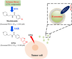
In the present study, using an antibody to recognize CD63 and an aptamer to recognize PD-L1, a homogeneous time resolved fluorescence (HTRF) based assay to quantify CD63 and PD-L1 double positive signaling was established using cell medium as the sample. In this assay, signaling of CD63 was considered to indicate the amount of exosomes, while signaling of PD-L1 was considered to indicate the amount of exosomal PD-L1. With the aid of this phenotype-based assay, our in-house compound library containing 2791 compounds was applied for the screening, and moclobemide was characterized to inhibit the generation of exosomal PD-L1 with an IC50 = 4.363 μM. To obtain more potent exosomal PD-L1 inhibitor, a series of biphenyl-based compounds based on moclobemide were designed and synthesized, and EP-16 was the most active one that can inhibit the generation of exosomal PD-L1 with IC50 = 0.108 μM. Additional study further confirmed that EP-16 can inhibit the generation of exosomal PD-L1 in cells, leading to activation of T cells as well as the sensitization of gastric cancer cells to anti-PD-1 promoted T-cell killing in vitro and in vivo. Our findings give a proof-of-concept that exosomal PD-L1 can be used as a target to sensitize the gastric cancer to T-cell killing, and EP16 may serve as a lead compound to promote the response of PD-1/PD-L1 targeted therapy.

留言 (0)