Cell culture
The MV4-11, Kasumi-1 human AML cell lines were obtained from the Chinese Academy of Sciences Cell Bank, and authenticated through Short Tandem Repeat (STR) analysis in 2023. Cells were cultured in RPMI1640 medium (22400089, Gibco, USA) supplemented with 10% fetal bovine serum (Biological Industries, CT, USA), and 1% penicillin–streptomycin (Millipore Sigma, MA, USA), cultured in a humidified incubator at 37 ℃ containing 5% CO2, and routinely tested for mycoplasma.
ChIP-Seq data collection and analysis
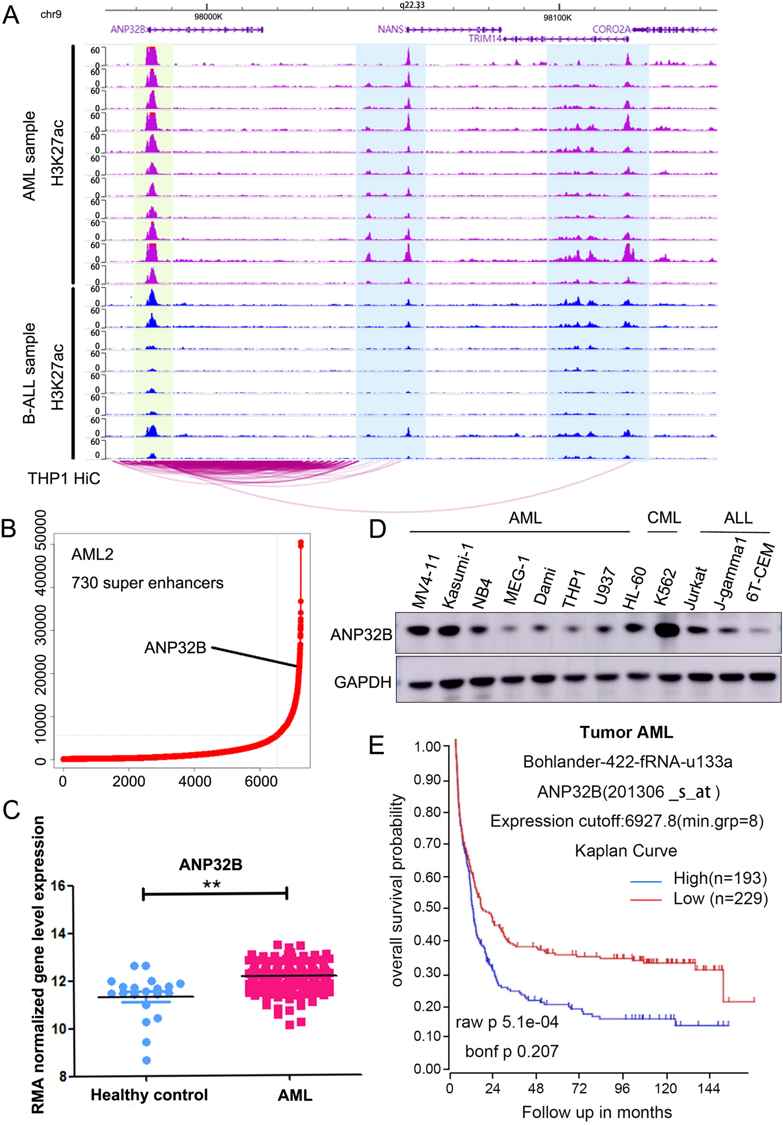
In the present study, the raw data of our previous ChIP-Seq datasets for AML samples (GSE188605) were aligned to UCSC hg38 (the reference genome) using Bowtie2 (v 2.3.5) [19], with the following parameters -p 4 -q -x. Peaks were identified using MACS2 (v2.0.9) [20], applying the parameters -g hs -n test -B -q 0.01. The bigwig files of these datasets were then visualized using Integrative Genomics Viewer (IGV) [21] and WashU tool (http://epigenomegateway.wustl.edu/browser/).
Public Hi-C data collection and analysis
The Hi-C data for THP-1 cell line (GSE126979) was obtained from the Gene Expression Omnibus database. Read mapping and loop calling were performed using HiC-pro (v.3.1.0) [22]. MboI restriction sites in the hg38 build were used for alignment with Bowtie2 as the mapping tool, specifying global options of –very-sensitive -L 30 –score-min L, − 0.6, − 0.2 –end-to-end –reorder and local options of –very-sensitive -L 20 –score-min L, − 0.6, − 0.2 –end-to-end –reorder during mapping procedure with 'GATCGATC' as the ligation site. The results were visualized and graphed using WashU tool (http://epigenomegateway.wustl.edu/browser/).
White slice assay
MV4-11 and Kasumi-1 cells stably transfected with sh-NC and sh-ANP32B were inoculated into T25 culture bottles for culture. On day 3/5/7 after purinomycin screening, the number and morphology of the knocked down cells were observed under the microscope, and white light microscopic images were taken by the light microscope.
Cell proliferation assay
Cells (2 × 10³/well) were seeded into 96-well plates with 1 µg/ml puromycin (ST551, Beyotime, China) added into the cultural medium. After 48 h and every 2 days, 20 µl CCK-8 solution (Dojindo Molecular Technologies, Tokyo, Japan) was supplemented and incubated for another 2 h. The absorbance at a wavelength of 450 nm was subsequently measured using an A Microplate Absorbance Reader (Thermo, USA).
Soft agar clone formation analysis
1.2% agarose gel was used to prepare the underlying gel. After curing at room temperature, 2 × 10³/ml with 100 µl of cells suspension AAA cell suspensions were added into 0.7% agarose gel to prepare the upper gel. Put six-well plate of soft agar in CO2 incubator at 37 ℃. After a 2-week incubation period, the cells were initially fixed with 100% methanol for a duration of 15 min, followed by staining with Giemsa solution for an hour. Subsequently, colonies were meticulously observed and quantified using an optical microscope (Leica).
Preparation and infection of lentivirus
As previously described [18], The backbone plasmids Sh-ANP32B (IGE Biotechnology LTD, Guangzhou, China) were co-transfected into 293FT cells along with packaging plasmids psPAX2 and pMD2G (pMD2.G: Cat. No. 12259; psPAX2: Cat. No. 12260; Cambridge, MA, USA). Screen stable cell lines with puromycin (Sigma-Aldrich).
The sequences of shRNA used were as follows:
ANP32B-shRNA-1:
5ʹ-CCGGGAAGAATTTGGACTTGATGAACTCGAGTTCATCAAGTCCAAATTCTTCTTTTTGAATT-3ʹ
ANP32B-shRNA-2:
5ʹ-CCGGGAGGGCTTAACAGCTGAATTTCTCGAGAAATTCAGCTGTTAAGCCCTCTTTT TGAATT-3ʹ
ANP32B-shRNA-3:
5ʹ-CCGGGCTTACCTACTTGGATGGCTACTCGAGTAGCCATCCAAGTAGGTAAGCTTTTT GAATT-3ʹ
RNA preparation and real-time PCR expression analysis
According to the manufacturer’s protocol, the cells were subjected to RNA extraction using TRIzol® reagents (Invitrogen, CA, USA). Subsequently, the extracted total RNA was reverse transcribed into cDNA using a high-capacity cDNA reverse transcription kit (Applied Biosystems, CA, USA). For PCR amplification, the reaction system was prepared with LightCycler®480 SYBR Green I Master mixture (cat. 04707516001; Roche, Penzberg, Germany), and real-time PCR was performed on the LightCycler 480 system (Roche). The primer sequences are listed in Additional file 1: Table S1.
Cell cycle analysis
Following the manufacturer's protocol, cells were collected and then fixed overnight in a – 20 ℃ refrigerator after the addition of pre-cooled 75% ethanol. On the following day, the cells were washed with cold phosphate buffered saline (PBS) and treated with PI dye and RNase A (cat. No. 550825; BD Pharmingen™, San Diego, CA, USA),. Subsequently, they were incubated at 4 ℃ for 30 min in darkness. The cell cycle was analyzed using Beckman Gallios™ flow cytometry (Beckman, Krefeld, Germany) following standard procedures, and the results were analyzed using FlowJo_V10.
Cell apoptosis analysis
Follow the manufacturer’s instructions. 1 × 10⁶ cells were collected, washed with pre-cooled 1xPBS, and then resuspended in 100 µl of 1 × binding buffer. Subsequently, fluorescein isothiocyanate (FITC)-AnnexinV (5 µl) and PI (5 µl) were sequentially added (cat. No. 556420; BD Biosciences, Franklin Lakes, NJ, USA). The mixture was incubated away from light for 15 min with an additional volume of 400 µl of 1 × binding buffer. Apoptosis analysis was performed using Beckman Gallios™ flow cytometry (Beckman, Krefeld, Germany) following standard procedures, and the results were analyzed using FlowJo_V10.
Western blotting analysis
The following antibodies should be utilized for Western blot analysis. ANP32B (cat. No. ab200836; Abcam, USA), ANP32A (cat. No. 15810-1-AP; Proteintech, USA) and C-MYC (cat. No. 9402S; Cell Signal Technology, USA), PLK1 (cat. No. ab17056; Abcam, USA), Anti-acetyl-Histone H3 antibody (cat. No. 06-599; Millipore, USA), Histone H3 (D1H2) XP® Rabbit mAb (cat. No. 4499S; Cell Signal Technology, USA), H3K27ac (cat. No. ab4729; Abcam, USA), Monoclonal ANTI-FLAG M2 antibody (Lot#SLBS3530V; Sigma, GER), CCNB1(cat. No. ab32053; Abcam, USA), CDK2 (cat. No. ab32147; Abcam, USA), BAX (cat. No. ab32503; Abcam, USA). The reference protein used was GAPDH (cat. No. MA3374; Millipore).
In vivo experiments
4–6 week-old NSG mice (Shanghai Model Organizations, China) were randomly allocated into sh-NC group and sh-ANP32B group. Each mouse was injected with 2 × 10⁶ AML cells expressing firefly luciferase through the tail vein. Bioluminescence signal values of mice in each group were monitored intermittently by small animal imaging (Berthold, Germany). Liver, spleen and bone were stained with immunohistochemistry and eosin (HE). Antibodies against Ki67(GB111499-100, Servicebio, China), C-MYC (GB13076-50, Servicebio, China), ANP32B (cat. No. ab200836; Abcam, USA) were employed in accordance with the manufacturer's instructions. The Animal Care and Use Committee at the Children's Hospital of Soochow University (CAM-SU-AP#:JP-2018-1) granted approval and licensing for all animal studies.
RNA sequencing and data analysis
The RNA-seq experiment was conducted following the recommended protocol provided by Novogene Bioinformatics Technology Co., Ltd. (Beijing, China). Collect the cell suspension containing one million cells and discard the supernatant after centrifugation. The cell pellet was preserved in 1 ml Trizol reagent, and stored at – 80 ℃ for RNA-seq sequencing at Novogene in Tianjin. The RNA-seq reads were aligned to the hg38 reference genome using HISAT2 (v2.0.5). Differentially expressed genes were identified through DESeq2 analysis, (p < 0.05 and |log twofold change |> 0.5). The RNA-seq data from this study has been deposited in the GEO database (https://www.ncbi.nlm.nih.gov/geo) under accession code GSE242850.
Cleavage under targets and tagmentation (CUT&Tag) assay
According to the manufacturer’s instructions, we performed the CUT&Tag Assay using the Hyperactive Universal Cut and Mark Analysis Kit (TD903-01, Vazyme,). Samples incubated overnight with H3K27ac antibody (Item No: AB4729, 2ug, Abcam, Cambridge, UK). The samples were then amplified using the TruePrep Index Kit V2 for Illumina (#TD202) and sent to Tianjin Novogene for sequencing. For CUT&Tag analysis, reads were aligned with hg38 using Bowtie2 (v2.2.5) and then PCR repeat reads were removed using Picard. The replication was merged using samtools merge (v1.15.1). Use MACS2 (v2.2.7) to call the peak based on the parameter -Q 0.01—bdg—SPMR.
Statistical analysis
All experiments were independently performed at least three times. All statistical analyses were performed using GraphPad PRISM 8.0.2 software (GraphPad Software Inc., La Jolla, CA, USA). The two groups were compared by double-tailed unpaired Student's t test for data analysis. A p value < 0.05 was considered statistically significant (*p < 0.05, **p < 0.01, ***p < 0.001, ****p < 0.0001), while NS indicated no significance."


留言 (0)