Animals
Adult male (weighing 270–320 g) and female (230–260 g) Wistar rats were used. Rats were fed on standard diet and water ad libitum and housed under controlled conditions (12-h light/dark cycle, 22 ± 2 °C, 50—70% relative humidity). The experiments were approved by the Ethical Committee for Animal Care of the University of Szeged (approval ID: XIV./1800/2021 and XIV./2368/2023) and carried out in accordance with the Directive 2010/63/EU of the European Parliament. All efforts were made to minimize the number of animals used and their suffering.
Administration of antibodies
Rats were anaesthetized in a plastic box with isoflurane at an increasing concentration up to 4% (Aerrane, Baxter Hungary Kft, Hungary). One group of animals received 30 mg/kg of the anti-CGRP antibody galcanezumab (in 10 mg/ml solution) via subcutaneous injection into the shaved area at the neck and shoulder region. The concentration of galcanezumab was chosen according to previously published data indicating morphological and functional changes in the rat trigeminal system following the administration of an anti-CGRP antibody at the same dose [25, 27, 28]. Galcanezumab was taken from the commercial injector containing 120 mg galcanezumab (Emgality, Eli Lilly Netherlands B.V., Utrecht, Nederland) and diluted with saline (0.9% NaCl). Control rats received equivalent amounts of the vehicle (0.9% NaCl).
To visualize the presence and the distribution of galcanezumab in the dura mater and the trigeminal ganglion, a fluorophore-labelled antibody was used (see below). For comparison, fluorophore-labelled bevacizumab (30 mg/kg in 10 mg/ml solution Avastin, Roche, Switzerland), a humanized monoclonal anti-tumor antibody targeting the vascular endothelial growth factor, was administered subcutaneously in some experiments.
Labelling of antibodies with fluorophore
As a first step in the fluorescent modification of galcanezumab and bevacizumab, the original buffer (not defined by the manufacturer) was exchanged to 110 mM NaHCO3/Na2CO3, pH 9.0 using desalting Zeba™ spin column (7 K MWCO; # 89,890 Thermo Scientific USA) according to the manufacturer’s protocol. Five equivalents of Cy3-active ester (succinimidyl ester, NHS-Cy3) was added to the IgG antibodies (galcanezumab: 120 mg/ml; 800 μM; bevacizumab: 25 mg/ml; 167 μM) in two portions, each time reacting at room temperature in the dark for 30 min. The excess reagents were removed, and buffer was exchanged with sterile and isotonic Salsol solution by using the above mentioned Zeba™ spin column. Reaction yields, IgG concentration, and purity were checked by two types of capillary electrophoresis methods and intact protein mass spectrometry analysis [29].
Immunohistochemical staining of the dura mater and the trigeminal ganglion
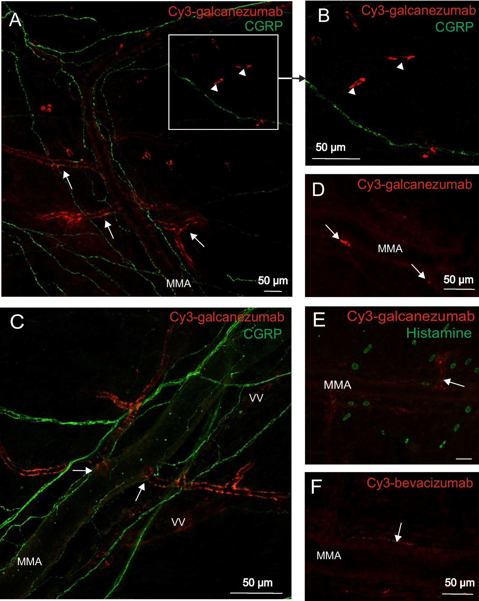
Rats injected with the fluorophore-labelled galcanezumab (Cy3-galcanezumab) or bevacizumab (Cy3-bevacizumab) 3 or 30 days prior to the experiment were deeply anaesthetized with thiopental sodium (200 mg/kg, intraperitoneally, Braun, Spain) and perfused transcardially with physiological saline followed by 4% paraformaldehyde in phosphate buffer (pH 7.4). The animals were decapitated, skin and muscles were removed and the skull was divided into halves along the sagittal suture. After removing the brain, the parietal dura mater and the trigeminal ganglia were dissected and postfixed for 2 h in the same fixative. Then trigeminal ganglia were placed in 0.1 M phosphate buffered saline (pH 7.4) containing 30% sucrose at 4 °C for 24 h and cut into 16 μm thick longitudinal sections using a cryostat (Leica CM 1950, Switzerland).
Whole mount preparation of the dura mater and sections of trigeminal ganglia of Cy3-galcanezumab-treated animals were processed for staining with the indirect immunofluorescence technique using a monoclonal mouse anti-CGRP antibody (1:500, Sigma-Aldrich, Germany), a monoclonal mouse anti-smooth muscle actin antibody (1:1000, Sigma Aldrich, Taufkirchen, Germany), a polyclonal rabbit anti-von Willebrand factor antibody (1:50, abcam, Cambridge, UK) or a polyclonal rabbit anti-histamine antibody (1:100, GeneTex, USA). IgG labelled with DyLight 488 or Alexa 488 was used as secondary antibody (both 1:500, Jackson Immunoresearch Laboratories, USA). Some sections of trigeminal ganglia were mounted with Roti-Mount FluorCare DAPI (Sigma Aldrich, Taufkirchen, Germany). Preparations of the dura mater and trigeminal ganglia were examined with a confocal fluorescence microscope (ZEISS LSM 700, Germany).
Ex vivo measurement of CGRP, SP and SOM release from meningeal afferents
Measurement of CGRP, SP and SOM release from the rat dura mater was performed by the method originally developed by Ebersberger et al [30]. Control male and female rats treated with galcanezumab or vehicle 7 days prior to the experiment were deeply anaesthetized with thiopental sodium (200 mg/kg, intraperitoneally) and decapitated. After removal of the skin and muscles, the skull was divided into halves along the midline and the cerebral hemispheres were removed. The skull preparations were washed with carbogen-gassed synthetic interstitial fluid (SIF, containing in mM: NaCl 135, KCl 5, MgCl2 1, CaCl2 5, glucose 10 and Hepes 10, pH 7.4) at room temperature for 30 min and then mounted in a humid chamber at 37 °C. The cranial fossa was filled with 300 μl of carbogen-gassed SIF solution. Samples of the superfusate were collected at periods of 10 min. Control samples were taken to determine basal peptide release in the presence of SIF, then the dura was stimulated for 10 min with the TRPV1 receptor agonist capsaicin at 100 nM in case of CGRP and SP or with 60 mM KCl in case of SOM release. 200 μl of samples diluted with 50 μl of enzyme-linked immunoassay (EIA) buffer were placed into Eppendorf cups and immediately frozen at -70 °C for subsequent analysis. The EIA method was used for the measurement of CGRP (Bertin Pharma, France), SP and SOM content (MyBioSource, USA) of the defrosted samples. For CGRP and SP measurements the same samples divided into halves (125 μl) were used. Peptide concentrations of the superfusates were expressed in pg/ml. Changes in peptide release were expressed as percentage changes relative to the basal release.
Ex vivo measurement of histamine release from meningeal mast cells
Skull halves of control rats and animals treated with galcanezumab 7 days prior to the experiment were prepared as described above for the measurement of peptide release. Control samples were taken in the presence of SIF for 10 min to determine basal histamine release, then the dura mater was stimulated for 10 min by application of 300 μl of CGRP (Sigma-Aldrich, Germany) at 10 μM or 2.5 μg/ml compound 48/80 (Sigma-Aldrich, Germany). The concentrations of CGRP and compound 48/80 used in the experiments were found effective in releasing histamine in previous experiments of our laboratory [31]. 100 μl of samples diluted with 25 μl of EIA buffer were placed into Eppendorf cups and immediately frozen at -70 °C for subsequent analysis. The EIA method was used for measurement of the histamine concentration (Bertin Pharma, France) in pg/ml. Changes in release were expressed as percentage changes relative to the basal values.
Statistics
Statistical analysis was performed using Statistica 13 software (StatSoft, USA). Following verification of the normal distribution of data, the Student’s t-test and analysis of variance (factorial ANOVA or one-way ANOVA) extended by the unequal N honest significant difference (HSD) test were used as specified in the results. All values were expressed as mean ± standard error of the mean (SEM). A probability level of p < 0.05 was regarded as statistically significant.


留言 (0)