Drugs and chemicals
In this study, doxorubicin 50 mg/25 ml vial (Dox, Adriamycin, Hikma specialized, USA) and cyclophosphamide (Cyclo, Endoxan 1gm IV vial, Baxter Oncology, USA) were used. Simvastatin and Pitavastatin were generous grants from EVA Pharma, Egypt. Other used materials include Dulbecco’s modified eagle’s medium high glucose enriched medium (DMEM, Lonza, Verviers, Belgium), fetal bovine serum (FBS, Sera laboratories international, Ltd., Brazil EU grade), phosphate buffer saline (PBS, Lonza, Verviers, Belgium) streptomycin and penicillin (Lonza, Verviers, Belgium), favor-PrepTM blood/cultured cell total RNA purification mini kit (Favorgen Biotech Corp., Ping-Tung, Taiwan), Revert Aid First Strand cDNA Synthesis Kit (Thermo Scientific, Waltham, MA, USA), HERAPLUS SYBR® Green qPCR Kit (Willowfort, Nottingham, UK) and propidium iodide (PI, ab14083, Abcam).
Experimental cell lines
Breast cancer cell lines (M.D. Anderson - Metastatic Breast 231 (MDA-MB-231) & Michigan Cancer Foundation-7 (MCF7)) were purchased from Nawah Scientific (Almokattam, Cairo, Egypt) and grown in DMEM medium enforced with 10% FBS and 1% streptomycin/penicillin incubated under standard conditions (37oC humidified air and 5% CO2 pressure).
Ethical approval
The ethical committee of the Faculty of Pharmacy, Mansoura University (Ref. No. 2020 − 176) approved this study.
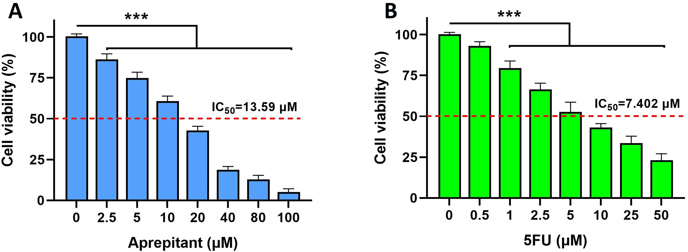
Cell viability analysis
MDA-MB-231 and MCF7 cells were seeded in 96-well plates with 20,000 cells/well under standard conditions. The plates were incubated to allow cell growth for 24 h before stimulation. The next day, cells were stimulated with doxorubicin (50, 25, 12.5, 6.25, 3.125 µM), cyclophosphamide (100, 50, 25, 12.5, 6.25 µM), Pitavastatin (200, 100, 50, 25, 12.5 µM), simvastatin (200, 100, 50, 25, 12.5 µM) in triplicates. Drugs were used with doxorubicin combination at concentrations around or below the resultant IC50; cyclophosphamide 100 µM, Pitavastatin 50 µM, or simvastatin 25 µM. The percentage of viable cells was detected using the crystal-violet assay technique 24 h after stimulation using a microplate reader (Bio Tek ELx800, USA) at a wavelength of 570 nm. The results are expressed as the percent of viable cells compared with the living control group (100% viability); cells grown with standard media without added drugs, negative control group (0%viablility); cells treated with a mixture of toxic compounds containing doxorubicin, dimethyl sulfoxide, sodium azide.
Quantitative real-time PCR (qPCR)
Cells were cultivated in 6 well plates with 1 × 10⁶ cells/well in triplicates, then incubating the cells in standard conditions for 24 h before stimulation. Cells were treated with doxorubicin (10 µg/ml), cyclophosphamide (100 µM), Pitavastatin (50 µM), simvastatin (25 µM), or their combinations for gene expression. After stimulation for 24 h, cells were washed twice using cold PBS, scraped from the flask, transferred into Eppendorf tubes, centrifuged to get the precipitated cells, and discarded the supernatant. Total ribonucleic acid (RNA) was extracted using the Favor-PrepTM Blood/Cultured cell total RNA purification mini kit (Favorgen Biotech Corp., Ping-Tung, Taiwan). The first-strand cDNA was formed using the Revert Aid First Strand cDNA Synthesis Kit (Thermo Scientific, Waltham, MA, USA). HERAPLUS SYBR® Green qPCR Kit (Willowfort, Nottingham, UK) was used in (qPCR) following the manufacturer protocol. Using the 2−∆∆ct method, gene expression fold changes were determined and presented as an average of three independent experiments (Livak and Schmittgen, 2001). Table 1. shows the primer sequences used in qPCR for caspase-3, Bax, BCL-2, and cyclin D1.
Table 1 Primer sequences used in qPCRCell cycle analysis
Cells were seeded into 6 well plates at a density of 1 × 10⁶ well, maintained in standard conditions for 24 h to allow their adhesion. Following centrifugation at 1800 rpm and removal of the supernatant, cells were permeabilized and fixed in 1 ml of cold 96% or absolute ethanol in ice, which was added dropwise, while vortexing to ensure the fixation of all cells with minimum clumping. The tubes stood for 15 min before centrifugation for 10 min at 1800, followed by aspiration of alcohol without disturbing the pellets [25]. Then, pellets were resuspended in 1 ml of propidium iodide (PI) buffer (25 µg/mL PI, 500 mg sodium citrate, and 0.5 ml Triton X-100 to 500 ml distilled water). Cells were suspended in 1 ml of the staining solution at 4 °C for 30 min and maintained in ice. Afterward, they were filtered through 30 μm nylon mesh to remove nuclear aggregates in another 5 ml tube. DNA content was measured using Accuri™ C6, and G0/G1, S, and G2/M cells were appropriately gated using Accuri™ C6 Software.
Statistical analysis
Data were expressed as mean ± standard error of the mean (SEM). One-way ANOVA followed by Tukey-Kramer, multiple comparison test, was performed using GraphPad Prism version 9.0.0 for Windows; “GraphPad Software, San Diego, California USA, www.graphpad.com”. The significance level was at a P-value of ˂0.05.


留言 (0)