Materials
The Pasteur Institute of Iran supplied the A-375 melanoma cell line. MTT powder (3-(4,5-dimethylthiazol-2-yl)-2,5-diphenyl tetrazolium bromide), citral, tripolyphosphate (TPP) and DPPH powder (1,1-diphenyl-2picrylhydrazyl) were purchased from Sigma Aldrich (USA). Chitosan’s low molecular weight, PBS tablet (phosphate buffered saline), and Tween 20 were purchased from Merck (Germany). L. citriodora EO was purchased by Giah Essence Phytopharm Co. (Iran). RPMI 1640 culture medium, trypsin, penicillin-streptomycin, and FBS were obtained from Shellmax (China). cDNA synthesis kits and TRIzol were purchased from Yektatajhiz (Iran). SYBR green Master Mix High ROX was purchased from Ampliqon (Denmark), and the Annexin-V/FITC apoptosis detection kit was purchased from Mab Tag (Germany).
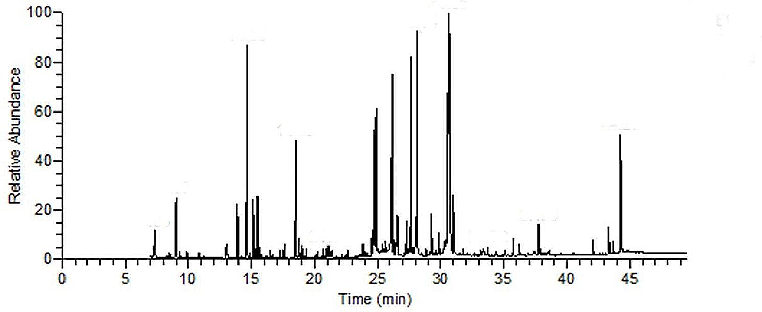
GC-MC analysis
L. citriodora EO was analyzed by an Agilent 6890 gas chromatography device (USA). First, the sample was diluted with n-hexane, and one microliter was injected into the BPX5-type column of this device with a length of 30 m, an inner diameter of 0.25 mm, and a layer thickness of 0.25 micrometers. The temperature program of the column was such that at first, the oven was stopped at 50 °C for five minutes, then it was increased with a thermal gradient of three degrees per minute until it reached 240 °C. Then, the temperature gradient was increased to 15 °C per minute until the temperature reached 300 °C and was stopped at this temperature for three minutes. The injection chamber was split from one to 35, and its temperature was 250 °C. The carrier gas of the injection chamber was helium at a rate of 0.5 mm per minute. An Agilent 5973 mass spectrometer with an ionization voltage of 70 electron volts, an ionization source temperature of 220 °C, and an EI ionization method were used in this device. The detectors used to scan the masses were set in the range of 40 to 500. CHEMSTATION software was used for the identification of compounds.
Preparation of nanoparticles
The ionic gelation method was used to prepare chitosan nanoparticles [26]. For this purpose, first, L. citriodora EO (0.16% v/v) and citral (0.16% v/v) were mixed separately with Tween 20 (0.24% v/v) for 4 min at 2000 rpm. Then, 4500 µl of chitosan solution (0.2% w/v chitosan dissolved in 1% acetic acid) was added dropwise to each sample and stirred for 45 min at room temperature at 2000 rpm. Then, 500 µl of TPP aqueous solution (1- 0.2% w/v) was added dropwise and stirred at room temperature for 60 min at 2000 rpm to obtain chitosan nanoparticles containing L. citriodora EO (L. citriodora-ChiNPs) and chitosan nanoparticles containing citral (Citral-ChiNPs). The above steps were also performed to make chitosan without anything (Free-ChiNPs).
The sizes of Free-ChiNPs, Citral-ChiNPs, and L. citriodora-ChiNPs were investigated using a DLS-type apparatus (K-One nano Ltd, Korea). To calculate particle size distributions (SPAN), D90—D10 ⁄ D50 was used. D is the diameter, and D10, D50, and D90 are percentiles of particles with a diameter lower than these values. In addition, the zeta potential of the nanoparticles was investigated using a DLS HORIBA SZ-100 (Japan) with the following settings: dispersion medium viscosity: 0.891 mPa·s, temperature of the holder: 25.2 °C, electrode voltage: 3.3 V and conductivity: 0.574 mS/cm. Moreover, to determine the morphology of Free-ChiNPs, Citral-ChiNPs, and L. citriodora-ChiNPs, a transmission electron microscope (TEM) with 100 kv accelerating voltage was used (Philips EM208S, Netherlands).
Confirmation of loading of L. citriodora EO and citral by chitosan nanoparticles
To investigate the chemical composition of nanoparticles and to prove the loading of L. citriodora EO and citral in chitosan nanoparticles. ATR-FTIR spectra of citral, L. citriodora EO, Free-ChiNPs, Citral-ChiNPs, and L. citriodora-ChiNPs were recorded in the range of 400–4000 cm− 1 using a spectrometer machine (Brooker, Tensor II, USA).
DPPH assay
The DPPH assay measured the antioxidant effect of citral, L. citriodora EO, Citral-ChiNPs, L. citriodora-ChiNPs, and Free-ChiNPs at 25–800 µg/ml concentrations. Ethanol (96%) was used to prepare a serial dilution of each sample. First, 50 µl of each sample and 50 µl of DPPH solution (0.3 mM) were poured into each well. In addition, six wells containing 50 µl of ethanol and 50 µl of DPPH solution were considered controls. Next, the plate was incubated away from light for 30 min to complete the reaction, and finally, the absorbance of the wells was read by a plate reader (Synergy HTX Multi-Mode Reader, USA) at a wavelength of 517 nm. Finally, the percentage of DPPH scavenging activity at each concentration was calculated by ([OD Control-OD Sample]/OD Control ×100).
MTT assay
A375 cells were cultured in RPMI 1640 complete medium containing 10% fetal bovine serum and 1% penicillin/streptomycin. The cells were grown at 37 °C and 5% CO2. Then, the cells were seeded in 96-well plates (10,000 cells per well) and incubated for 24 h. Next, the culture medium of each well was replaced with 50 µl of RPMI complete culture medium. After that, the cells were treated with citral, L. citriodora EO, Citral-ChiNPs, L. citriodora-ChiNPs, and Free-ChiNPs at concentrations of 25, 50, 100, 200, 400, and 800 µg/ml and incubated for 24 h.
After 24 h, 100 µl of MTT solution (0.5 mg/ml in PBS) was added to the wells and then incubated for 4 h at 37 °C. Then, 100 µl of DMSO was added to each well and shaken for 5 min, and the produced purple crystals were dissolved. After that, the absorbance of each sample and control (untreated with sample) well was read using an ELISA reader (Synergy HTX Multi-Mode reader, USA) at a wavelength of 570 nm. Finally, the cell viability at each concentration was calculated by (OD sample/OD control) ×100.
qPCR technique
The qPCR technique was employed to evaluate the expression of apoptotic-involved genes, including Bax pro-apoptotic and Bcl-2 anti-apoptotic genes. Initially, 50,000 A375 cells per well were seeded in 6-well plates. The cells were then treated with Free-ChiNPs (200 µg/mL), L. citriodora-ChiNPs (200 µg/mL), L. citriodora EO (200 µg/mL), citral (100 µg/mL), and Citral-ChiNPs (100 µg/mL) followed by incubation for 24 h at 37 °C. Next, total RNA was extracted using the TRIzol RNA extraction kit (Yektatajhiz). The wells were washed with PBS, and the liquid content was discarded.
The cells were centrifuged for 10 min at 700 g, and then 500 µl of TRIzol was added to the pellet and shaken for 6 min at room temperature. Then, 100 µl of chloroform was added, and after staying at room temperature for 3 min, the sample was centrifuged for 10 min at 15,000 g. Next, 500 µl of isopropanol was added to the supernatant in a new microtube, kept at -20 °C for 10 min, and then centrifuged for 10 min at 15,000 g. Next, 200 µl of 75% alcohol was added to the residue and centrifuged for 5 min at 15,000 g; the ethanol was discarded, and the obtained pellets were dried at 50 °C (two times). After that, the extracted RNA was dispersed in DEPC water and assessed for quality and quantity using a Nanodrop apparatus (Synergy HTX Multi-Mode Reader, USA). The purity of RNA and protein contamination was determined by measuring the absorbance (OD) ratio at 260 and 280 nm. A ratio of > 1.8 was considered indicative of a pure sample.
A cDNA synthesis kit (Yektatajhiz, Iran) was used to synthesize cDNA. Initially, the extracted total RNAs were mixed with oligo dT and DEPC water and incubated for 5 and 1 min at 70 °C and 4 °C, respectively. Subsequently, 5X strand buffer, dNTPs (10 mM), RNasin (40 U/µL), and M-MLV were added to the mixture and subjected to the Bio-Rad Thermocycler apparatus. The thermal program was set at 60 min at 42 °C, and the resulting cDNAs were stored at − 20 °C. Amplification was performed using a qPCR machine (StepOnePlus, Applied Biosystems, USA) and RealQ Plus 2x Master Mix Green high ROX™ (Amplicon, Denmark). The master mix (Green High Rox), forward and reverse primers for each gene (Pishgam Biotech Co., Tehran, Iran, see Table 1), and cDNA template were combined to a final volume of 20 µl using DEPC water. Amplification reactions were then carried out under the following conditions: 10 min at 95 °C, 40 cycles of 95 °C for 15 s, 55 °C for 30 s, and 72 °C for 30 s. The relative fold changes in the expression of target genes (Bax and Bcl-2) with β-actin as an internal control were normalized using the 2˗ΔΔCT method, where ΔCT = CT target ˗ CT reference, ΔΔCT = ΔCT test sample ˗ ΔCT control sample, and relative expression = 2˗ΔΔCT.
Table 1 Primer sequences of genesApoptosis detection using flow cytometry
To confirm the induction of apoptosis, A375 cells were seeded in 6-well plates and treated with different formulations of Free-ChiNPs (200 µg/mL), L. citriodora-ChiNPs (200 µg/mL), L. citriodora EO (200 µg/mL), citral (100 µg/mL), and Citral-ChiNPs (100 µg/mL) for 24 h at 37 °C. The Annexin-V/FITC/PI Apoptosis Detection Kit protocol (MabTag, GmbH, Germany) confirmed apoptosis. The cells were harvested, washed with PBS, and resuspended in 1x Annexin-V binding buffer. Then, Annexin-V conjugate and propidium iodide solution were added to the cell suspension and incubated for 20 min in the dark at room temperature. After incubation, 1x binding buffer was added, mixed gently, and analyzed using flow cytometry (BD FACSCalibur, USA). FlowJo software (BD, Becton, Dickinson, and Company) was used to determine the numbers of viable cells, cells undergoing necrosis (positive for PI), early apoptosis (positive for Annexin-V/FITC), and late apoptosis (double-positive for Annexin-V/FITC and PI).


留言 (0)