Chemicals
Andrographolide (purity ≥ 98%, B20207), neoandrographolide (purity ≥ 98%, B21275) and deoxyandrographolide (purity ≥ 98%, B21392) were purchased from Yuanye bio-technology Co.,Ltd.(Shanghai, China); Aluminum chloride hexahydrate (AlCl3, 97% purity) and maltol (99% purity) were purchased from Aladdin (Shanghai, China); D-gal(purity 99%) was purchased from Biosharp (Shenzhen, China); Donepezil hydrochloride (98%, D859456) was purchased from Macklin biochemical technology Co., Ltd (Shanghai, China). The preparation method of Al(mal)3 referred to our previously published literature [27].
Preparation of Andrographis paniculata ethanol extract
Andrographis paniculata (AP, lot number:18,006) were purchased from Huadong Medicine Co., Ltd (Hangzhou, China). The extraction processes and analytical techniques used to acquire and standardize AP have been described in detail here. Ultrasonic extraction with 95% ethanol for 1 h, and the ratio of material to liquid was 1:15. Then the filtrate was spun dry with a vacuum rotary evaporator. Ethanol extract of AP (refer to AP extract in this study) was diluted to different mass concentrations and stored at 4 °C, and it would be filtered through a 0.22 μm micropore filter to use in vitro experiment.
UPLC-ESI-qTOF-MS analysis of the constituents from Andrographis paniculata
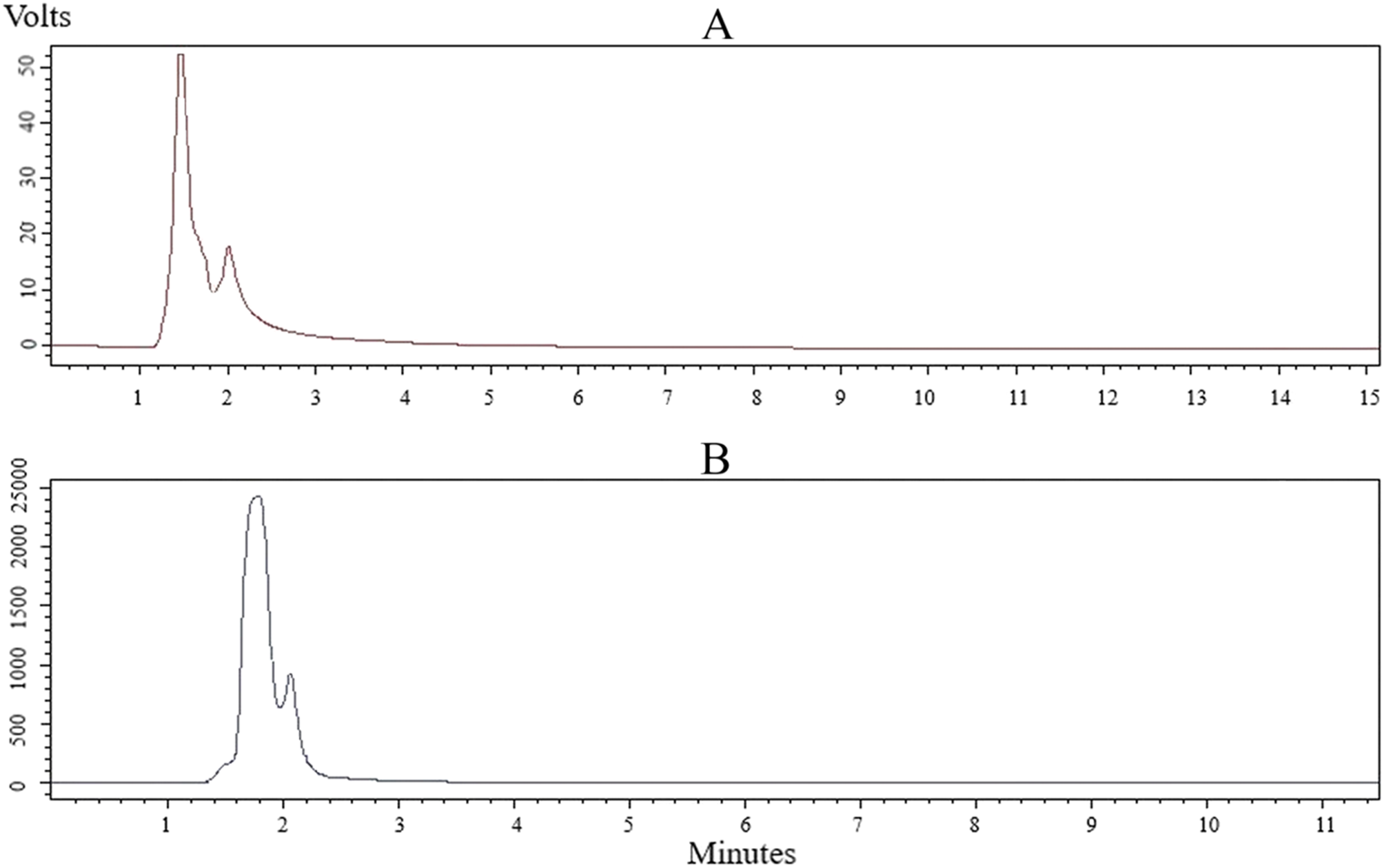
AP extract (10.9 mg) was solubilized in methanol (10 mL) under an ultrasound. The sample solution was filtered with a 0.22 μm membrane and transferred into a sample bottle for testing. Andrographolide (10.5 mg), neoandrographolide (10.6 mg) and deoxyandrographolide (10.6 mg) were solubilized in methanol (10 mL) under an ultrasound. UPLC-MS/MS analyses were performed using the UPLC system (Waters) with an ACQUITY UPLC BEH C18 column (100 mm×2.1 mm, 1.7 μm). The column temperature was 40℃, injection volume was 5 µL and the flow rate was 0.35 mL/min. A 0.1% formic acid-water (v: v) solution was used as mobile phase A and acetonitrile as mobile phase B. The gradient elution conditions were set as follows: 0-0.2 min: 10% B; 0.2–15.0 min, 10-40% B; 15.0–22.0 min, 40–85% B; 22.0–24.0 min, 85% B; 24.0-24.5 min, 85 − 10% B; 24.5–28 min, 10% B.All mass spectrometric data were acquired by a Waters Synapt G2 Q-TOF system (Waters Manchester, UK) equipped with an electrospray ionization source that operated in both the positive ion mode (ESI+) and negative ion mode (ESI−). The mass spectrometric data were collected from m/z 100 to 1,000 in the positive and negative ion modes under the centroid mode. Data acquisition and processing were performed using MassLynx V4.1 software (Waters Corporation).
Animal treatment
Male ICR mice (18–20 g) were purchased from Zhejiang experimental animal center (license No. SYXK 2019-0011). All the animal experiments were approved by the Institutional Animal Care and Use Committee of Zhejiang Center of Laboratory Animal (Approval No.ZJCLA-IACUC-20,070,022). All animals were acclimatized in the room with humidity of 30–55% and temperature of 22–25℃ for one week. The mice (n = 100) were randomly divided into normal (n = 10) and AlCl3/D-gal-treated (n = 90) group. The AlCl3/D-gal-treated mice were assigned into 6 experimental groups randomly (n = 15 in each group): [1] AD model control (vehicle-treated); [2] AD + Andro 2 mg/kg/2 days; [3] AD + AP low (200 mg/kg/day); [4] AD + AP middle (400 mg/kg/day); [5] AD + AP high (600 mg/kg/day); [6] AD + donepezil 3 mg/kg/day (reference or positive control group). First, mice except the normal group were given AlCl3 orally at 20 mg/kg/d for 4 weeks. The normal group was given equal volume of double distilled water by intragastric administration. Then, mice except the normal group were intraperitoneally injected with D-gal 120 mg/kg/d and given AlCl3 orally 20 mg/kg/d for 2 weeks. After that, mice except the normal group were administrated with AlCl3/D-gal in the morning. And in the afternoon, mice from treatment group were intraperitoneally injected with andrographolide or donepezil or given AP extract orally for 5 weeks. Normal and AD model group mice were given equal volume of double distilled water by intragastric administration. (volume-matched).
Morris water maze
The Morris water maze test is a widely-used behavioral task for investigating spatial learning and memory in animals. The test involves placing mice in a circular pool of water with specific visual clues to help them locate a hidden platform. The platform is submerged one centimeter below the water surface and measures ten centimeters in diameter. The pool is divided into four quadrants of equal size. Mice are given four daily acquisition training sessions with 5-min intertrial intervals over four consecutive days. During each session, mice are allowed to locate the platform for 60 s and rest for 5 s upon reaching it. If a mouse fails to locate the platform, it is placed on the platform for a 10-second rest period. After the final acquisition trial, a single 60 s probe trial is conducted to evaluate spatial memory retention. During the trials, the swim latency, path length, swimming speed, and frequency of entry to the target area (during the probe trial) are all recorded using the ANY-maze Video Tracking System (Stoelting, Wood Dale, IL, USA). All collected data are used to evaluate the performance of the water maze task. The escape platform remains in the same location throughout the training trials, while it is moved away during the probe test.
Sample collection
Subsequent to the behavioral tests, the mice were administered anesthesia and subjected to blood collection before being humanely euthanized. The brain was promptly placed on ice and the hippocampus was meticulously dissected, snap frozen in liquid nitrogen, and securely stored at a temperature of -80℃ for future processing needs.
Hematoxylin & Eosin (HE) staining
Histopathological studies were performed in the hippocampus. The brain tissues were fixed with 10% buffered formalin at room temperature for 48 h. Then the brain was paraffin-embedded and cut into 4-µm-thick coronal sections. Sections through the hippocampus were deparaffinized, rehydrated, stained with hematoxylin and eosin, and visualized under a light microscope (Olympus,Japan).
Biochemical assay
The brains were processed into tissue homogenate using a 0.9% saline solution in order to assess protein concentration using a BCA protein assay kit (Beyotime, Shanghai, China). The levels of malondialdehyde (MDA) and activity of superoxide dismutase (SOD) in both brain samples and serum were measured utilizing commercial MDA and SOD kits (Nanjing Jiancheng Bioengineering Institute, China), following the manufacturer’s instructions.
Cell culture and MTT assay
PC12 cells are a rat adrenal pheochromocytoma cell line, a monoclonal cell line transplanted from rat adrenal medulloblastoma by Greene and Tischler in 1976 [28]. However, PC12 cells differentiate into sympathetic nerve-like cells under the induction of nerve growth factor (NGF), which are close to neurons in terms of morphology, physiological and biochemical functions, such as growing cell protrusions, forming synapse-like structures, and having electrical excitability properties. Furthermore, under the action of NGF, they can synthesize acetylcholine and form neurite structures [29]. Additionally, the PC12 cell membrane has IV-methyl-D-aspartic acid (IV-methyl-D-panic acid, NMDA) receptors (NMDARs, as excitatory amino acid receptors in the central nervous system) that regulate synaptic plasticity, memory, and cognitive ability. The weakened nerve conduction function mediated by NMDARs can lead to brain aging, neuroplasticity damage, and cognitive dysfunction. In addition, NMDARs can interact with amyloid β-peptide/amyloid precursor protein and tau protein [30]. Therefore, PC12 cells are generally used as an ideal cellular model to study pathological molecular mechanisms of AD.
The highly differentiated PC12 cells were obtained from the cell center of the Chinese Academy of Medical Sciences, located in Beijing, China. These cells were maintained in Dulbecco’s Modified Eagle’s Medium (DMEM) supplemented with 10% fetal bovine serum (FBS) and 0.1% penicillin/streptomycin under optimal conditions of 37 °C and 5% CO2. An AD cell model was established using Al(mal)3, and cell viability was assessed using the MTT assay. The cells were seeded in a 96-well plate at a density of 100 µL/well and categorized into normal, Al(mal)3 treatment and Al(mal)3 + AP co-treatment groups for 24 h. Then, MTT solution (20 µL of 5 mg/mL) was added, and after 4 h of incubation, the culture medium was removed, and DMSO (150 µL/well) was added. The absorbance was determined at 570 nm using a Cytation 1 imaging reader (BioTek, USA).
MRFP-eGFP-LC3 transfection
Cells were inoculated into 6-well plates at a density of 2 × 10⁵ cells/mL, and transfection began when the growth fusion approached 70–90%. Add 3.75 µL Lipofectamine™ 3000 (Invitrogen, USA) into 125µL FBS-free DMEM medium and mix well. Add 2.5 µg LC3 plasmid into 125µL FBS-free DMEM medium, then add 5µL P3000TM reagent and mix thoroughly. Then the two tubes were blended and hatched at room temperature for 10 min. The above mixture was evenly dropped into the cell culture plate and placed in an incubator for 48 h. PtfLC3 was a gift from Tamotsu Yoshimori (Addgene plasmid#21,074; http://n2t.net/addgene: 21,074; RRID: Addgene_21074)). After 24 h with AP extract or Al(mal)3 treatment, the changes of red and green LC3 bright spots in the cells were observed under an inverted fluorescence microscope. The ptfLC3 could express mRFP-EGFP-LC3 fusion protein, in which mRFP emits red fluorescence and eGFP emits green fluorescence. The bright yellow spots represent autophagosomes and the bright red spots represent autophagolysosomes in the red and green merge images.
Transient gene silencing by small interfering RNAs (siRNAs)
Cells were inoculated into a 6-well plate for 24 h prior to transfection. p62 siRNA (20 µM) was transfected into PC12 cells using Lipofectamine™ 3000 transfection reagent (Invitrogen USA) according to the manufacturer’s instructions. The p62 siRNA duplexes were synthesized, and sequences were as follows: 5′-UAUCAGUUGUACUAAUCCCUU-3′ (sense), 5′- GGGAUUAGUACAACUGAUAGU-3′ (antisense). After 12 h, cells were grouped and were subjected to AP extract or Al(mal)3.
Quantitative PCR (RT-qPCR) analysis
Total RNAs were extracted from cells by using the RNAprep pure cell/bacteria kit (DP430, Tiangen, China). Then reverse transcribed into cDNA by Fasting gDNA Dispelling RT SuperMix (KR116, Tiangen, China). The RT-qPCR analysis was conducted using ABI 7500 fast system (Applied Biosystems, USA) with SuperReal PreMix Color (SYBR Green, FP205). The primers were synthetized from Sangon Biotech (Shanghai, China) and shown as follows: β-actin: 5′-GCAGGAGTACGATGAGTCCG-3′ (forward), 5′-ACGCAGCTCAGTAACAGTCC-3′ (reverse); p62: 5′-GTCAATTTCCTGAAGAATGTGGG-3′ (forward), 5′-GAGTTCACCTGTGG ATGGGTC-3′ (reverse); Nrf2: 5′-GCCCTCAGCATGATGGACTT-3′ (forward) and 5′-GTTTGGGAATGTGGGCAACC-3′ (reverse); Keap1: 5′-TGGGTCAAATACGACTGCCC-3′ (forward) and 5′- TGGCTCATATCTCTCCACGC-3′ (reverse). Analysis of melting curve data to identify the specificity of PCR. Relative fold expressions were analyzed using the 2-ΔΔCt method and using β-actin Ct values as the internal reference in each sample.
Western blot analysis
The samples from hippocampus tissues or cells were lysed and centrifuged, the supernatant was collected and mixed with 5×loading buffer at a volume ratio of 1:4, and denatured at 100℃for 30 min. Each sample was isolated by SDS-PAGE and then transferred onto membranes. The membranes were then blocked with 5% (w/v) fat-free milk for 1 h, followed by incubation overnight at 4 °C with primary antibodies at 1:1000 dilution ratio: Nrf2(AF7623, Beyotime, Shanghai, China); Keap1 (sc-514,914, Santa Cruz); Tau (ab32057, Abcam); phospho-Tau (ab109390, Abcam); p62 (AF5384, Affinity, USA); LC3B (L7543, Sigma); GAPDH (FD0063, Fudebio, China). After cleaning, the membranes were incubated with horseradish peroxidase (HRP)-conjugated secondary antibodies for 1 h. The membranes were washed in the western lighting plus-ECL solutions, and lastly the immunoreactive bands were measured using a Chemiluminescence imaging system (Amersham Imager 800, Cytiva). The related optical density of the digitized image was analyzed by ImageQuant TL 8.1 software.
Statistical analysis
Statistical analysis was conducted using GraphPad Prism 8.0 statistical software (Graphpad Inc, San Diego, CA, USA). All data were displayed as the mean ± SD. The P-values were calculated using a one-way analysis of variance (ANOVA) or Tukey’s multiple comparisons test. A P-value of < 0.05 was regarded as indicating a statistically meaningful result.

Comments (0)