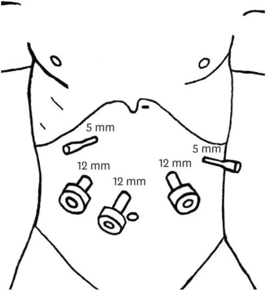
In this study, six patients successfully underwent 3D laparoscopic nephrectomy combined with bench surgery and autologous transplantation, which achieved tumor resection and renal function preservation without significant intraoperative and postoperative complications, providing a new option for the treatment of patients with highly complex renal tumors. In terms of the choice of surgical access for radical nephrectomy, although Ju et al. 's study suggested that retroperitoneal access could reduce the interference of the intestine to the operation and provide a better intraoperative field of vision, we adopted the transperitoneal access because the kidney extraction incision and autologous kidney transplantation could be classified as the same incision for reducing postoperative pain [19]. During radical nephrectomy, none of the patients reported damage to the renal pedicle vessels and renal pelvic structure. We attribute this primarily to the accurate depth representation provided by the 3D laparoscopic system, which enhances spatial perception, particularly during the separation of the renal pedicle structure and grasping of the edge of clipping tissue. These inherent advantages significantly enhance surgeon confidence and surgical efficiency, while reducing intraoperative complications; these findings align with those observed in Nguyen et al.’s study [20]. The warm ischemia time was controlled within 2 min. This time was similar to the results of Prudhomme et al., who suggested that 3D laparoscopic kidney extraction was significantly superior to 2D laparoscopic system in terms of total operation time (80.9 ± 10.2 vs. 114. 1 ± 32.3 min in 3D and 2D, p = 0.0002) and warm ischemia time (1.7 ± 0.6 vs. 2.3 ± 0.9 min in 3D and 2D, p = 0.02), these findings might be attributed to the improved speed of the 3D laparoscopic system in terms of vessel separation and ligation [21]. The tumor resection and the transplanted kidney repair were all performed after immersion in cryogenic perfusion fluid. Compared with in situ nephrectomy, sufficient operative time is acquired for tumor removal and wound suturing under external cold ischemia conditions, while the operation under direct vision in vitro ensures a favorable negative incision margin and the collection system closure because some small lesions that were not indicated on imaging can be found with the naked eyes [22]. Intraoperative ultrasound can also be used to visualize the kidney to avoid the residual lesion, especially if the tumor is located in the renal portal. Hence, sufficient operative time and an appropriate surgical perspective allow complete tumor removal without damaging the renal pelvis and renal vessels. Although orthotopic autologous kidney transplantation is also performed, the probabilities of postoperative complications and renal graft resection or even loss due to renal artery pseudoaneurysm, renal artery embolism, or renal artery hypoperfusion are higher when compared with ectopic autologous kidney transplantation. Therefore, we adopted ectopic autologous kidney transplantation, which is routinely performed by most surgeons [23]. Autologous kidney transplantation and repair of graft blood vessels before transplantation were performed by experienced surgeons in our hospital. During the operation, an emphasis was placed on anastomosis of the renal blood vessels and ureter with the pelvic blood vessels and bladder, respectively. After anastomosis completion and resuming the blood supply to the kidney, the tension was carefully observed, and the presence of blood infiltration sites and ischemia was confirmed. Consequently, urine was produced from the transplanted kidney after the blood supply was restored, the surface tension and the pulse of the kidney were normal, the local bleeding sites were relieved, and no ischemia was observed. In this study, the duration of cold ischemia was 121 ± 26 min, compared with the kidney obtained from other hospitals, the cold ischemia time is significantly shorter, which helps to protect the function of the transplanted kidney. Although had no impact on graft survival, a prolonged cold ischemia time was associated with more incidence of delayed graft function and lower graft function [24]. After the blood supply was restored, one patient underwent an autologous blood transfusion because of bleeding. This can be due to an incomplete closure of the dorsal side of the anastomosis between the renal artery and the external iliac artery, owing to the deep position of the iliac artery in the patient. No significant bleeding requiring blood transfusion occurred in other patients during or after the surgery, which can also be proved by the fact that there was no significant decrease in MAP after surgery compared with that before surgery. In a study by Janssen, the most common complication was bleeding, with 50% (6/12) of patients requiring intraoperative and postoperative blood transfusions while two patients required temporary hemodialysis, owing to increased surgical trauma of the open procedure [13]. In our study, although all patients experienced a temporary decline in renal function postoperatively, they gradually recovered without any temporary dialysis and the renal function almost returned to preoperative levels 1 month after the surgery, which due to short warm ischemia period and reduced reperfusion injury. In contrast to the temporary decrease in the SCr level, the 24-h urine output of all the patients increased to varying degrees after surgery, which we considered to be due to the increase in the volume of intravenous fluid after surgery as compared with that before surgery. And none of the patients had renal failure requiring dialysis during the follow-up period, and their ultrasound examination showed that the pulsatility and resistance indexes of the transplanted kidney were normal. However, due to the longer lesion resection and wound repair time under cold ischemia, renal cell carcinoma patients experience chronic renal graft failure more frequently than other patients undergoing this surgery for renal aneurysm or ureteral stenosis, which reminds us not to neglect the monitoring of renal function [25, 26]. Although our pathology results indicated that the surgical margins were negative in all the patients, one patient (patient 3) had a recurrence of the transplanted renal tumor 20 months after surgery and underwent transplanted kidney resection in the same month; the postoperative pathology diagnosis was clear cell carcinoma. Therefore, constant monitoring for tumor recurrence is very crucial, even though the other patients had no tumor recurrence and distant metastasis during the follow-up period. Another study by Tran showed that 50% of the patients (4/8) still had tumor recurrence despite negative incisal margins; thus, this might be related to the high aggressiveness of central tumors [27]. Similar to the study by Cowan, we observed that in addition to temporary renal failure, the most common complication was urinary tract infection (4/6), followed by postoperative pain (3/6). In their study, the most common postoperative complications were infection and bleeding, while two patients underwent renal graft resection for renal artery dissection and renal graft failure, respectively, thereby suggesting that cold ischemia time significantly correlates with the incidence of postoperative complications [28]. We suggest that the higher incidence of urinary tract infections is related to a patient’s lower immunity due to the prolonged operation time and postoperative induration of the double-J tube, which can be alleviated after symptomatic antibiotic treatment. With the gradual recovery of patients’ baseline conditions, recurrent and multiple infections did not occur. Although the surgical wound area has been minimized, 50% of the patients still need analgesic drugs to relieve pain after surgery because of the surgical wounds involving both the renal fossa and iliac fossa. Fortunately, a gradual wound repair alleviated long-term chronic pain, and no postoperative incision infection and incisional hernia occurred as in other studies [29, 30]. This is mainly due to the use of 3D laparoscopic nephrectomy, a minimally invasive technique, which reduces the incidence of incision-related complications compared with open nephrectomy [31]. There were no serious postoperative complications in this study, but the monitoring and nursing work of patients during the perioperative period should not be slacken. For some studies have shown that the incidence of postoperative complications such as intestinal obstruction, venous thromboembolism, and pulmonary infection is high, especially for obese patients and patients with chronic renal insufficiency, the incidence of postoperative complications is higher than the general population [12, 32]. The short duration of postoperative hospitalization and the utilization of cost-effective 3D laparoscopic surgery rather than expensive robot-assisted surgery, have effectively maintained the overall expenses associated with this study at a modest level that is affordable for most patients.
The main limitation of this article is the patient population was too small to carry out an effective comparative study with other surgical procedures, so drawing any comparative conclusion was difficult. For example, studies have shown that robotic technology can benefit patients in terms of postoperative complications and recovery time by providing surgeons with a better view of the intra-operative field, enhanced precision and flexibility, and a smaller surgical incision than traditional open surgery [33, 34]. Hence, increasing sample sizes and comparative studies with robotic surgery are future research directions. Furthermore, as this was a single-center retrospective study, our results were greatly influenced by the medical level of the center. Subsequent multicenter studies are needed to evaluate the efficacy and safety of this surgical intervention. Thirdly, limited by the hospital conditions, we solely relied on the SCr level to evaluate the overall renal function in this study. In future research, we will accurately evaluate the differential renal function by radiorenogram or other examinations in other hospitals. In addition, our study’s follow-up time was short, especially for the preservation of transplanted kidney function and tumor recurrence; hence, it is not possible to draw long-term conclusions.


留言 (0)