
Remember me


A total of 567 abstracts were retrieved following the searches, when duplicates had been removed (Fig. 2). Of these, 125 did not meet the inclusion criteria and were excluded, leaving 442 articles that were read in full text. After a more detailed scrutiny, a further 406 articles were excluded, resulting in 36 studies to be included in the review (Table 1).
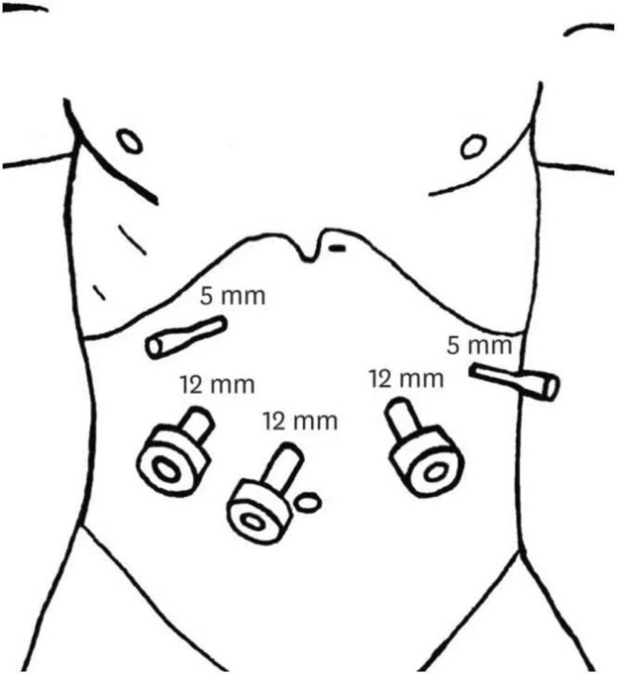
Fig. 2 Study characteristics
Effects of venous supercharging were studied mainly in non-randomised cohorts with controls, where venous supercharging was performed in case of signs of venous congestion intraoperatively or postoperatively. Three studies report ‘routine’ use of venous supercharging and performed it prophylactically in patients who did not have clinical signs of venous congestion. One of the studies randomised patients to venous supercharging or not supercharging (29 vs. 23 pats) [31], one reported two consecutive series comprising 30 pats in each group [58] and one did not state how the patients were allocated to the two groups [35]. For the randomised controlled trial (RCT), a sample size calculation was not performed and primary and secondary end points as well as outcome measures were not defined.
Flap complicationsSeventeen studies report on flap complications [27,28,29, 31, 32, 35, 37, 38, 42, 43, 46, 49, 51, 55,56,57,58], of which one is an RCT [31] and two are controlled cohort studies [35, 58] including prophylactic venous supercharging in patients without clinical signs of venous congestion (Additional file 1). The groups of the RCT [31] and the study comparing two consecutive cohorts [58] have groups that are comparable with regards to age, body mass index (BMI) and comorbidities (Additional file 1). The three studies share the methodological weakness that they have not defined complications and how, when and by whom they were diagnosed. The RCT [31] demonstrated statistically significant lower complication rates in the intervention group. The findings were similar in the two cohort studies [35, 58]. Nonetheless, the magnitude of the effect on complications of a SIEV must be interpreted with caution as the samples are small and there is a clear heterogenicity in the frequency of complications. Venous congestion was 13% in the intervention group and 55% in the control group and partial flap loss 9% in the intervention group and 45% in the control group in the RCT [31]. Total flap loss was 0 in all three intervention groups and ranged from to 0% [35, 58] to 17% [31] in the control groups. The numbers of the studies comparing two incomparable groups, patients with and without clinical signs of venous congestion, are given in Additional file 1.
The overall certainty of evidence for the effect of a SIEV anastomosis on flap complications, in patients without clinical signs of venous congestion, is low (GRADE ⊕ ⊕ ⊝ ⊝). The evidence was downgraded three levels due to a high risk of bias, imprecision and inconsistency in the magnitude of effect and upgraded one level as magnitude of the effects of venous supercharging on flap complications seems to be large and is consistent across studies.
Donor site complicationsThree retrospective non-randomised studies with controls reported donor site complications (Additional file 2) [45, 54, 63]. One of the studies [55] reported that there were zero donor site complications, whereas the other [45] stated that the rate of abdominal seromas requiring drainage was higher in the group where the SIEV had been harvested, especially if it had been harvested bilaterally. However, the study did not control for other factors that might increase the rate of seromas. The third study reported that the usage of the cephalic vein as a recipient vessel does not seem to increase the risk for arm lymphoedema [54].
The overall certainty of evidence for the occurrence of donor site complications after SIEV harvesting is very low (GRADE ⊕ ⊝ ⊝ ⊝). The evidence was downgraded three levels due to a very high risk of bias, indirectness, and imprecision.
Length of hospital stayFive retrospective non-randomised controlled cohort studies reported LOS [28, 29, 42, 45, 46]. Most of the studies stated a longer LOS in the venous supercharging group than among the controls (Additional file 3). Nonetheless, the studies did not have comparable groups and did not control for confounders that might affect LOS, such as both patient related and surgical factors as well local tradition.
The overall certainty of evidence for the effect of venous supercharging on LOS is very low (GRADE ⊕ ⊝ ⊝ ⊝). The evidence was downgraded three levels due to a high risk of bias, indirectness, and imprecision.
Operative timeSeven studies reported operative time, of which one was an RCT and six were retrospective non-randomised controlled cohort studies [28, 30,31,32, 37, 46, 49]. Most of the studies stated a longer operative time in the venous supercharging group than among the controls (Additional file 4). The RCT demonstrated that the mean time increased from 405 to 510 min [31]. However, all the included studies had methodological flaws such as lack of information on learning curves and techniques used and in case of the retrospective studies, different populations among the interventions and the controls and no adjustment for confounders that might affect operative time. As only patients with clinical signs of congestion were included in the venous supercharging group, it is likely that aspects, other than the SIEV dissection and anastomosis itself, also affected operative time. Moreover, none of the studies took possible savings in re-operation times into considerations.
The overall certainty of evidence for the effect of venous supercharging on the operative time is very low (GRADE ⊕ ⊝ ⊝ ⊝). The evidence was downgraded three levels due to a very high risk of bias, indirectness, and imprecision.
Surgical takebacksSeven studies reported surgical take backs of which one was an RCT [31] and six retrospective non-randomised controlled cohort studies (Additional file 5) [31, 35, 38, 42, 55, 57, 58]. In the RCT [31] and the study with two consecutive series [58], there were considerable differences in takebacks between the groups, 13% vs. 55% and 1% vs. 10%, respectively. The other studies also consistently showed that the takeback rates were lower in the intervention groups. However, the magnitude of the decrease of takebacks is unclear as intervention and control groups were completely different in some studies which could result in an underestimation of takebacks. In brief, the magnitude of the effect of venous augmentation on surgical takebacks is unclear due to uncompilable groups in many studies and small samples in the more high-quality studies.
The overall certainty of evidence for the effect of venous supercharging on surgical takebacks is low (GRADE ⊕ ⊕ ⊝ ⊝). The evidence was downgraded three levels due to a high risk of bias, imprecision and indirectness and upgraded one level as magnitude of the effects of venous supercharging on surgical takebacks seems to be large and is consistent across studies.
Strategies for the usage of venous superchargingTwenty-one studies presented data on strategies for when venous supercharging should be performed (Additional file 6) [9, 18, 30, 33, 34, 36, 39,40,41, 44, 46,47,48, 50, 52, 53, 55, 58,59,60]. Nine [18, 33, 36, 40, 41, 50, 52, 59, 60] of them present radiological signs that could predict venous congestion, four intra-operative measurements [30, 34, 47, 53] and six [9, 39, 46, 48, 50, 55] clinical signs that could predict the need for venous supercharging. Two studies [44, 58] merely give recommendations based on their clinical experience. The studies investigation predictors generally compare patients who have had clinical venous congestion with those that have not and can thereby be classified as non-randomised observational studies with controls.
Regarding radiological findings predictive of venous supercharging, a few studies have investigated the role of the SIEV diameter. One study concluded that a big SIEV diameter or deep inferior epigastric vein (DIEV) diameter and a high SIEV/DIEV diameter ratio (no cut off values are given) [36] and another that SIEV size > DIEV size at origin (5.2 vs 3.5 mm, p = 0.007) [59] were predictive of the need for supercharging, whereas two other found that the diameter seems to be negatively correlated to the need for venous supercharging [40] and that the was no correlation between SIEV diameter and the need for supercharging [50]. Four studies investigated connection between the deep and superficial system on computed tomography (CT) and concluded that venous supercharging is needed when there are no direct [52, 59] or atypical connections (in terms of caliber, tortuosity or superficial path) [
Comments (0)