Animals
Male C57BL/6 mice (6–8 weeks old) were obtained from Beijing Vital River Laboratory Animal Technology (China). The mice were placed in controlled environments (12-h light/dark cycle; 22 °C; 50–60% humidity) and had free access to bacteria-free water and food. All animal housing and experiments were conducted in accordance with the ethical guidelines formulated by the Animal Experimental Committee of the First Affiliated Hospital of Wenzhou Medical University.
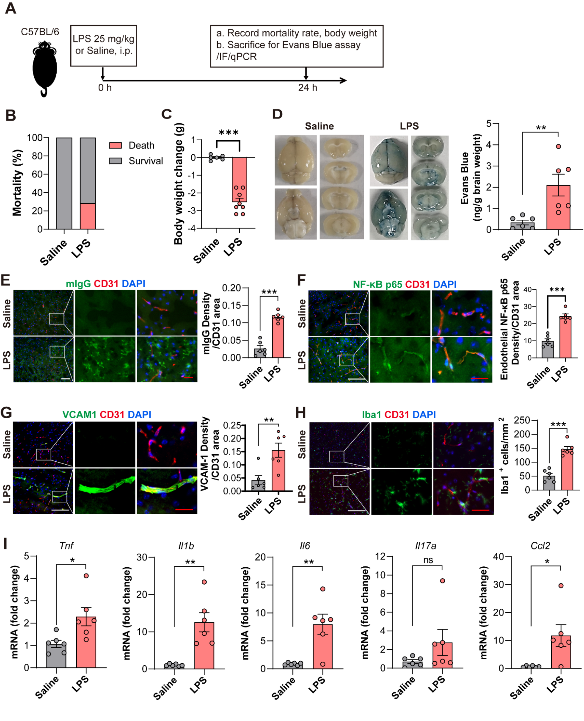
Sepsis model
The sepsis mouse model was established by intraperitoneal injections of LPS (10 mg/kg; from Escherichia coli 055:B5, L2880, Sigma-Aldrich), and mice in the Sham group received intraperitoneal injections of an equal volume of PBS. Twenty-four hours later, the mice were anaesthetized and perfused with normal saline until the lungs were whitened. The hippocampus were collected for histological analysis or frozen in liquid nitrogen for rapid cryopreservation.
Behavioural testsMorris water maze (MWM) test
Ten days after the intraperitoneal injection of LPS, the MWM test was performed to evaluate the spatial learning and memory abilities of the mice. First, the mice were trained for 4 consecutive days, and then the experiment was carried out on the fifth day. The MWM consisted of a round steel pool (1.2 m in diameter and 0.6 m in height) with a hidden platform (0.1 m in diameter). The water in the pool was maintained at 23 °C, and the hidden platform was located in the southwest quadrant of the pool, approximately 1 cm under the surface. Propylene dye was used to make the water opaque. During the training period, each mouse was randomly placed in a different quadrant each day. The mice were allowed to find the platform within 60 s, and the time to reach the platform was recorded. If the platform was not found within 60 s, to the animal was placed on the platform to rest for 10 s. On the fifth day, the platform was removed, and the mouse was placed in the water in the quadrant opposite the platform. Each mouse swam freely for 60 s, and the number of passes and time spent in the target quadrant were recorded.
Nissl staining
Nissl staining was performed to evaluate neuronal damage and loss. After paraffin embedding and sectioning (4 μm), brain tissues were stained with a 1% toluidine blue solution (Beyotime, C0117, China).
Immunocytochemistry
BV2 cells were treated, soaked in 4% paraformaldehyde for 30 min and then permeabilized for 5 min with 0.2% Triton X 100. After being sealed with 5% BSA for 1 h, the cells were incubated with the indicated antibodies at 4 ℃ overnight. After being washed with PBS three times, the cells were incubated with Daylight 488-coupled secondary antibodies (1:500, CL488-10,594, Proteintech) or 594-coupled secondary antibodies (1:500, CL594-10594, Proteintech) for 1 h, and the nucleus was stained with DAPI. Images were recorded with a Leica confocal microscope.
Frozen embedded brain tissue sections were prepared. After the tissue sections were blocked with 5% bovine serum albumin for 1 h, 4-μm-thick brain slices were prepared and treated with rabbit anti-Gsdmd (1:100, A10164, ABclonal) or mouse anti-NLRP3 (1:100, AG-20B-0042, AdipoGen) overnight. Then, the sections were incubated with Daylight 488 -coupled secondary antibodies (1:500, CL488-10594, Proteintech) or 594-coupled secondary antibodies (1:500, CL594-10594, Proteintech) at room temperature for 1 h. After the sections were washed with PBS 3 times, the nuclei were stained with DAPI. Images were recorded with a Leica confocal microscope.
Cell culture and treatments
The mouse microglia BV2 line, mouse astrocytes MA line and mouse hippocampal neuronal HT22 cell line were purchased from the Cell Bank of the Chinese Academy of Sciences (Shanghai, China) and cultured in DMEM (Gibco, USA) supplemented with 10% FBS and 1% penicillin/streptomycin at 37 °C in a humidified atmosphere containing 5% CO2. Lipopolysaccharide (LPS, L2880, Sigma‒Aldrich) and adenosine triphosphate (ATP, A3377, Sigma‒Aldrich) were used to activate the NLRP3 inflammasome in the cell model. For NLRP3 inflammasome activation, BV2 cells were treated with LPS (1 µg/mL) for 6 h and then treated with ATP (5 mM) for 30 min. DMSO was used as a vehicle control for the treatment conditions.
Plasmids and siRNA transfection
Small interfering RNAs against TRIM45 were purchased from Hanbio Biotechnology (Shanghai, China). Small interfering RNAs against Atg5 (sc-41446, Santa Cruz) were purchased from Santa Cruz Biotechnology. Flag-Atg5, HA-TRIM45, HA-Ub, HA-Ub-K48 and HA-Ub-K63 were purchased from Limibio Biotechnology. These agents were transfected into BV2 cells using Lipofectamine RNAiMAX transfection reagent (13778075, Thermo Fisher Scientific) or Lipofectamine 2000 transfection reagent (11668500, Thermo Fisher Scientific). After 48 h, the cells were used for further experiments. The following sequences were used: siTRIM45 sense: 5′-GGTGGAGTGAAGGCTTTAACG-3′ and negative control siRNA (siNC) sense: 5′- UUCUCCGAACGUGUCACGUTT-3′.
Determination of ROS by flow cytometry
ROS were examined by an ROS assay kit (Beyotime, S0033S, China). A total of 1 × 10⁶ BV2 cells were plated in 6-well plates and treated with LPS (1 μg/mL) for 6 h and ATP (5 mM) for 30 min. Then, the cell culture fluid was discarded, and the cells were carefully washed three times with DMEM. The cells were incubated with DMEM containing 10 µM dichlorodihydrofluorescein diacetate (DCFH-DA) for 30 min at 37 °C. Then, the cells were washed with DMEM three times to eliminate the excess DCFH-DA and collected in centrifuge tubes. Finally, the DCF fluorescence intensity was measured by flow cytometry at wavelengths of 485 nm and 535 nm. ROS levels were analysed by FlowJo Software.
Detection of apoptosis in HT22 and BV2 cells by flow cytometry
Apoptosis in HT22 and BV2 cells were tested by Annexin V-FITC/PI Apoptosis Kit (AP101, MULTI SCIENCES). Adherent cells were collected by 0.25% EDTA digestion and centrifugation (4 °C, 1000 g, for 3–5 min), and 105–106 cells were collected and centrifuged (4 °C, 1000 g, for 3–5 min). Cell pellets were resuspended with 0.8–1 mL of cell staining buffer. Then, 5 μL of Annexin V-FITC staining solution was added, and 10 μL of PI staining solution was added. The mixture was mixed well and incubated at 37 °C for 5 min. Red fluorescence and blue fluorescence were detected by flow cytometry.
JC-1 staining
The mitochondrial membrane potential in BV2 cells was determined via a JC-1 fluorescent probe (Beyotime, C2003S, China), and these cells were incubated with JC-1 working solution for 20 min at 37 °C. After being treated with LPS (1 μg/mL) for 6 h, the cells were treated with ATP (5 mM) for 30 min. Then, JC-1 buffer solution was used to wash the cells at least three times. The results were determined by calculating the rate of the green/red fluorescence intensity, which represented the level of mitochondrial disruption.
PBMC collection, cytokine detection, antibodies, cell staining, and flow cytometry
Mouse eyeball blood (PBMC) was collected after isoflurane anaesthesia. Leukocytes were isolated from whole blood with red blood cell lysis buffer, and the remaining cells were stained. The antibodies and reagents used for flow cytometry are listed in Resources Table 1. Surface staining was performed in PBS containing 2% BSA or FBS (w/v). To detect cytokine production (IL-6 and TNF-α), lymphocytes were stimulated for 5 h in the presence of Cell Stimulation Cocktail (plus protein transport) (00-4975-93, Thermo Fisher). Intracellular cytokine staining (ICS) of IL-6 and TNF-α was performed with the Cytofix/Cytoperm Fixation/Permeabilization Kit (554714, BD Biosciences). Flow cytometry data were acquired with a BD Fortessa (BD Biosciences) and analysed using FlowJo (Tree Star).
Table 1 Flow antibody catalogueTRIM45 adeno-associated virus infection
To downregulate the expression of TRIM45 in the mouse brain, we used an AAV9 vector carrying shRNA targeting mouse TRIM45 mRNA (Shanghai Genechem Co., Ltd.) and the core sequence of AAV-shTRIM45 was 5'-GGTGGAGTGAAGGCTTTAACG-3'. Stereotactic surgery to transfer the AAV vector was performed on male mice aged 11–12 weeks (25–30 g) that were anaesthetized with 350 mg/kg 4% chloral hydrate (i.p.). The mice were fixed on a stereotactic instrument, the skull was pierced by a drill, and the microsyringe was driven by a stepper motor. A total of 500 nL (2.5 × 10¹² vg/mL) of virus solution was injected into the hippocampus. The speed was 50 nL/min. Two weeks later, the model was established in transfected mice.
Reverse transcription real-time quantitative polymerase chain reaction (qRT-PCR)
Total RNA was extracted from BV2 cells with TRIzol reagent (Invitrogen, USA). RNA was reverse-transcribed to complementary DNA using RT-qPCR Fast Master Mix (Vazyme, China). Real-time fluorescence quantitative PCR was performed according to the manufacturer’s instructions. β-Actin and gapdh were used as internal controls for IL-1β, IL-18, TRIM45, Atg5, P62 and beclin1 mRNA expression analysis. Gene expression was quantified using the 2−ΔΔCt method. The gene primer sequences are listed in Table 2.
Table 2 Base sequence of each geneELISA
The levels of IL-1β and IL-18 in medium of BV2 cells and peripheral blood of mice were determined using an ELISA kit (MEIMIAN) according to the manufacturer’s instructions. The absorbance of the samples at a wavelength of 450 nm was measured with a BioTek microplate reader.
Western blot analysis
RIPA lysis buffer was used to extract protein from hippocampal tissue or cultured cells, and a BCA protein detection kit was used to measure the concentration. Approximately 25 mg of protein was boiled for 5 min at 100 °C, separated by 10% SDS-PAGE, and transferred to PVDF membranes (1620177, Bio-Rad) The PVDF membranes were then blocked with 5% skim milk for 2 h at room temperature and incubated with primary antibodies (TRIM45 (ab169036, Abcam), NLRP3 (ab263899, Abcam), Gsdmd (ab219800, Abcam), IL-1β (ab16288, ABclonal), Atg5 (ET1611-38, HuaBio), LC3 (#4108, CST), SQSTM1/p62 (#23214, CST), beclin1 (JE59-31, Huabio), β-actin (HRP-60008, Proteintech), HA (51064-2-AP, Proteintech), Flag (80010-1-RR, Proteintech), Caspase3 (Abmart, T40044), cl-Caspase3 (Affinity, Asp175)) overnight at 4 °C. Then, the membranes were washed three times with TBST and then incubated with the appropriate horseradish peroxidase-labelled secondary antibodies. An enhanced chemiluminescence reagent was used to view the reaction. We measured the signal intensity using ImageJ AQ7. Standardization was performed using β-actin.
Coimmunoprecipitation
The supernatants were incubated with Atg5 primary antibodies overnight at 4 °C, followed by the addition of 30 µL of protein A/G PLUS-agarose. After that, the solutions were incubated at 4 °C for 6–8 h. Then, the protein A/G PLUS-agarose was washed three times, and the protein attached to the agarose and linked to keap1 was extracted. We discarded the supernatant and resuspended the pellet in 45 µL of 2 × PAGE loading buffer, and boiled it for 5 min at 100 °C.
Human sepsis specimens
To observe cell activity in the peripheral blood of normal subjects and patients with septicaemia, all septic patients hospitalized in the ICU from January 2023 to June 2023 were selected. Sepsis was determined according to the third internationally recognized definition of sepsis and septic shock, and the patients were minors, pregnant or had type 1 diabetes, aplastic diseases or immunosuppressive diseases or patients receiving immunosuppressive therapy were outside the scope of our study. Ethical review of human studies (KU2022-126) was performed by the Ethics Committee of the First Affiliated Hospital of Wenzhou Medical University, and the study was performed in accordance with the Helsinki Declaration and federal policy to protect human subjects. Each participating patient provided informed consent. For further study, peripheral blood mononuclear cells were obtained from blood by density gradient centrifugation.
Statistical analysis
GraphPad Prism 8.3.0 was used to analyse the data and construct the graphs. The data are expressed as the mean ± SEM. Experiments with only 2 groups were analysed by the unpaired two-tailed Student’s t test. Single-factor experiments with > 2 groups were analysed with one-way analysis of variance (ANOVA) with Dunnett’s post hoc test. P < 0.05 was considered statistically significant.


Comments (0)