Animals
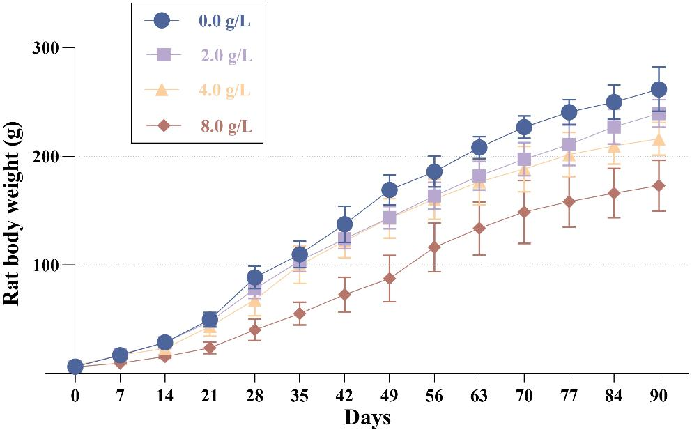
Adult Long–Evans rats (150–200 g) were used for the experiments; they were randomly divided in control and diabetic groups. Diabetes was induced by a single intraperitoneal STZ administration (90 mg/k, ip.) in buffer citrate, pH 4.5 [14]. Blood glucose levels from the tail-vein blood samples were measured using ACCU-CHEK test strips (Roche Diagnostics), 24–48 h after STZ administration and at sacrifice. Age-matched citrate buffer-injected rats were used as the control group. Rats were maintained (4–5 per cage) at 21 °C ± 1, 12 h light–dark cycle, and food and water provided ad libitum. Diabetes was confirmed by measuring blood glucose concentration and loss of gain body weight (Table 2). Animals were considered diabetic if blood glucose levels were higher than 250 mg/dl; insulin was not administered.
Previous studies demonstrated that lower STZ doses (60 mg/kg) has an efficacy of 60% in the induction of the hyperglycemic condition, while higher doses (90 mg/kg) have an efficacy of 95%. These studies also indicated that 12–15% of males did not became hyperglycemic after STZ treatment, while in only 2–5% of females we observed this STZ resistance. Therefore, in the present study, we used the relative high STZ doses and female rats. Animals were sacrificed at 7, 20, and 45 days after diabetes induction (7D; 20D; and 45D), along with non-treated animals (controls). A different set of control and STZ treated rats were used for biochemical or licking behavior. The lumbar spinal cord (L5 and L6, 0.2–0.25 g) was isolated for mRNA and Western blots (WB) assays.
Ethics
This study was conducted in strict accordance with the recommendations of the Mexican Institutes of Health Research (DOF. NOM-062-Z00-1999). The protocol was approved by the Institutional Laboratory Animal Care and Use Committee of the Cellular Physiology Institute of the National Autonomous University of Mexico (CICUAL, Comité Institucional para el Cuidado y Uso de los Animales de Laboratorio del Instituto de Fisiología Celular de la Universidad Nacional Autónoma de México). Protocol number: RSS190-22 and RSS110 (43)-17. All efforts were made to minimize animal suffering and to reduce the number of rats used.
Nocifensive Test
The capsaicin-evoked nocifensive response was evaluated in non-treated animals (control) and STZ-induced diabetic rats at 20 days and 45 days [15]. The animals were placed in individual plastic containers 1 h before the experiment. The stock solution of capsaicin was resuspended in ethanol (10 µg/µl) (Sigma-Aldrich). The injection and vehicle solutions were respectively prepared by diluting capsaicin (0.19 µg/µl) in saline solution (0.19 µg/µl) and in 1.9% ethanol. Both, control and diabetic rats were first intraplantarly injected on the left paw with 10 µl of saline solution, using a 30 G needle; then, the animals were placed in the containers and the licking behavior was quantified for 10 min. After 30 min adaptation, rats were intraplantarly injected on the right paw with 10 µl capsaicin solution, were placed in the containers, and the licking behavior was quantified for 10 min. The cumulative licking time (seconds) was reported as paw licking time (PLT).
Synaptosomes Preparation
Lumbar spinal cord was dissected and synaptosomes were isolated by the procedure described by Hajos [16] and slightly modified by Pérez-Léon and Salceda [17]. Tissue was homogenized in 0.3 M sucrose (10% w/v)—Tris 10 mM, pH 7.4 and centrifuged at 1500×g for 10 min. The supernatant was centrifuged at 9000×g for 20 min. The obtained pellet (crude synaptosomal fraction) was used for Western blot or qPCR.
RNA Extraction
Total RNA was extracted with TRIZOL (Ambion Life Technologies, Thermo Scientific Inc.), as previously described by [18]. cDNA was synthesized with the RevertAid H Minus First Strand cDNA Synthesis Kit (Thermo Scientific) following the manufacturer’s instructions. RNA integrity and concentration were verified by spectrophotometry (NanoDrop1000, Thermo Scientific) and 2% agarose gels.
qPCR
qPCR was performed under the same conditions previously described by Sánchez-Chávez et al. [19]. Primers were design with Primer 3 [20, 21], purchased from T4 Oligo (Irapuato, Guanajuato, Mexico). The sequence of each pair of primers is shown in Table 1. Data were analyzed by following the Livak and Schmittgen method [22] using the 18 gene as a reference. For each sample was determined the expression of the α1–α3 and β GlyR expression, and in parallel the 18S expression (reference gene). As described by Livak and Schmittgen [22], the Ct value obtained for the 18S gene—for each sample—was subtracted from values obtained for each of the GlyR subunits in each sample (∆Ct). Therefore, ∆Ct values obtained for the controls were subtracted from ∆Ct values obtained for the GlyR subunits in each sample (∆∆Ct). The fold-change values (2−∆∆Ct) were relative to the control condition.
Western Blotting
Spinal cord homogenates or synaptosomes were resuspended with lysis RIPA buffer containing proteases and phosphatases inhibitors (Tris–HCl 10 mM, H 7.5, EGTA 2 mM, NaCl 158 mM, Na2MoO4 10 mM; NaF 25 mM, EDTA 1 mM, bacitracin 1 mg/ml, benzamidine 2 mM, soybean trypsin inhibitor 0.1 mg/ml, pepstatin 10 μg/ml, aprotinin 1.2 μg/ml, leupeptin 4 μg/ml, Triton X-100 2%, SDS 0.2%) for 1 h at 4 °C under constant shaking. Total protein (30 μg) was loaded in 10% acrylamide gels and run for 2 h at a constant voltage. Afterwards, proteins were transferred to polyvinylidene fluoride (PVDF) membranes, which were blocked (3 h) with 1% albumin-delipidated milk (5%) dissolved in buffer TBS-Tween (Trizma 20 mM, NaCl 136 mM, Tween-20 0.1% pH 7.6). The transference efficiency was corroborated by staining the membranes with Ponceau S solution. Membranes were incubated with the respective primary antibody (anti-α1 GlyR (1:2500; 146,003, Synaptic systems; RRID:AB_2108989); anti-α3 GlyR (1:1000 ab118924, Abcam; RRID:AB_10903015); anti-α2 GlyR (1:1000, ab97628, Abcam; RRID:AB_10680442); anti-GlyRβ (1:2000, ab136239, Abcam; RRID:AB_2939031), and α-actin (1: 2000, ab3280, Abcam; RRID:AB_303668). Later, membranes were incubated for 1 h in the presence of the secondary antibody coupled to horseradish peroxidase (anti-Rabbit-HRP (1: 15,000, NA934, Cytiva; RRID: AB_772206); anti Mouse-HRP (1: 15,000, NA931, Cytiva; RRID: AB_772210)). The signal was visualized with chemiluminescence using the Hyperfilm ECL reagent (Immobilon Western Chemiluminescent HRP Substrate, Millipore Corp.) and digitized with the DigicDoc Rt Alfa software (Alpha INNOTECH). Relative values of each GlyR subunit were normalized with respect to α-actin (Supplementary Fig. S1).
Statistical Analysis
All data were analyzed with the GraphPad Prism 5 software and statistical significance was determined by the One-way ANOVA analysis, followed by Tukey’s post hoc test.

留言 (0)