
記住我
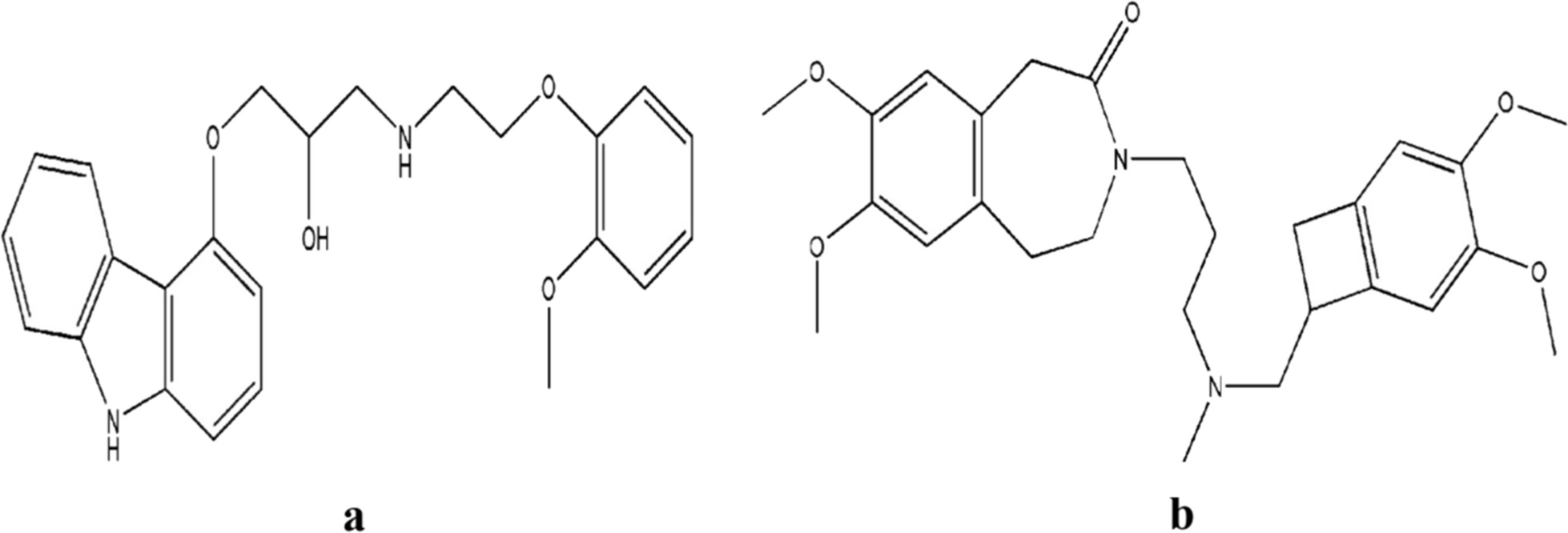


The λmax for LTB and BCA was found at 248 nm and 260 nm when scanned individually in UV–Vis spectrophotometer (UV-2600, Shimadzu, Japan). The isosbestic point for both drugs was found at 253 nm (Additional file 1: Fig. S1), giving maximum absorbance. All the measurements in the RP-HPLC were made at 253 nm (isosbestic point).
RP-HPLC method developmentThe quadratic design models from the Box–Behnken design type (Version 13.0.5.0) were employed for all the resulting responses. The model was decided based on p-value, F-value, and adjusted R2 values for each response (R1—area of LTB, R2—area of BCA, R3—peak resolution, R4—tailing factor of LTB, and R5—tailing factor of BCA). The p-values, F-value, and R2 values for R1, R2, R3, R4, and R5 were found significant . The quadratic model was validated employing normal plots of residuals and residual vs predicted plots (Additional file 1: Fig. S2). The plots depicted no such significant deviation from the center line of the normal plot explaining the linearity. The residue is not only randomly distributed on each side of the zero line but also falls within the range of 2 (even lower than the permissible range of 3). Therefore, the finding of such distribution suggested linear model is appropriate, devoid of systematic error and suitable for use in optimizing the RP-HPLC method.
The analysis of variance was employed to study the significant terms (factors) using p-value and F-value. The ANOVA tables (Additional file 1: Tables S1–S5) for each response resulted from variation in significant factors R1(A, B, AB, B2), R2(A, B, C, AB, A2, B2), R3(A, B, AB, B2, and C2), R4(A, C, AC, B2, C2), and R5(A, C, AC, B2, C2) with p and F-values for each factor < 0.05 and > 5. To explain the unit correlation of each factor with their responses, the coded equations were obtained with high-level factors (+ 1) and low levels of factors (− 1).
$$\begin }1 = \, & + \,1021 + 233.37A - 502.04B 55.81C \\ & \, \,350.20AB + 12.75 AC + 87.63 \\ & \, \,111.39 A^ + 325.84 B^ + C^ \\ \end$$
(3)
$$\begin }2\user2\, = & \, + \,1423 + 366.42A - 549.8 B 133.35C \\ & \, - \,450 AB - 4.05 AC + 177.75 BC \\ & \, \,227.04A^ + 275.10B^ - 74.85C^ \\ \end$$
(4)
$$\begin }3 = & \, + \, 7.65 - 1.96A 4.04 B 0.123C \\ & \, + \,1.84 AB + 0.015AC 0.107BC \\ & \,\, 0.826A^ + 0.98 B^ - 1.03C^ \\ \end$$
(5)
$$\begin }4 = & \, + \, 1.16 0.0875 A - 0.0112 B + 0.243C \\ & \, + \, 0.0175AB 0.137AC + 0.070BC \\ & \,\, 0.075A^ + 0.102B^ + 0.407 \\ \end$$
(6)
$$\begin }5 = & \, + \,1.21 - 0.078A 0.0513B + 0.225C \\ & \,+\, 0.0300AB - \,0.1375AC + 0.0325BC \\ & \, +\, 0.040A^ \, 0.105 B^ + 0.422C^ \\ \end$$
(7)
The coded equation depicted a correlation among the responses (R1–R5) where positive sign (+) displayed factors are directly correlated to responses and negative sign (−) displays indirectly correlated values. The increase in mobile phase ratio increases the area of LTB and BCA as shown in the (Eq. 3) owing to more solubility of LTB and BCA in ACN than water. An indirect correlation between flow rate, pH of an aqueous phase, and area of both drugs was displayed (Eqs. 3, 4). The lower flow rate gives enough time for the analyte to interact with the stationary phase, thus producing more retention with more area and inverse in case of higher flow rate. It also affects the peak resolution based on their solubility differences with the mobile phase. As given in Eqs. 3–6 negative correlation was established among flow rate, pH, and area of LTB, BCA, and peak resolution. The pKa of LTB and BCA was 5.05 and 6.5; thus, according to the rule of pH ± 2 from pKa the pH of the aqueous phase was selected. At pH closer to the pKa of the drugs, both drugs were in ionized fraction and thus reduction in pH up to 4 offered a higher area for both drugs. However, more reduction in < pH 4 makes BCA ionized, thus produced with a lesser area. The higher pH was found to produce peaks with more tailing factors than the lower pH. Thus, pH of the aqueous phase affects the area and tailing factors of LTB and BCA significantly (Eqs. 6, 7).
Visualization of dataThe correlation of each factor with responses of RP-HPLC parameters was established. Though, the additive correlation was established by employing two factors at a time and response. The significant factors were taken in such response surface methodology (RSM) studies. Five different RSM plots were obtained explaining the additive correlation among factors and responses. As shown in Fig. 2A, B, a steep increase in the slope of the graph can be observed depicting a positive correlation with factors A and B. Figure 2C depicts a positive correlation among mobile phase, flow rate, and peak resolution. As already described, a decrease in pH and a higher mobile phase ratio of ACN reduces the tailing factor of both peaks as depicted in Fig. 2D, E.
Fig. 2
3D-Response surface plots depicted the correlation between the significant factors and responses A. Area of LTB (R1) versus AB, B. Area of BCA (R2) versus AB, C. Peak resolution (R3) versus AB, D. Tailing of LTB (R4) versus AC, and E. Tailing of BCA (R5) versus AC
OptimizationAfter variations in chromatographic conditions, several chromatograms were obtained with different retention times, resolution, separation factors, asymmetrical factors, and areas according to the solutions generated from BBD. The overlay plots from factors A, B, and C were obtained in Fig. 3A, B, exhibiting probable solutions according to our desirability. The chromatograms with the higher area, more resolution (> 4), lesser asymmetrical factor (< 1.5), and more theoretical plates number (> 5000) were considered for method development among all solutions [29]. The desirability of each response can be achieved using a mobile phase ratio between 83:17 and 86:15, flow rate 0.35–0.4 mL/min, and pH of aqueous phase 3.5–4.
Fig. 3
Optimization of factors (A–C) for their desired responses A. Overlay plot of factor AB for their solutions B. Overlay plot of factor BC for their solutions
The finally optimized chromatograms for BCA and LTB (Fig. 4A) were obtained at conditions suggested using BBD as mobile phase ratio (Acetonitrile: Acidified water pH 4) 85: 15, flow rate 0.4 mL/min at 30 °C column temperature conditions. The responses were recorded as (R1) area of LTB 1071 ± 53.66 and (R2) area of BCA 1422 ± 16.28, (R3) peak resolution 6.61 ± 0.007, (R4) Tailing factor of LTB chromatogram 1.153 ± 0.011 and (R5) Tailing factor of BCA chromatogram 1.116 ± 0.02. The 10 µl volume of both drugs in different concentrations was injected that retained on the stationary phase (YMC-Pack Pro C18, 150 × 4.6 mm L.D. S-5 µm,12 nm) at Rt 4.09 min (BCA) and 5.15 min (LTB) to give a straight line for quantification of known concentrations.
Fig. 4
A. Chromatograms of BCA and LTB at retention time 4.09 min and 5.15 min, respectively B. Standard calibration curves of BCA and LTB
Assay validationLinearity and rangeThe standard calibration curve was plotted for both drugs and found to be linear over the concentration range of 0.5–32µg/mL. The coefficient of linear regression (R2) for LTB was found to be 0.9998 with its linear equation y = 153.43x-15.202 (Fig. 4B). However, linear regression analysis of BCA resulted in an R2 value of 0.997 with its linear line equation y = 93.987x + 11.254 (Fig. 4B).
AccuracyAccuracy is the degree of closeness between observed values and standard values and is generally expressed as percentage recovery. The quality control samples (LQC, MQC, and HQC) for LTB and BCA exhibited higher % recovery in the range of (94–103%) as given in Table 3. The % average bias (difference from standard value) results depicted acceptable values, i.e., ± 5%, thus confirming the accuracy of the proposed method for the quantification of both drugs simultaneously.
Table 3 Accuracy values at different levels of the validated analytical methodPrecisionThe precision can be assessed utilizing % RSD values that determine the existence of random error on repeatability. The data from quality control samples (LQC, MQC, and HQC) of both drugs were analyzed for % RSD, % accuracy, and % average bias for their intra-day and inter-day precision determination. Table 4 explains the intra-day precision values that were found to be in the acceptable range of < 2% RSD and ± 5% average bias.
Table 4 Intra-day precision values at different levels of the validated analytical methodThe values from inter-day (3 consecutive days) precision calculations in Table 5 depicted lower values of % RSD (< 2) and % average bias (± 5). The overall findings from precision determination (intra-day and inter-day) demonstrated the proposed RP-HPLC method is more precise for determining LTB and BCA simultaneously [29, 31, 32].
Table 5 Inter-day precision values at different levels of the validated analytical methodSensitivityThe standard error (SE) intercept for LTB and BCA from their calibration curve was found to be 0.164 and 0.085, respectively. The values were further calculated to give LOD and LOQ for LTB at 0.01 µg/mL and 0.030 µg/mL, while the LOQ and LOD for BCA were determined to be 0.008 µg/mL and 0.025 µg/mL, respectively. The measured values are sufficient for detecting and quantifying LTB and BCA with accuracy and precision.
$$\begin & }\, = \,3.3 \times \left( }\,\,}\,\,}\,\,}} \right) \\ & }\, = \,10 \times \left( }\,\,}} \right) \\ \end$$
RobustnessThe results shown in Table 6 confirmed the robustness of the proposed RP-HPLC method with no significant difference in resolution, tailing factor, and theoretical plate number values. The finding from the study reflects the reliability of the robustly developed method for estimating two drugs concurrently.
Table 6 Robustness of the validated analytical methodSystem suitabilityAfter 10 µg/mL injection of both drugs, the well-resolved chromatograms with their Rt values at 4.09 min for BCA and 5.15 min for LTB were obtained, having a resolution value of 6.61 ± 0.007. The tailing factor played an important role in explaining the symmetry of the peak and was found to be within acceptable limits of < 1.5, according to USP. All the other parameters of RP-HPLC, i.e., theoretical plates and height equivalent to theoretical plates (HETP), were found to be acceptable limits as shown in Table 7. The results from such observation depicted the system suitability of the validated analytical method.
Table 7 System suitability parameter of the validated analytical methodApplication of RP-HPLC method in drug degradation studiesQuantification of force-degradant products using RP-HPLC and HRMSThe degradation behavior for both drugs under acidic, basic hydrolytic, and oxidative conditions was reported through % recovery, as shown in Table 8. The individual drugs were accessed for each degradant by use of RP-HPLC. In the case of LTB, major degradation was found in basic hydrolytic and oxidative conditions with percentage recovery of 57.79 ± 7.9 and 59.25 ± 3.9%. The degradants found in LTB were observed to be retained on the stationary phase of RP-HPLC (Fig. 5A1–D1) explains the specificity of the proposed method for LTB.
Table 8 Percentage recovery of LTB and BCA after different stress conditionsFig. 5
Chromatograms obtained after A1-2. Acidic (0.1N HCL) B1-2. Basic (0.1N NaOH), C1-2. Neutral (H2O) and D1-2. Oxidative (3% H2O2) treatment of LTB and BCA at 80 °C for 2 h
However, BCA undergoes basic and oxidative degradation to a more considerable extent. The results from the oxidative degradation study showed that BCA undergoes an oxidative coupling reaction. The observed degradants of BCA were found to be quantified through the developed RP-HPLC method with a peak resolution of more than 1.5 (Fig. 5A2–D2), thus supporting the specificity of the method.
HRMS studies of LTB, BCA, and their DPsThe study employed to find the major degradants of LTB and BCA after stress conditions. LTB and BCA showed protonated ions at m/z 427.11 and 285.07, respectively, depicting the characteristic m/z according to the literature. Further, HRMS analysis of respective LTB and BCA was performed. The LTB shows (Fig. 6A) a base peak at m/z 427.11, with its major fragments (m/z 410.34, 392.06, 370.05, 355.0, 338,327, and 312.02) [40]. The prediction model (Zeneth) depicted the total eighteen degradants of LTB, namely LD1–LD18, under different stress conditions, as shown in Table 1. However, experimentally, seven major degradants were quantified through their m/z charge ratio i.e., related to the degradant average mass (Fig. 6A–H). The mass accuracy error was found to be < 10 ppm for each degradant.
Fig. 6
HRMS spectra of (A) LTB (B) LD1, (C) LD4, (D) LD5, (E) LD6, (F) LD11, (G) LD14 and (H) LD16
The observed degradants were LD1, LD4, LD5, LD6, LD11, LD14, and LD16 having m/z ratios 58.06, 102.05, 445.12, 227.05, 144.02, 418.04, and 595.08 respectively. Briefly, all the degradants were formed due to the loss or gain of molecular mass from parent ions (LTB) or daughter ions (degradants) (Additional file 1: Fig. S3). The FDS caused parent ions to generate four major degradants with their loss in the molecular formula from LTB as LD1 formed due to loss of—C18H12ClN3O4 from LTB, similarly LD4;—C4H5NO, LD5;—HN, and LD6;—C11H8N2O2 . The formation of LD11, LD14 and LD16 were driven by the loss of–C4H6N2 from LD3,–C10H10ClN3 from LD5, and addition + C7H3NO from LD6. More studies using NMR analysis need to be conducted in the future to further characterize each degradant [34, 41].
A total of twenty-four BCA degradants (BD) were obtained with their mass error value < 10 ppm. The BCA degrades owing to hydrolysis or coupling reactions between the degradants. Different isoforms consisting of similar molecular average mass or formulas and different structures were examined. The average mass of degradants was found for each degradants as BD4: 566.51, BD5: 566.51, BD6: 566.51, BD14: 848.76, BD15: 848.76, BD16: 1131.1, BD17: 1131.1, BD18: 848.76, BD19: 1131.1, D20: 564.5, BD21: 564.5, BD22: 580.5, BD23: 580.5, BD24: 580.5, BD25: 580.5, BD26: 580.5, BD27: 848.76, BD28: 1131.01, BD29: 564.5, BD30: 580.5, BD31: 1131.01, BD32: 564.5, BD33: 564.5, and BD34: 564.5 (Fig. 7B–F).
Fig. 7
HRMS spectra of (A) BCA (B) BD4, BD5, and BD6 (C) BD14, BD15, BD18, and BD27 (D) BD16, BD17, BD19, BD28, BD31 (E) BD20, BD21, BD29, BD32, BD33, and BD34 (F) BD22, BD23, BD24, BD26, and BD30
The types of degradants were formed due to coupling reactions among the degradants, as also illustrated in the in silico prediction study. A better understanding of each degradant for its isomers can result from an NMR study in the future.
In this study, to explain the underlying mechanism of each degradant, major common degradants with their average masses were considered from their parent moieties as depicted in (Additional file 1: Fig. S4). The degradants D4, D5, and D6 with molecular mass at m/z ratio 566.51 were formed due to the addition of +C16H10O5 in the parent BCA moiety. The degradants with an average mass of 848.76 m/z, i.e., BD14, BD15, BD18, and BD27, were formed owing to oxidation coupling of phenol (C32H20O10) with parent BCA and degradants (D4, and D5). The degradants (BD16, BD17, BD19, BD28, BD31) with an average mass of 1131.01 were formed owing to the oxidation coupling between C48H30O15 and degradants D4, D5, and D6 individually. An average mass of 564.5 at their m/z resulted in degradants (BD20, BD21, BD29, BD32, BD33, and BD34) due to the oxidation of biphenyldiol to biphenyldione with a gain of molecular formula (C16H8O5) in D4, D5, and D6. Finally, degradants (BD22, BD23, BD24, BD26, and BD30) with an average mass of 580.5 at m/z were formed owing to oxidation coupling between C16H8O6 and degradants D4, D5, and D6 individually[33, 34, 41].
In-silico degradation toxicityThe in-silico and experimental observations reported different degradants for the LTB and BCA. The Derek analysis using different animals models resulted in numerous toxicities for drugs and their derivatives. A total of eighteen drug alerts were found from in silico toxicity studies for BCA. However, LTB possesses only two drug alerts for their derivatives (Additional file 1: Table S6).
Development of drug nanocarriersSelection of solventThe saturation solubility of BCA and LTB was determined to select the best possible solvent to formulate the PLGA NPs. The solubility of BCA in water, methanol, acetone, acetonitrile, DMF, ethanol, DCM, and DMSO were found to be 0.007 ± 0.003 mg/mL, 9.90 ± 0.15 mg/mL, 26.48 ± 0.15 mg/mL, 13.27 ± 0.19 mg/mL, 24.83 ± 0.32 mg/mL, 12.03 ± 1.047 mg/mL, 1.34 ± 0.003 mg/mL, and 33.87 ± 1.74 mg/mL respectively (Fig. 8). However, the solubility of LTB in the same solvents was determined as 0.57 ± 0.146 mg/mL, 4.93 ± 0.44 mg/mL, 1.19 ± 0.049 mg/mL, 0.17 ± 0.244 mg/mL, 3.33 ± 0.33 mg/mL, 0.72 ± 0.075 mg/mL, 0.63 ± 0.08 mg/mL, and 10.56 ± 0.209 mg/mL respectively (Fig. 8).
Fig. 8
Solubility determination of BCA and LTB in different solvents
The cosolvents acetone and methanol were used to prepare the PLGA NPs owing to the higher solubilities of drugs and polymers. However, methanol was adopted as a solvent to dissolve the lipid, LTB, and BCA during the production of CBs.
Particle size, PDI, and zeta potentialThe particle size and zeta potential for PLGA NPs were found to be 185.3 ± 12.3 nm and − 13.3 ± 0.35 mV Fig. 9A1, B1.
Fig. 9
Particle size and zeta potential of A1-2 PLGA NPs and B1-2 CBs
The particles were found to be of uniform size with a PDI value of 0.183 ± 0.06. However, CBs were observed to exhibit the 182.4 ± 16.3 nm aerodynamic particle size with a slight deviation in uniformity with a PDI value of 0.213 ± 0.05. The CBs were examined to possess − 10.8 ± 0.39 mV zeta potential. The results from such findings depicted that the PLGA NPs and CBs were of uniform nanosized range particles.
% EE and %DL of LTB and BCA in PLGA NPs and cubosomesThe PLGA is well known to encapsulate the hydrophobic and hydrophilic drugs inside the matrix. The % EE of the LTB and BCA in PLGA NPs were found to be 53.64 ± 4.81% and 61.29 ± 4.67%, respectively. The higher % EE of BCA was examined owing to the inherent hydrophobic nature of the former. Additionally, it was discovered that both types of drugs were more effectively captured by the CBs, for that %EE of BCA was found to be 72.88 ± 5.52% and LTB was 55.62 ± 7.73%. The housing that BCA and LTB were able to hold inside the carrier was provided by the liquid crystalline structure. The % DL was found to be 5.42 ± 1.10% and 4.43 ± 0.77% for BCA and LTB-embedded CBs, whereas PLGA NPs exhibited lower % DL with 3.7 ± 0.46% and 4.63 ± 0.48% content of BCA and LTB, respectively.
In-vitro drug release and kinetics studiesThe quantification of LTB and BCA release from coarse suspension, polymeric, and lipidic nanocarriers at different time intervals was studied using RP-HPLC. The coarse LTB and BCA were found to show significant differences in release performance owing to the hydrophilic or hydrophobic nature of both drugs, as shown in Fig. 10A, B.
Fig. 10
In-vitro drug release A. Cumulative percentage of LTB release from Coarse-suspension of LTB, LTB- PLGA NPs, and LTB-CBs, B. Cumulative percentage of BCA release from Coarse-suspension of BCA, BCA- PLGA NPs, and BCA-CBs
A long-sustained drug release effect for up to 72 h was observed from PLGA and CBs. However, a slight difference was observed in release performance and release kinetics of the LTB and BCA from PLGA NPs and CBs (Fig. 10A, B). BCA was found to exhibit a longer release pattern (92.80 ± 9.34%) due to the entrapment of hydrophobic content in the hydrophobic polymer (PLGA). It follows the first-order release kinetics mechanism from PLGA NPs with its highest regression coefficient (R2) of 0.991 and AIC value of 40.46 with a 0.038 release rate constant (K). A faster LTB release (73 ± 4.9% in 12 h) was observed from PLGA than BCA (36 ± 4.0% in 12 h) owing to more hydrophilicity of the LTB than BCA. An LTB was found to obey the First-order release kinetics mechanism with R2 0.934, AIC 59.23, and K = 0.82.
The CBs are well known to entrap hydrophilic and hydrophobic moieties in their core compartment region. LTB and BCA exhibited sustained release patterns from CBs with 95.30 ± 6.6% and 90.308 ± 2.09% release of BCA and LTB after 72 h, respectively. The release kinetics of BCA follows 1st order release mechanism with R2 ;0.98, AIC ;46.28, and K ;0.055 values. However, LTB was found to follow the Higuchi model with the highest R2 ;0.973, AIC ; 51.19, and 11.53 release constant values (Table 9).
Table 9 Release rate kinetics of BCA and LTB release from PLGA NPs and CBs
留言 (0)