
Remember me
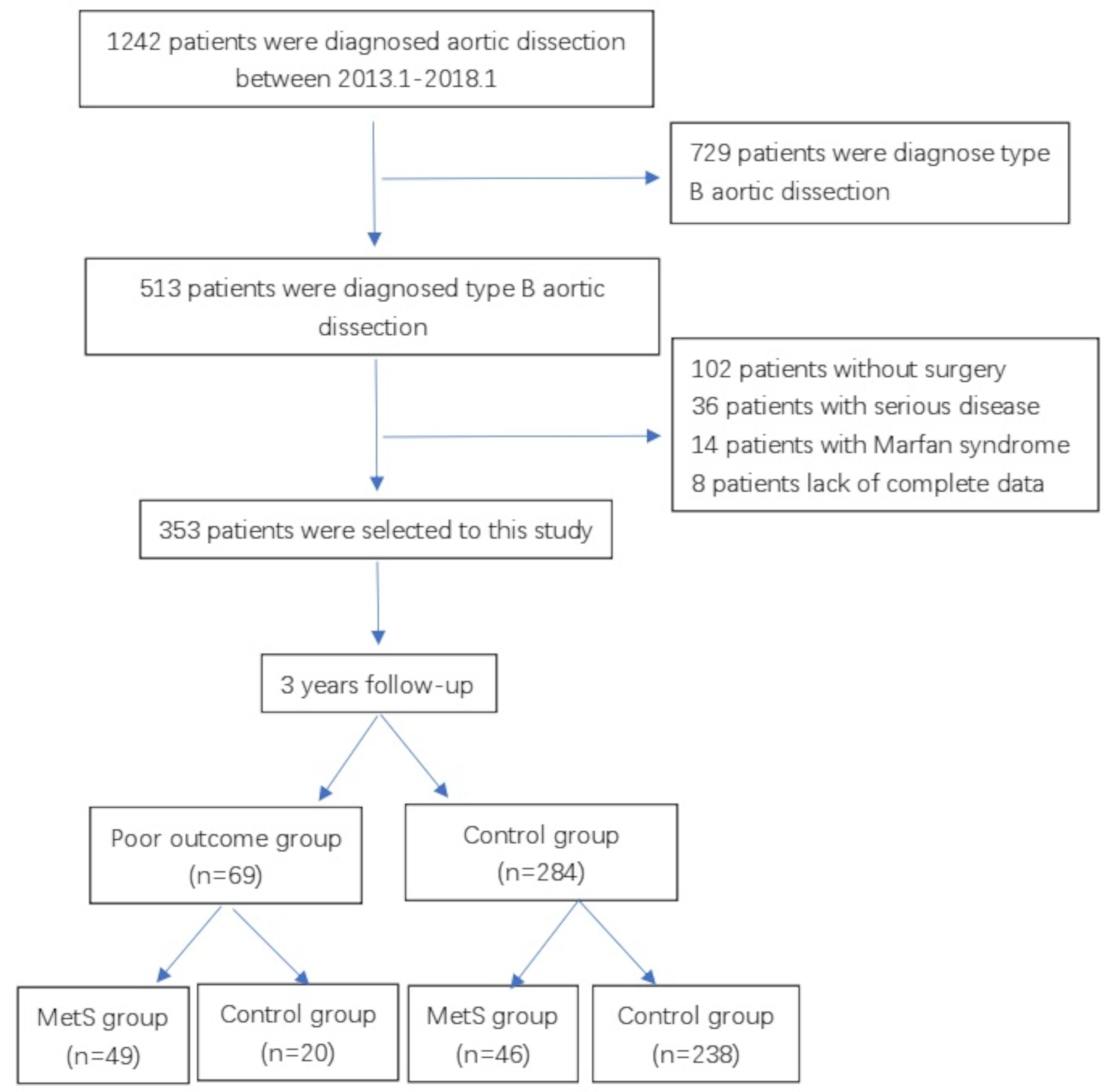


This retrospective study included 249 consecutive patients who underwent cardiac surgery at Toda Chuo General Hospital between April 2017 and December 2019. The following patients were excluded: 2 patients who underwent minimally invasive cardiac surgery, 1 with chest closure using SternaLock Blu (Biomet Microfixation CO. Ltd., Jacksonville, FL), 26 with chest closure using Fixsorb wave (Teijin Medical Technologies CO. Ltd., Osaka, Japan), and 2 who could not be withdrawn from cardiopulmonary bypass. Ultimately, the study included 218 patients.
For basic sternal fixation, conventional wire cerclage was used. Between July 2018 and December 2019, we used a three-piece mesh plate for full sternotomy (group M, n = 109). For comparison, group P (n = 109) included patients who underwent chest closure using a pin between April 2017 and February 2019. We selected patients based on the expectation that external fixation with mesh would be effective for group M due to the presence of sparse sternal marrow and internal fixation with sternal pins would be effective for group P. Data on the postoperative course, bleeding, infection, and computed tomography (CT) were compared between the groups. This study was approved by the Institutional Review Board of Toda Chuo General Hospital (approval number: 0517; received on November 29, 2021).
Preoperative managementOff-pump coronary artery bypass grafting was performed with oral aspirin, and surgery using a cardiopulmonary bypass was performed 7, 5, 5, and 2 days after discontinuation of aspirin, clopidogrel, warfarin and novel oral anticoagulant, respectively. This preoperative management was routine practice for patients taking anticoagulant medication at our institution. It was not specifically discontinued for the purpose of this study.
Surgical procedureAll patients underwent sternal closure with conventional wire cerclage without bone wax that was performed by a single surgeon. Six simple single wires were used; two transsternal and four parasternal wires were placed at the manubrium and sternal body, respectively. The mesh plate and all corners of each plate were cut with scissors (Fig. 1A). The schema of the mesh plate implantation method is shown in Fig. 1B. The mesh plate was fixed by passing two transsternal and parasternal wires (first and third) through the holes on the mesh plate. Subsequently, each wire was tied, and two mesh plates were implanted on the posterior surface of the sternum. The sternum was closed using three mesh plates; two on the posterior surface and one on the anterior surface. Figure 2 shows the procedure for sternal closure with a three-piece mesh plate. From deep to superficial, the pectoral fascia was closed with 0 Vicryl sutures in a continuous pattern. The subcutaneous tissue was closed with 2 − 0 Vicryl sutures in a continuous pattern after placement of a 10-Fr subcutaneous drain, and the skin was closed with 5 − 0 polydioxanone clear sutures in a continuous pattern. In group P, the above procedure was also performed except that instead of a mesh, one pin each was inserted into the sternal manubrium and body. All patients received prophylactic antibiotic treatment with cefazolin (dosage based on individual patient weight) for 48 h postoperatively.
Fig. 2
How to perform sternal closure with sandwiched three-piece mesh plate. A) The first mesh plate is threaded through the holes on the plate with the first manubrium wire. B) The second mesh plate is threaded through the holes with the third parasternal wire. C) The sternum is closed with six wires; two transsternal wires at the manubrium and four parasternal wires at the sternal body. D) When the sternum is closed, two mesh plates are implanted on the posterior side of the sternum. E) When closing the wire, the silk is towed so that the mesh plate fits on the posterior side of the sternum. F) The chest is closed using three mesh plates with the sternum sandwiched in between, with two mesh plates located posteriorly and another anteriorly
Postoperative managementOnce admitted to the postoperative intensive care unit (ICU), activated clotting time was measured hourly and maintained at < 120 s by administering protamine as needed. Postoperative blood glucose levels were monitored hourly and maintained at < 180 mg/dL by continuous intravenous insulin administration. Drains were placed in the pericardial and thoracic cavities as well as retrosternally. The effluent volume was measured hourly with milking of the drainage as needed. Sedation was discontinued when the drainage rate was < 30 mL/h, and the patient was extubated from the ventilator when he was fully conscious and oriented. The criterion for leaving the ICU was having a hemodynamically stable condition and being able to walk 50 m. All patients underwent chest CT on postoperative day 5 to assess the sternum displacement and check for substernal hematoma.
Definitions and endpointsThe primary endpoint was the postoperative sternum displacement. The secondary endpoints included the amount of bleeding, length of ICU and hospital stays, rate of deep sternal wound infection, degree of substernal hematoma (Fig. 3), sternal healing, and dehiscence using CT data on postoperative day five. After cardiac surgery, because of the incision of the pericardium, blood from the sternum may flow into the pericardial cavity and be aspirated. Therefore, we used the total drainage volume, including bleeding from the sternum, as an indicator to more accurately assess the amount of bleeding from the sternum. To evaluate sternal healing, we adopted methods from previous studies [10, 11]. Deep sternal wound infection was defined according to the following criteria: (1) bacteria could be isolated from cultures of mediastinal tissue or fluid; (2) evidence of mediastinitis was observed during surgery; and (3) presence of chest pain, sternal instability, or fever (> 38℃) with either purulent mediastinal discharge or bacterial isolation from a blood culture of drainage fluid originating from the mediastinal area [12].
Fig. 3
Grade of substernal hematoma. A) Substernal hematoma is not present on CT. B) Slight substernal hematoma is seen on CT. C) Substernal hematoma equal to or larger than the size of the sternum, up to twice the size. D) Substernal hematoma larger than twice the size of the sternum. CT, computed tomography
Statistical analysesContinuous and normally distributed data are presented as means ± standard deviations, while categorical data are expressed as percentages. All analyses were performed using JMP Pro version 15 (SAS Institute, Cary, NC, USA). Statistical significance was set at p < 0.05. Between-group differences were evaluated using the Chi-square test for categorical variables and the t-test or Mann–Whitney U test for continuous variables. We compared the baseline characteristics, operative data, and postoperative complications of groups P and M using the Pearson 2 test for categorical variables, the t-test for continuous variables, and the Mann–Whitney U test for continuous skewed data. We evaluated independent predictors for preventing substernal hematomas using logistic regression analysis. We categorized CT-detected substernal hematomas as ‘hematoma present’ if they were Grade 2 or higher, and as ‘hematoma absent’ if they were Grade 1.
Comments (0)