
記住我
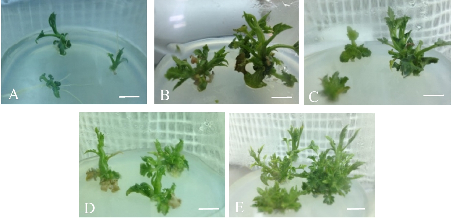
Multiple shoot proliferation was observed on explants in both experimental groups (Fig. 2). Shoot proliferation occurred via indirect somatic embryogenesis with callus formation preceding shoot formation in groups. However, the extent of callus formation varied between all groups before giving rise to shoot formation. For the BAP:Kinetin group, there was a significant difference between treatment groups (N = 249, P = 0.0436) with 59.26% explant shoot response occurring on MS media containing 0.5 mg L−1 BAP (Table 2). Analysis using the Analyze Binary Response for Factorial Design tool in Minitab (2023) was done to determine main and interaction effects of PGRs on response. An ANOVA showed that only BAP had a significant effect on response (P = 0.018; Table 3A) with no significant effects from kinetin or interaction with BAP. Kruskal–Wallis analysis was used due to non-normal distribution of size data. One-way ANOVA showed that only BAP had a significant effect on plant size (P = 0.0058; Table 4).
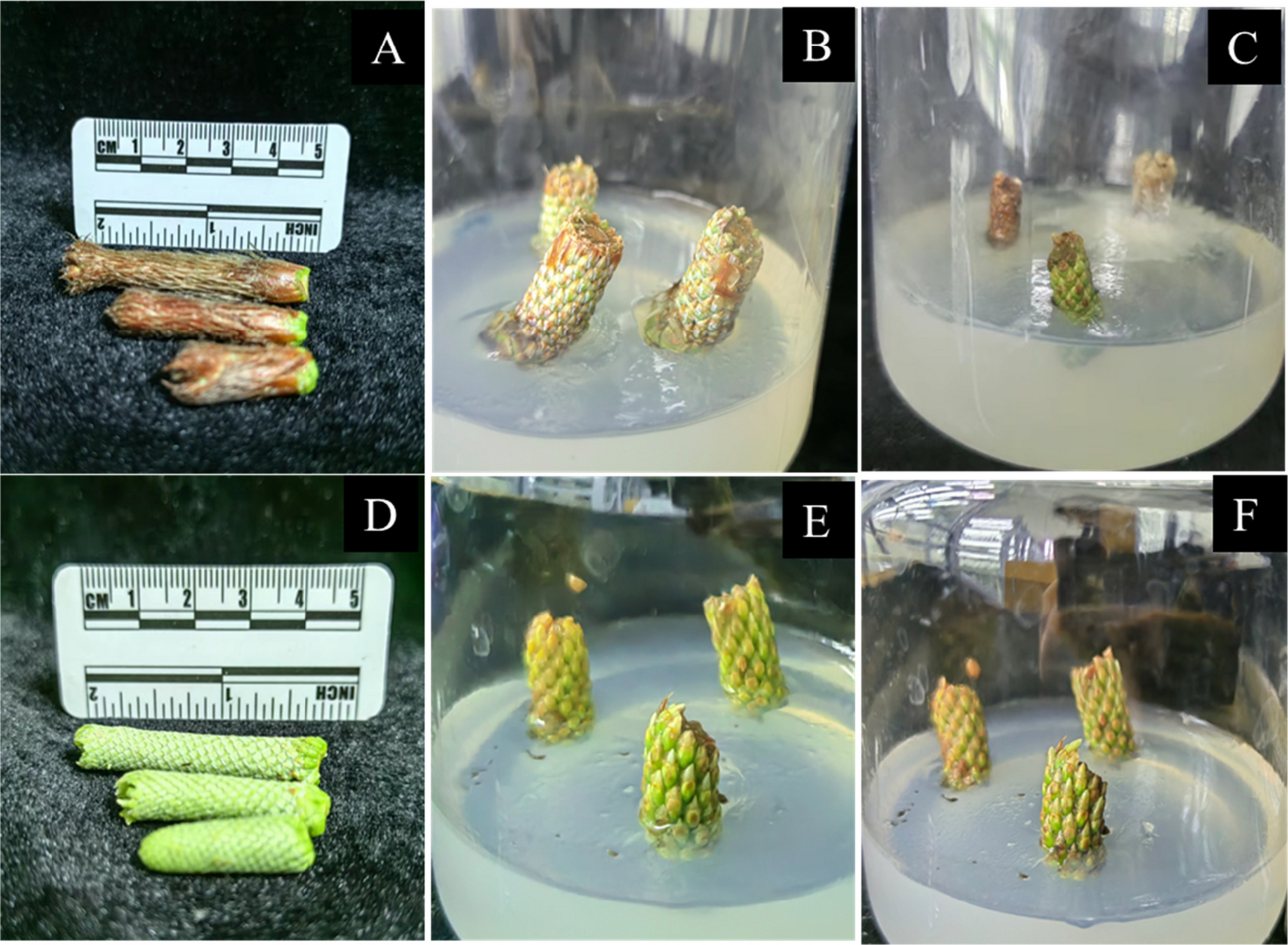
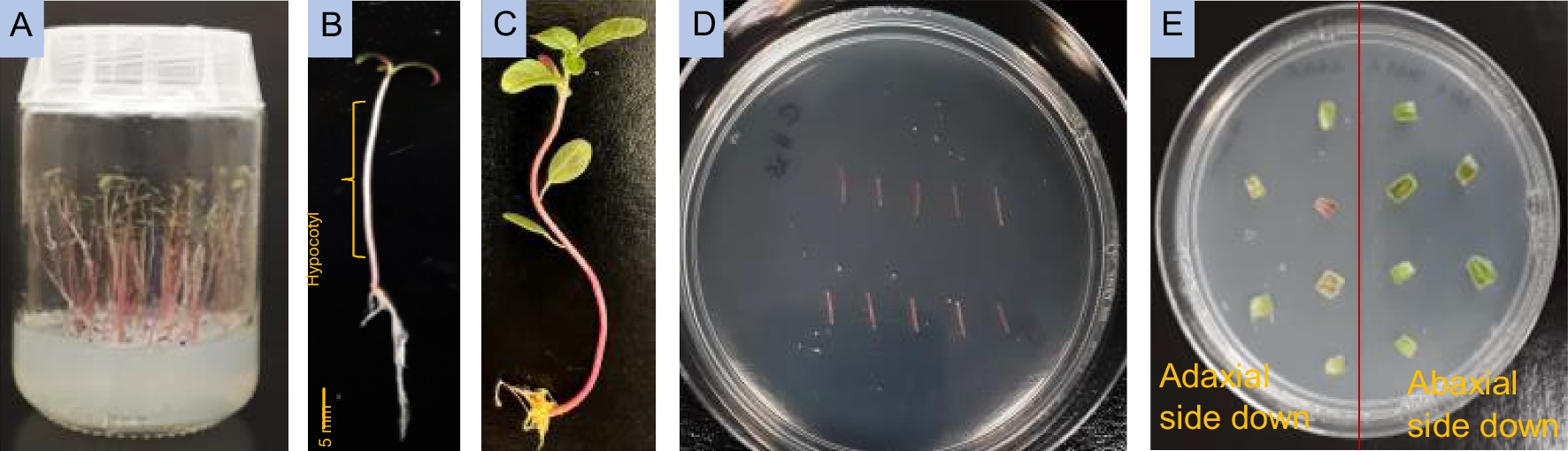
Figure 2.
Lepidium ostleri S.L. Welsh and Goodrich plantlet displaying shoot organogenesis from leaf tip explants 17, 24, 57, and 71 d after culture on Murashige and Skoog medium containing 0.5 mg L.−1 BAP (6-Benzylaminopurine).
Table 3. ANOVA tables for main and interaction effects of (A) BAP (6-Benzylaminopurine), and kinetin and (B) BAP and IAA (indole-3-acetic acid) on inducing shoot response in Lepidium ostleri S.L. Welsh and GoodrichTable 4. One-way ANOVA tables for analysis of the effect of BAP (6-Benzylaminopurine), and kinetin on plantlet size in Lepidium ostleri S.L. Welsh and Goodrich using the Kruskal–Wallis testResponse and vigor peaked at the upper limit of BAP concentrations in the BAP:Kinetin group (0.5 mg L−1), which indicated that the organogenic response may continue to rise with an increase of BAP concentration in culture media. The response was also relatively high both in the control (T1) and in treatment 2 containing 0.2 mg L−1 kinetin without the addition of BAP. While BAP has the largest effect on response rates, the interaction between two cytokinin hormones at lower concentrations may be detrimental to optimal growth in L. ostleri. Although previous studies examined Lepidium response to a combination of two cytokinin hormones, it is recognized that both cytokinin and auxin signaling are required for callus and shoot apical meristem development (Schaller et al. 2015). However, there was no interaction effect seen in analysis (Table 3A).
In the proceeding BAP:IAA experiment, the response was still substantially higher at the upper limit of tested BAP concentrations (N = 180, P < 0.001). Interaction effects of PGRs on response could not be analyzed by the same model as the BAP:Kin group due to quasi-complete separation (see ANOVA in Table 3B). Analysis of main effects showed that only BAP had an effect on response frequency (P < 0.001; Table 3B) and on plant size with Kruskal–Wallis tests (P < 0.0001; Table 5). At the highest concentration of BAP (5.0 mg L−1), response declined as IAA concentration increased. While the development of shoot meristems is dependent upon auxin-cytokinin interactions, redirection of auxin transport, and gene expression controlling shoot meristem organization (Motte et al. 2014), shoot response still seemed to be primarily driven by BAP.
Table 5. One-way ANOVA tables for analysis of the effect of BAP (6-Benzylaminopurine), and IAA (indole-3-acetic acid) on plantlet size in Lepidium ostleri S.L. Welsh and Goodrich using the Kruskal–Wallis testThe source and developmental conditions of explant tissue may account for notable differences between the two experimental groups. Specifically, the response observed in the BAP:Kinetin control group, which was absent from the BAP:IAA control group, may have resulted from potentially increased endogenous PGRs in the source tissue. Because this tissue was derived from a plant grown in vitro on MS containing BAP and IAA, it is likely that PGRs remained relatively elevated in tissue even after several weeks of culture on plain MS preceding experimental use. In contrast, the source tissue used in the BAP:IAA experiment was not exposed to exogenous PGRs during its development.
Rooting and acclimatizationApproximately 61% of plants across treatment groups survived the rooting and acclimatization process. These plantlets proved to be photosynthetically capable, responded well to transfer from nutrient-rich media to soil, and displayed strong overall root development (Fig. 3). Treatments with 200.0 and 400.0 mg L−1 IBA had the highest survival rates, achieving 83% and 75% survival, respectively. Treatments with 100.0 and 300.0 mg L−1 had the lowest overall survival rates at 33% and 50%, respectively (Table 6). These results suggest that pulse treatment of L. ostleri shoots with 400 mgL−1 IBA was most effective in promoting root development, though no statement of significance can be made using logistic regression due to small treatment size. The control group achieved a 58% survival rate, indicating adequate levels of endogenous phytohormone needed for rooting. It is unclear without further experimentation whether these levels are typical of L. ostleri or are the result of hormone absorption of the in vitro-cultured plant used as an explant source.
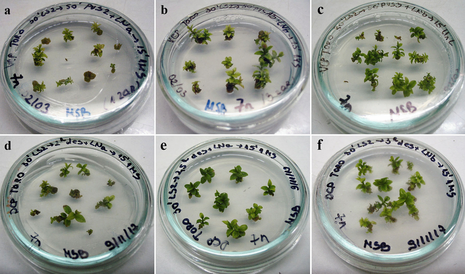
Figure 3.
Strong root development displayed in Lepidium ostleri S.L. Welsh and Goodrich plants (T6) 28 d after pulse treatment with IBA (indole-3-butyric acid) and transfer to sterile soil.
Table 6. Percentage of plantlets that formed roots, through de novo development or through root reformation, after pulse treatment with IBA (indole-3-butyric acid)Approximately half of the plants utilized for the rooting experiment developed roots in vitro prior to treatment. Although all previously developed roots were removed along with the culture media, 83% of these plants reformed their roots after transfer to soil (Table 6). Alternatively, only 46% of the plants lacking roots in vitro developed roots following treatment and transfer to soil. This observation represents a confounding factor and makes it difficult to determine the full effectiveness of the IBA pulse treatments without further experimentation. This observation also supports the practice of selecting L. ostleri plantlets featuring in vitro root development for the ex vitro rooting phase, regardless of whether these roots are preserved for the rooting phase.
The use of autoclave-sterilized soil and aseptic technique was highly effective in preventing contamination among healthy plantlets during the high-humidity phases of root induction and initial acclimatization. Sterile conditions during initial root induction have been used by other researchers when employing ex vitro rooting approaches (Arya et al. 2003; Shekhawat et al. 2015a; Shekhawat et al. 2015b; Phulwaria et al. 2013; Sharma et al. 2017). These conditions allowed for necessary high humidity levels to be maintained during the 4 wk of root induction and for humidity to be lowered gradually thereafter without microbial proliferation and plant decay.
Plantlets developed in vitro from both treatment groups displayed variation in morphology during shoot and root development. When utilizing plants developed in vitro, it is important to consider the extent of phenotypic variation arising from in vitro regeneration. While differences were observed throughout different experimental phases, no formal assessment was performed to quantify the extent of somaclonal variation. During the acclimatization process, plantlets displayed more typical morphology in leaves, color, and growth habit.
留言 (0)