
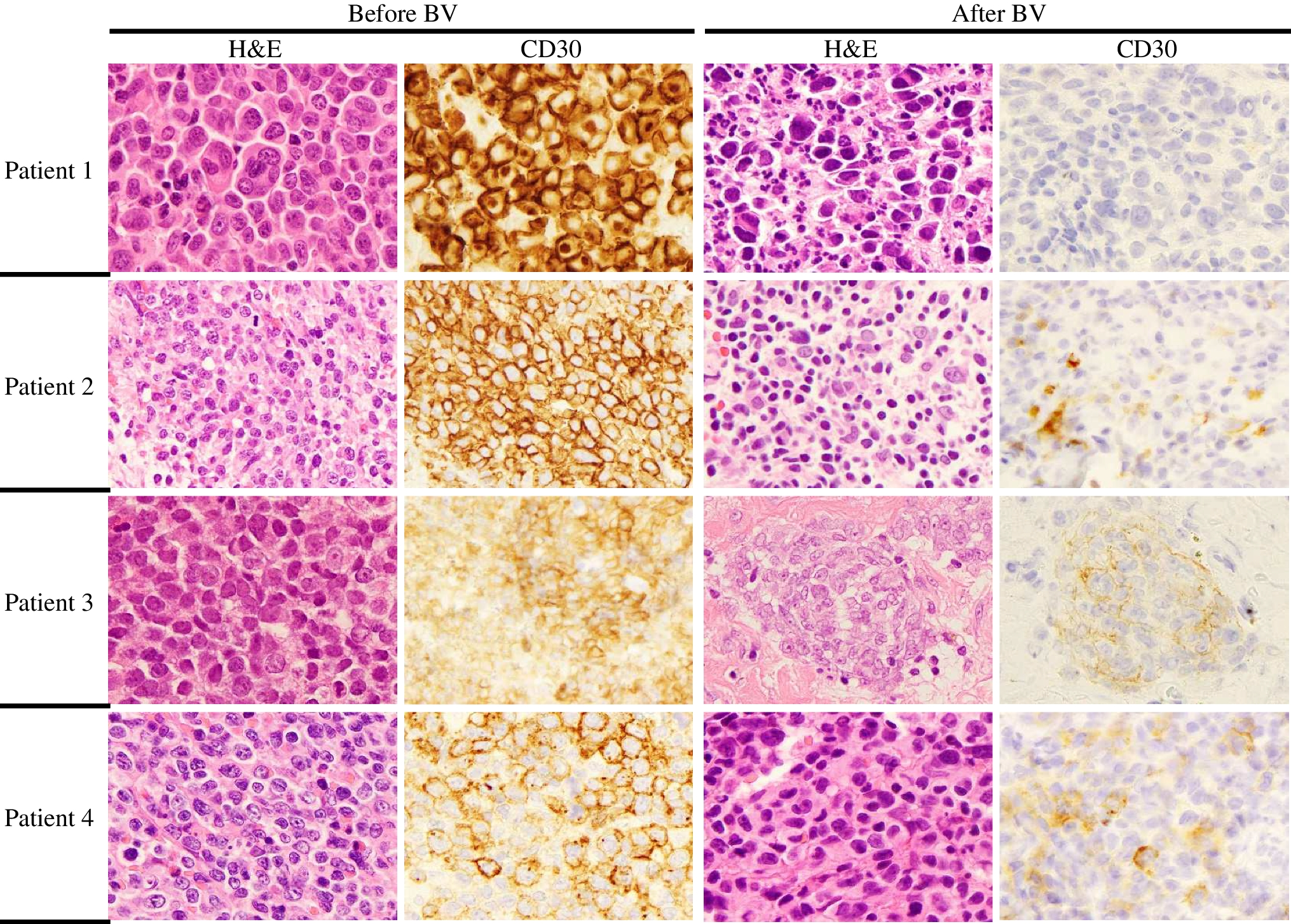
記住我

A cohort of FFPE BC biopsies (n = 170) with at least 30% histologically confirmed tumor cell content was re-investigated for its HER2 status to establish the ddPCR CNV ERBB2 assay (Bio-Rad, Hercules, CA, USA). Inclusion criterion was a known HER2 status, determined during routine diagnostics, independent of women’s age, tumor classification, tumor size, nodal status, and ER, PR, or Ki67 values (Table 1). The cohort included 3 groups: (1) HER2 negative (0/1 + ; n = 82), (2) HER2 equivocal (2 + ; n = 52) with positive amplification result by ISH (n = 16) and with negative amplification result by ISH (n = 36), and (3) HER2 positive (3 + ; n = 36) cases (Table 1).
Table 1 Summary of FFPE breast cancer samples used for establishment of the ddPCR CNV ERBB2 assay including distribution of HER2 groupsImmunohistochemistry and in situ hybridizationIHC staining and additional ISH of HER2 2 + positive samples were performed during routine diagnostic evaluations at the Department of Pathology, University Hospital and Paracelsus Medical University Salzburg, using standardized operation procedures. Scoring and interpretation of the results were performed by experienced breast pathologists according to ASCO/CAP guidelines [14]. The tissue samples were routinely processed, including formalin-fixation and paraffin-embedding (FFPE). Immunohistochemical staining was performed fully automated on a Dako Omnis Immunostainer (Agilent, Santa Clara, CA, USA) using the polyclonal Rabbit Anti-Human c-erbB-2 Oncoprotein antibody (1:600, Agilent, Santa Clara, CA, USA) [24]. For silver-enhanced in situ hybridization (SISH), the Ventana® HER2 Dual ISH DNA Probe cocktail (Roche, Basel, Switzerland) was applied on the VENTANA BenchMark ULTRA System (Roche, Basel, Switzerland) as described by Lim et al. [25].
Droplet digital PCR CNV ERBB2 assayWe used the Maxwell® RSC DNA FFPE Kit (Promega, Madison, WI, USA) with Maxwell® RSC Instruments for automated purification of the genomic DNA from FFPE tissue samples. DNA quantification was performed using the Quantus™ Fluorometer together with QuantiFluor® Dye System (Promega, Madison, WI, USA).
For copy number analysis, we used the ddPCR CNV ERBB2 assay (Bio-Rad, Hercules, CA, USA) and WSB1 as standard reference gene for its genetic location close to centromere region of chromosome 17. Both are 20 × concentrated, ready-to-use primer–probe mixes optimized for use with ddPCR Supermix for Probes (no dUTP). The two probes are labeled with the fluorophores FAM (ERBB2) and HEX (WSB1). Mastermix for ddPCR was set up as recommended in the instructor’s manual. We performed validation steps for DNA concentration and enzyme digestion. Droplet generation was performed in the QX-200 Droplet Generator (Bio-Rad, Hercules, CA, USA). The oil-PCR reaction mixture was transferred to a ddPCR 96-well plate, and PCR was performed in a Bio-Rad cycler with a deep-well block. PCR conditions were as follows: 10 min at 95 °C for enzyme activation, 40 cycles of 30 s at 94 °C for denaturation and 1 min at 60 °C for annealing/extension, 10 min at 98 °C for enzyme deactivation, and final hold at 10 °C. After PCR cycling, the plate was placed into a QX-200 Droplet Reader for signal counting.
Results for copy number (CN) data were analyzed using the QuantaSoft software (Bio-Rad, Hercules, CA, USA) by calculating the ratio of ERBB2 concentration to WSB1 concentration. As the centromeric region of chr17 where WSB1 is located may be amplified, this in fact is not by definition a true ERBB2 copy number but a ratio, as in many dual-FISH tests. Since this commercial software refers to the end result as CN, we prefer to follow this but further refer to this as “CN.”
To define DNA concentration used for ddPCR analysis and additionally to demonstrate sensitivity of the method, a serial dilution of a small test cohort of n = 5 HER2-positive BC samples was performed using 8 dilution steps: 35 ng, 20 ng, 15 ng, 10 ng, 5 ng, 2.5 ng, 1 ng, and 0.5 ng (Fig. 1a, b). Finally, we used a concentration of 30 ng of DNA in our setup; each sample was investigated in duplicate.
Fig. 1
Adjustment of DNA concentration and enzyme digestion to the basic Droplet Digital™ polymerase chain reaction protocol. a, c The droplet counts (ERBB2: blue bars; WSB1: green bars) and b, d the corresponding copy number calculation. a A serial dilution of a HER2-positive breast cancer sample with a range from 0.5 to 35 ng DNA in 8 dilution steps. Copy number is comparable for b all concentrations. c Four samples performed with (all samples with +) and without HaeIII digestion (all samples with −). Difference between ERBB2 (blue bars) and WSB1 (green bars) droplets was higher with digestion, leading to a change in d copy number ratio. For sample 4 the interpretation of the result changes from negative to positive as with digestion the copy number elevates from 2.39 to 3.5 and therefore over the threshold of 3 (d, sample 4 +). The error bars represent a 95% confidence interval
To separate closely linked ERBB2 copies [4] in case of a HER2 gene amplification, DNA of the test cohort (n = 5) was digested with restriction enzyme HaeIII (New England BioLabs, Ipswich, MA, USA). Two modes of digestion were performed and compared for their diagnostic utility and convenience. First, 10 units of the restriction enzyme HaeIII were directly added to the PCR master mix and incubated for 15 min at room temperature. Second, sample DNA was digested with HaeIII for 1 h at 37 °C before merging with the PCR master mix. In addition, n = 97 samples were investigated with and without HaeIII digestion to further address the necessity of the digestion step.
Statistical methodsData were checked for consistency using McNemar’s test to analyze cross tabulations and to compare probabilities for HER2-positive cases assessed by both methods. Hodge-Lehman confidence intervals were computed for difference of both probabilities and Pearson-Clopper confidence intervals for sensitivity, specificity, negative, and positive predictive values. An univariate logistic regression model was applied to the relation between ddPCR ERBB2 copy number and risk for IHC/ISH positive HER2 status. All reported tests were two-sided, and p-values < 0.05 were considered statistically significant. All statistical analyses in this report were performed with the software tool STATISTICA 13 [26].
留言 (0)