
記住我
Our OTC program was open to women from the Oncology Department of our hospital and from other referral centers nationwide. On arrival, women completed a detailed anamnesis comprising menstrual data and pregnancy history, medical conditions, lifestyle factors, and potential infertility. A clinical assessment was performed, including anthropometric parameters (height and weight), pelvic examination, and endo-vaginal ultrasound to explore the uterus and ovaries. Blood samples were taken between 08.00 and 10.0 h after 12-h fasting. The analytical study included parameters as per protocol for surgical intervention under general anesthesia, together with hormonal measurements. Premenopausal status was confirmed by a regular menstrual cycle together with premenopausal hormonal levels of FSH (range 2.4–6.6 IU/L) and E2 (range 21–251 pg/mL), both in the follicular phase.
Ovarian decortication was performed by laparoscopy using a previously described technical procedure [12]. Briefly, the cortex from one of the ovaries was removed with scissors, and the hemostasis of exposed medullary tissue was ensured with electrocautery. The extracted ovarian cortex was placed in Ham F10 medium and cut into strips whose thickness did not exceed 2 mm, to allow the action of the cryoprotectants.
Our database was searched for women who were premenopausal at the time of surgery (≤ 38 years) living in the metropolitan area of Valencia. The two inclusion criteria, obtained from their corresponding electronic medical record (EMR), were an interval of at least three years since surgery and completed chemotherapy schedule. Exclusion criteria were recent (≤ 4 weeks) use of any hormonal drugs, pregnancy or lactation, menstrual irregularities prior to decortication or a history of any pathology other than cancer linked with ovarian dysfunction, such as polycystic ovarian syndrome, hyperprolactinemia, thyroid disease, Cushing disease, infertility for at least 6 months, a previous diagnosis of a disease linked with premature ovarian insufficiency (POI), or another previous cancer diagnosis, with or without chemotherapy or radiotherapy.
Patients meeting the conditions were sent a letter of invitation to participate. Respondents were scheduled an appointment at our service, in which the objective of the study was explained and additional clinical information was retrieved, including the status of the malignant disease from EMR, accessible through the platform that interconnects all public hospitals in our autonomous region.
The clinical and analytical assessments prior to decortication were repeated in this post-treatment visit, rescheduling patients with regular menstruation who were not at the follicular phase (days 2–5) of the cycle. Menstrual data were categorized into regular (all cycles between 23 and 35 days), irregular if outside this range, and amenorrhea if no bleeding during the last 6 months. Diminished ovarian reserve (DOR) was defined when AMH values were < 1.0 ng/mL [2], and POI in the presence of amenorrhea, elevated serum FSH levels of > 35 IU/L, and age < 40 years. Occurrence of any previous pregnancies and presence of menopausal symptoms were also recorded.
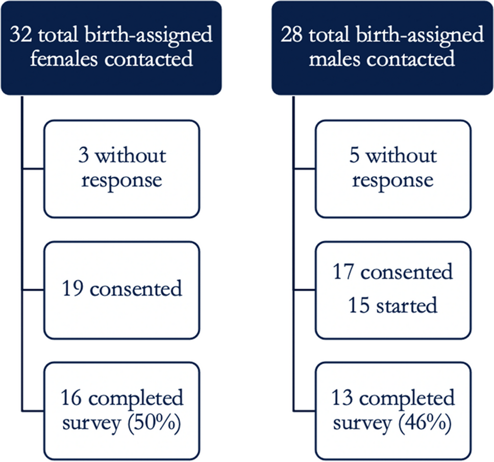
Two cohorts of normal-ovulatory healthy women were matched 1 : 1 to the age the women with cancer were at the predecortication stage (stage 1 group, n = 40) and at the post-treatment visit (stage 2 group, n = 40). The purpose was to verify that women had normal values of AMH before starting treatment (predecortication, stage 1) and what was the impact of cancer treatment on ovarian reserve (stage 2), since the comparison with healthy normo-ovulatory coetaneous females in the post-treatment stage would reveal the confounding effect of age. Figure 1 shows the design of the study, which was approved by the Research Ethics Committee of our center (approval code 44.12). Informed written consent was obtained from all participants.
Fig. 1
Women with previous cancer, who had undergone ovarian decortication for fertility preservation, were invited to participate in the study and evaluated at our center (evaluation visit). Current clinical and hormonal parameters were measured. In this step, predecortication clinical and hormonal parameters were retrieved from the EMR. In addition, two groups of volunteers with regular menstruation were recruited. These women were age-matched 1 : 1 to the cancer group at each stage, prior to decortication (stage 1, n = 40) and at the assessment visit (stage 2, n = 39, as one woman withdrew consent after enrollment). EMR: electronic medical record; FP: fertility preservation; OTC: ovarian tissue cryopreservation
Analytical assessmentTime-resolved chemiluminescent microparticle immunoassay (ARCHITECT, Abbott Laboratories) was used for assessment of FSH (analytical sensitivity 0.05 IU/L) and E2 (analytical sensitivity ≤ 10 pg/mL) in serum. Serum levels of AMH were analyzed with specific ELISA kits according to manufacturer’s instructions (AMH Gen II ELISA, Beckman Coulter, Inc., analytical sensitivity: 0.05 ng/mL).
Statistical analysisThe Chi-square test of independence was performed to assess the relationship between categorical clinical variables (alcohol intake, smoking, obstetric history, previous use of hormonal contraceptives, post-cancer gestation, POI, and menstrual cycle pattern) and cancer type (breast cancer vs. Hodgkin’s lymphoma). For continuous clinical variables (age, body mass index, and menarche), Student’s t-test was carried out to study mean differences in both cancer groups. In case of deviations from the assumption of normal distribution according to Shapiro’s test, the non-parametric Wilcoxon rank-sum test was used.
To study the change in the mean hormone levels (AMH, FSH, and E2) before and after chemotherapy treatment in the cancer cohort, we used paired samples t-test or Wilcoxon signed-rank test. For healthy normo-ovulatory women, an independent t-test (or Wilcoxon rank-sum test) was applied as neither prechemotherapy nor post-chemotherapy groups were dependent. Finally, logistic regression was used to identify the independent predictors related to binary outcomes, DOR, POI, or menstrual cycle.
Results were considered statistically significant when p values fell below the 5% significance level. All statistical analyses were performed using the R software (version 4.2.2).
留言 (0)