
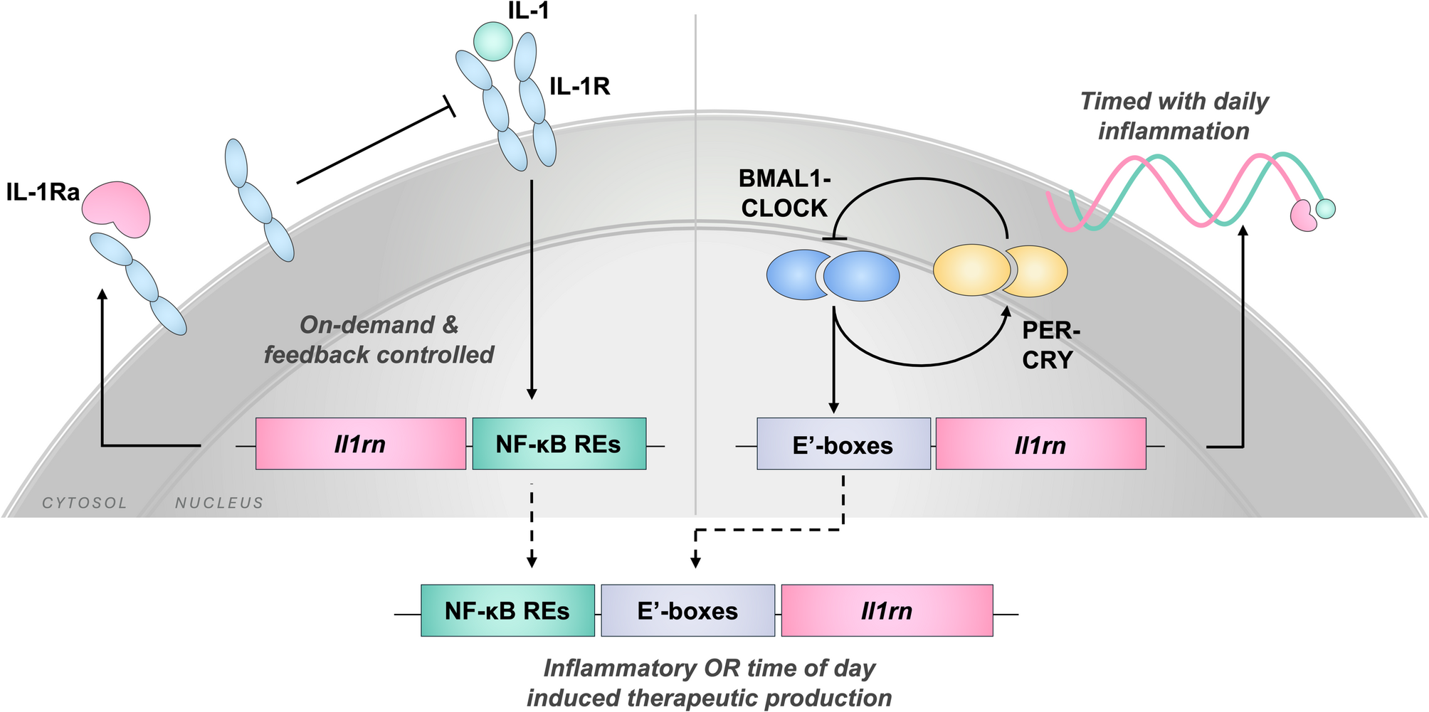
Remember me


To construct the chitosan-based magnetic hydrogel nanocomposites, magnetic montmorillonite nanoparticles were first synthesized through in-situ co-precipitation or the Fe2+/Fe3+ ions in the presence of the mMMt nano-clay. The adsorption of the Fe ions on the anionic centers of MMt and the intercalation between the MMt layers accordingly resulted in the formation of the magnetic NPs not only on the clay surface but also between the MMt layers. To reach a cross-linked magnetic chitosan-based hydrogel nanocomposite, the purified mMMt was then transferred into the HA solution. Furthermore, citric acid (CA) as a cross-linker was added to chitosan, forming the cross-link points through the ester or amino functional groups. During the cross-linking reaction, the mMMt NPs and HA were in the CA-chitosan hydrogel nanocomposites. A simple scheme, including the synthesis of the magnetic Chito/HA-mMMt nanocomposites hydrogels, is illustrated in Fig. 1.
Fig. 1
A simple scheme giving the process to synthesize Chito/HA-mMMt nanocomposite hydrogels
FT-IR studyThe possibility of cross-linking through the ester formation, originating from the reaction between the hydroxyl (–OH) groups on chitosan and the –CO2H groups of CA, was examined by the FT-IR, whose results are displayed in Fig. 2a. In the FT-IR spectrum of MMt, the broad peak at 3626 cm−1 was related to the stretching vibration of the –OH groups on the MMt clay, accompanied by the stretching vibration of the Al-O and the functional group of MMt. The stretching vibration modes of the Si-O-Al and Si-O-Si groups also had distinguishing peaks at around 794 and 1042 cm−1, respectively. The peaks appearing at 522 and 460 cm−1 were further associated with the bending vibration modes of the Si-O-Al and Si-O-Si groups, respectively [43]. In the chitosan spectrum, an overlapped and broad peak at 3443 cm−1 owing to the N-H and O-H groups was further observed. The appearance of the characteristic bands at 1697 and 1599 cm−1 in the chitosan spectrum could be thus assigned to the amide I and II stretches. As well, CA exhibited the IR peaks at 3370 and 3030 cm−1, attributing to the stretching vibration of the O-H and C-H bonds, respectively [44]. The symmetric and asymmetric stretches of the –CO2H groups on CA were also evident from the peaks in the FT-IR spectrum, appearing at 1398 and 1568 cm−1, respectively. Considering the FT-IR spectrum of the mMMt, the characteristic peaks of MMt at 1636 cm−1 (to 1328 cm−1) and 1042 cm−1 (to 1034 cm−1) were relatively shifted to the lower frequency, possibly due to the interaction between MMt and the immobilized Fe3O4-NPs [45]. The Fe3O4 IR peaks at around 400–600 cm−1 also overlapped with the MMt ones, so they were not detectable. The FT-IR spectra of hydrogel further showed the characteristic peaks of chitosan, Hyaluronic acid, mMMt, and citric acid. The intensity of all peaks for Chito-HA was thus higher than that of the Chito/HA-mMMt1 and Chito/HA-mMMt2. In the Chito/HA-mMMt2 sample, the peak at 1750 with low intensity also appeared, mostly assigned to the –CO2H groups of CA. Any peak related to the ester or amide formation due to the reaction between chitosan and CA was not visible. The overlapping of the IR peaks of the ester-amide functional groups with those of other ingredients might be thus responsible for the non-appearing new peaks.
Fig. 2
a FTIR spectra and b XRD patterns of raw materials and synthesized hydrogels
XRD studyFigure 2b shows the XRD pattern of the MMt, mMMt, Chito/HA, Chito/HA-mMMt1, and Chito/HA-mMMt2. The XRD pattern of the MMt clearly revealed its distinctive peak around 2θ = 7.5, 20, and 28.1 degrees. Upon the immobilization of the Fe3O4-NPs on the MMt plates to reach mMMt, the characteristic MMt peak disappeared, possibly associated with the exfoliation of the MMt plates. This might be further attributed to the cation exchangeability of MMt in which the Fe ions could be intercalated between the MMt layers, and subsequently exfoliate by making the Fe3O4-NPs on the MMt layers. The creation of the Fe3O4-NPs in the presence of MMt was further confirmed by the diffraction peaks of (220), (311), (400), (422), (511), and (440) at 2θ = 30.0, 35.5, 43.2, 53.5, 57.0, and 62.6º, respectively. The XRD pattern of the neat chitosan powder also displayed two distinctive peaks at 2θ = 10.5 and 20.1º, which were related to the semi-crystalline structure of chitosan and hydrate, and the increasing hydrogen bonding and flexibility of the chitosan chain, respectively [41].
The crystallinity was the outcome of the presence of intermolecular hydrogen bonding in chitosan. These peaks demonstrated a regular structure formed as a result of hydrogen bonds between the amine and –OH groups, which were responsible for preventing chain movement. After the cross-linking of the Chito/HA-mMMt mixture with CA, the intensity of the chitosan peaks diminished, indicating the presence of amorphous chitosan in the composition of the nano-carriers. In the nano-carrier patterns, the presence of the Fe3O4-NPs was further established by the characteristic peaks at 2θ = 35.7 and 63º. Considering the high content of the mMMt, the intensity of the Fe3O4 peaks in sample 2 was sharper as compared with sample 1.
Morphology studiesThe SEM analysis was done to evaluate the influence of the mMMt on the surface morphology of the magnetic carriers and the hydrogel structure. Figure 3 shows the morphology of the hydrogels using SEM. It became apparent that all hydrogels have a porous structure.
Fig. 3
SEM and EDX image of a Chito/HA, b Chito/HA-MMt1, and c Chito/HA-MMt2
Compared with the Chito/HA-mMMt hydrogels, the structure of the Chito/HA-mMMt1were more porous, and the size of the pores increasedincreases with the growth in the mMMt content. Moreover, these synthesized hydrogels had excellent interactions between chitosan, HA molecular chains, and the mMMt NPs.
The SEM image of the Chito/HA-mMMt also showed a rough surface with spherical particles. The corresponding formed particles could be accordingly ascribed to the magnetic NPs. According to this image, the size of the NPs was small. When the mMMt was introduced into the Chito/HA hydrogels to produce magnetic nano-carriers, a rough surface with spherical particles appeared with a larger size. The results might be devoted to the covering effect of the cross-linked Chito/HA on the mMMt, leading to a rise in the NPs size. Accordingly, the spherical NPs on the Chito/HA-mMMt2 were much more compared with the Chito/HA-mMMt1. The mMMt content utilized to prepare the Chito/HA-mMMt2 was also higher than that of the Chito/HA-mMMt1, and subsequently, the chitosan ratio dwindled. In other words, the low content of the mMMt in the Chito/HA/mMMt1 was embedded in the cross-linked chitosan carrier, and the spherical particles tended to drop.
The elemental analysis of the samples was further investigated by the EDX spectroscopy. In this line, the EDX of the mMMt displayed the Si, Al, and Ca elements, which were in agreement with previous research. In addition to the main MMt elements, the appeared Fe peak indicated the presence of the magnetic Fe3O4-NPs. In the EDX of the magnetic chitosan-based nano-carriers, the C and N peaks along with the mMMt elements, also appeared because of chitosan and HA ingredients.
TEM studyFigure 4 displays the TEM images of the mMMt, Chito/HA-mMMt1, and Chito/HA-mMMt2. According to the mMMt image, the MMt clay existed in the exfoliated platelets or intercalated tactoids. The formed Fe3O4-NPs could be further seen on the surface or among the MMt plates. By adding the mMMt to the chitosan/HA solution to generate the magnetic carriers, the mMMt-NPs were dispersed inside the chitosan/HA, resulting in the covering of the mMMt NPs by the chitosan ingredient. Unlike the mMMt, the MMt plates did not clearly appear that could be related to the interweaving of the chitosan chains into the MMt layers to produce the exfoliated MMt clay [46].
Fig. 4
TEM image of a mMMt, b Chito/HA-MMt1, and c Chito/HA-MMt2
VSM studyThe magnetization behavior of the samples was investigated using the VSM technique at an applied field of ± 10 kOe at 298 K to compare the saturation magnetization variation (SMV) by different contents of the mMMt. The SMV of the mMMt, Chito/HA-mMMt2, and Chito/HA-mMMt1 were accordingly about 34.2, 13.4, and 8.2 emu/g, respectively (Fig. 5a). The magnetization curves of the mMMt, Chito/HA-mMMt1 and Chito/HA-mMMt2 also showed their superparamagnetic property with no remanence or coercivity [47]. Compared with the mMMt, the SMVs of the magnetic samples were in lower values. The drop in the SMVs of the hydrogels compared with the mMMt could be thus attributed to the non-magnetic behavior of chitosan and HA applied to prepare hydrogels. The SMV was also reported per g of the magnetic matter, so the combination of the non-magnetic ingredients with the magnetic ones resulted in a reduction in the SMV. Moreover, lower SMV corresponded to the amount of the magnetic NPs in the hydrogels. On the other hand, combining chitosan and HA with the mMMt components led to a decline in the amount of the magnetic NPs because of the non-magnetic behavior of these neat biomaterials, which decreased in the SMV. Of note, the magnetic saturation of the Chito/HA-mMMt was enough to discrete it from the hydrogels via an outside magnetic force.
Fig. 5
a Hysteresis loops and b TGA thermograms of raw materials and hydrogels
TGA studyTo investigate the thermal stability of the chitosan and chitosan-based hydrogels, the TGA technique was performed by heating the samples under a nitrogen atmosphere in a range of temperatures, 25–700 oC (Fig. 5b). The neat mMMt, chitosan, and the magnetic carriers accordingly had a weight loss up to 200 oC, which was due to the evaporation of the free and adsorbed water by the samples [48]. The mMMt further showed no special weight loss up to 700 oC, indicating its high thermal stability. About 70 wt% of the Chito/HA was lost from 250 to 700 oC, which was assigned to the chitosan decomposition of chitosan. Introducing the mMMt correspondingly had a significant impact on the thermal stability of the chitosan-based nano-carriers. The weight loss of 60 and 51 wt% also occurred in the Chito/HA-mMMt1 and Chito/HA-mMMt2, respectively. After 300 oC, no significant weight loss was observed in the magnetic nano-carriers, originating from the introduced mMMt with high thermal stability. The weighted residual of the magnetic samples was further found to be more than that of the Chito/HA, representing the high thermal stability of the samples owing to the mMMt introduced for this purpose to attain the magnetic nano-carriers. The difference in the weighted residual of chitosan and the magnetic nano-carriers might be thus related to the amount of the mMMt used to synthesize them. According to the TGA curves, the weighted residual of the Chito/HA-mMMt2 (42.5 wt% more than chitosan) was more than that of the Chito/HA-mMMt1 (32.5 wt% more than chitosan) because of the high amount of the mMMt applied to prepare the Chito/HA-mMMt2.
Particle size and zeta-potential studyThe particle size and zeta-potential values of the synthesized hydrogels were explored using the DLS analysis, in which the Stokes-Einstein equation was used to measure the Z-average size of the particles in this instrument. It was assumed that the mMMt NPs were spherical. Figure 6(a to c) shows the mean particle size and zeta-potential of the mMMt, Chito/HA-mMMt1, and Chito/HA-mMMt2.
Fig. 6
Particle size distribution of a mMMt, b Chito/HA-mMMt1, c Chito/HA-mMMt2, and d zeta-potential distribution of samples
The smallest particle size and the relatively high net values of the zeta-potential of the samples accordingly revealed the high surface active functionality and stability of the given obtained hydrogels.
Furthermore, the zeta-potential value as indicative of the stability of the nanoparticles and their surface charge was explored. In designing the DDS, it was thus essential to determine the zeta-potential values, so the carriers with high zeta potential were preferred. The high value in the zeta potential could thus stop the aggregation of the NPs. The zeta-potential of the mMMt was further found to be different from the magnetic carriers. The mMMt, Chito/HA-mMMt1, and Chito/HA-mMMt2 also showed the zeta-potential values of -42.3, + 39.8, and + 33.7 mV, respectively (Fig. 6d). The negative value for the mMMt also originated from the negative centers on the MMt. Upon combining the mMMt with chitosan and HA to attain the magnetic NPs, the zeta-potential values tend to elevate and reach positive values. In fact, the results demonstrated the coating effect of the mMMt by the chitosan containing the primary amine (–NH2) functional groups. For the Chito/HA-mMMt2, the increase in the mMMt ratio resulted in a reduction in the zeta-potential value, which was associated with the electrostatic interaction between MMt and some –NH2 groups of chitosan, leading to a drop in the zeta-potential value.
Swelling testAs rehydration was necessary to release any coated drug or bioactive material from dried beads, controlling release in DDS could depend on the SF. For this reason, the SF of all hydrogels was done at pH = 7.4 and 5.5 (Table 1). These results showed that the SFs at pH = 7.4 were higher than pH = 5.5 and 1.2. Therefore, the Chito/HA hydrogel was suitable to use in DDS for wound healing. The Chito/HA also displayed high SFs and water uptake percentages compared to other hydrogels because of their high porosity and mechanical properties. On the other hand, the SF rose by entering the water molecules into the hydrogels via the pores. Therefore, these hydrogels consisted of large pores or volumes of water for a long time, thereby providing a wet situation for the wound to be treated quickly. As a result, they could be effective as wound dressings. Moreover, the SF of all hydrogels varied on the basis of the mMMt doses in the hydrogels [49]. The presence of the mMMt-NPs slightly decreases the porosity of the Chito/HA-mMMt hydrogels. The extraordinary porosity of the hydrogels as dressings was thus helpful for absorbing more exudate from the wound surface and reducing wound infection caused by exudate. A wet wound medium could thus help improve the wound and minimize scar formation, while the dressing could be removed without pain.
Table 1 The swelling tests and factors of all synthesized hydrogels at pH equal to 7.4 and 5.5 Drug releaseThe CIP and CUR release from the Chito/HA, Chito/HA-mMMt1, and Chito/HA-mMMt2 hydrogels was examined by measuring their concentrations during the dipping in the media at different pHs (5.5 and 7.4) and at the temperature of 37 ºC. Figure 7 displays the CIP and CUR release from the hydrogels at different pHs (5.5 and 7.4). Not only the pH of the releasing media could have an impact on the amount of CIP and CUR release, but also the amount of MMt could influence it. The increasing amount of the MMt in the hydrogels considerably impacted drug release.
Fig. 7
Ciprofloxacin and Curcumin release behavior of Chito/HA, Chito/HA-mMMt1, and Chito/HA-mMMt2 hydrogels at pH = 5.5 and 7.4
According to the study results, the cumulative release of CUR from the Chito/HA, Chito/HA-mMMt1, and Chito/HA-mMMt2 hydrogels were 89.9, 53.9, and 35.7%, at pH = 7.4 and 48.7%, 24.6%, and 21.8% at pH = 5.5, respectively, after 6 h. The Chito/HA, Chito/HA-mMMt1, and Chito/HA-mMMt2 hydrogels accordingly showed the CIP release of about 78.8, 26.9, and 21.1%, at pH = 5.5 and 86.1%, 43.4%, and 34.6% at pH = 7.4, respectively, after 6 h, possibly due to the electrostatic interactions.
According to the high solubility of CIP under the acid media and the CUR degradation in the base ones, it was expected that the CIP and CUR release at pH = 5.5 would occur higher than that at pH = 7.4. However, the results were inconsistent with the CIP solubility, originating from hydrogels. Figure 7 displays that the release rate is lower at pH = 5.5 than pH = 7.4, because the –CO2H groups show more mutual effects with buffer media at pH = 5.5, allowing the network to be tauter; therefore, the entrapped drug molecules hardly get out of the network. Based on the CIP pKa values (pKa1=6.18 and pKa2=8.8) [50], at pH = 5.5, the amine group on CIP was present in an ionic status (= NH2+), while the carboxylic acid presented in its protonated form (-COOH). At pH = 5.5, free amine pendants were also created by dissociating chitosan and cationic amine groups on it. Furthermore, chitosan was soluble in the acidic media, and its cationic structure could facilitate drug diffusion. The chitosan–NH3+ groups could thus boost the diffusion of the drug molecules through electrostatic repulsive force. According to Wu et al. [51], the maximum adsorption of CIP on MMt could occur at pH = 5.5. In fact, the high tendency of the cationic CIP to get adsorbed on the negative centers of the mMMt layers at pH = 5.5 could prevent its easy diffusion into the media. On the other hand, there was a stronger interaction between the group (–CO2H) and the negative surface charges in the acidic solution after the CIP adsorption. Additionally, the coating effect of the cross-linked chitosan on the mMMt was not negligible. The protonation of the chitosan biopolymer (pKa ~ 6.5) at pH = 5.5 might thus restrict the diffusion of the cationic CIP from the surface of the mMMt owing to the repulsive forces. The positive charges on the surface of the nano-carriers were also evident from the zeta-potential data. While the mMMt comprised of a negative surface charge after being coated with chitosan, the surface charge could be shifted to a positive charge.
Once the pH of the releasing media increased to 7.4, the physical interactions of the drug and the nano-carriers were changed, resulting in a high release of CIP and CUR. The deprotonation of chitosan at pH = 7.4 also made the cationic nature of coating on mMMt disappear. Thus, the CIP and CUR from the mMMt surface through the chitosan layer could occur quickly without any repulsive forces. While the drug molecules were in the cationic form at pH = 5.5 and 7.4, the dissociation of the carboxylic acid groups to produce anionic carboxylate by maintaining ammonium charges on the drug could make the drug a zwitterion carrying the anionic and cationic charges simultaneously. A repulsive force between the carboxylated groups on the drug and the anionic centers on the mMMt accordingly encouraged the desorption of the drug molecules from the mMMt surface, leading to the drug diffusion through the chitosan layer without any electrostatic interactions.
The decrease in the cumulative drug release from the Chito/HA-mMMt with the high content of the mMMt could be thus related to the interaction between chitosan and the mMMt. It has been also reported that the cross-link density could be augmented due to the MMt used to prepare the hydrogels. The physical interactions through the hydrogen bonding could also lead to a growth in the cross-link points, and subsequently, the pore sizes tended to decrease. The decline in the pore sizes could further result in the restriction in the mobility of CIP to diffuse into the media. The effect of the MMt on the release of the tanshinone IIA (Chemical drug) from chitosan and the chitosan/MMt had been similarly investigated by Luo et al. [52]. The release of tanshinone IIA from the chitosan/MMt microspheres was thus lower than that of the neat chitosan microspheres.
Release kineticsTo study the prediction of the mechanism for the drug release, Higuchi and Korsmeyer-Peppas models were utilized with reference to Eqs. 4 and 5, respectively.
where Rr, KKP, and KH are the drug release rate as well as, the drug release rate constants of the Korsmeyer-Peppas and Higuchi kinetic models, respectively. Moreover, n is the diffusion exponent, by which the release mechanism is determined by it. In the Korsmeyer-Peppas model, the n value can have different values, of which the values of ≤ 0.45, that between 0.45 and 0.89, and ≥ 0.89 demonstrates the predomination of the Fickian diffusion phenomenon, the anomalous transport (namely, the non-Fickian or diffusion kinetic and polymer relaxation kinetic), and the case-II transport mechanism, respectively. The release mechanism was also affected by the pH of the releasing media, indicating the effect of surface charge on the mechanism of CIP and CUR release. Therefore, the release kinetic was investigated in two media (pH = 5.5 and 7.4).
However, the Higuchi model was defined by plotting the drug (CIP and CUR ) release (DR%) versus the square root of time (t). The outcomes showed a linear relationship for all hydrogels in both media (pH = 7.4 and, 5.5) with R2 > 0.95 for CIP and CUR, which suggested the diffusion process for the CIP and CUR release from the hydrogel (Table 2).
Table 2 The calculated kinetic parameters of Curcumin and Ciprofloxacin release according to Higuchi and Korsmeyer-Peppas modelsFurthermore, the intercept of log (DR) against log t plots could determine the KKP and n for the Korsmeyer-Peppas model. Table 2 displays the results of the Korsmeyer-Peppas model fitting. The achieved correlation coefficients of Korsmeyer-Peppas (R2) were also higher than 0.98, indicating a linear relationship. The release data of the hydrogels at pH = 5.5 additionally denoted the best fitting to the Korsmeyer-Peppas model, evidenced by the higher R2 values (~ 0.99). According to the results in Table 2, the n value for the CIP release from the Chito/HA-mMMt1 and Chito/HA-mMMt2 hydrogel and the CUR release from the Chito/HA-mMMt2 were lower than 0.45 at pH = 5.5, showing the release of CIP and CUR from the hydrogels through the Fickain diffusion [53]. Moreover, the R2 values for CUR and CIP were higher in the Korsmeyer-Peppas model than in the Higuchi model, indicating the CIP and CUR release from the hydrogels followed by the Korsmeyer-Peppas model. The results accordingly confirmed that the release of CIP and CUR from the hydrogels was dominated via diffusion control [54]. In fact, the release of the drugs from these hydrogels through swelling or dissolution did not occur [55]. Besides, the n values at pH = 7.4 (n < 0.45) demonstrated the Fickian diffusion of the drug from the hydrogels. Overall, the release of CIP and CUR from the samples through the diffusion process was established by the release kinetic Lajevardi et al. [55] studied the release of cephalexin from the Fe3O4/silica/MIL/100(Fe)-β/CD. They showed that the mechanism of the cephalexin release from the respective carriers could be altered by changing the pH of the releasing media.
Effect of external magnetic field on releaseAlthough the main problem facing DDS is the lack of tissue selectivity, the mMMt can deal with this limitation by directing drugs to the targets using an external magnetic field. With regard to the use of the mMMt with magnetic properties, there was an attempt to investigate the release of CIP and CUR from the Chito/HA-mMMt2 by applying an external magnetic field. The impact of the external magnetic field on the CIP and CUR release profiles at pH = 5.5 is illustrated in Fig. 8. Accordingly, the content of the CIP and CUR release under the applied magnetic field was higher than that without it. In the absence of a magnetic field, the cumulative release of CIP and CUR from the Chito/HA-mMMt2 was about 36% and 27% after 15 min, respectively. In contrast, the release of CIP and CUR from the Chito/HA-mMMt2 was accelerated as the magnetic field was applied [41]. Therefore, DDS could be controlled using an external magnetic field to operate the drug release. In this vein, Perera et al. [56] found a comparable outcome that utilizing an external magnetic field had speeded up the release of acetaminophen from the polymer-magnetic composite fibers. Their study was further clarified using the position of the mMMt-NPs in exposing the hydrogels under an external magnetic field. The motion of the mMMt-NPs could thus expand the network because of the relaxation of the polymeric chains [57].
Fig. 8
The influence alternating magnetic field on the drug release profiles of Curcumin and Ciprofloxacin from Chito/HA-mMMt2 hydrogel at pH 5.5
Antibacterial experimentsThe bactericidal activity of the Chito/HA, Chito/HA-mMMt1, and Chito/HA-mMMt2 against Gram-positive and -negative bacteria was further examined here (Fig. 9; Table 3). To reach a better comparison, the effects of CIP and CUR on the antibacterial activity of CIP and CUR-loaded hydrogels were compared with the antibacterial activity of pure chitosan and CIP-CUR. Although the antibacterial activity of chitosan was not negligible, no inhibition zone was observed against Chito/HA-mMMt hydrogel without drug. The bactericidal activity of chitosan also originated from the mobility of chains, which made an inhibition zone appear for bacterial growth. After the cross-linking process using HA and magnetic nanoparticles, the mobility of the chitosan chains became limited, leading to no inhibition zone of bacterial growth. In this line, Li et al. [58] investigated the bactericidal activity of chitosan against Gram-positive and -negative bacteria and found that the cross-linking chitosan scaffold had a significant effect on a decrease in the chitosan bactericidal activity. The CIP and CUR-loaded samples had further exhibited an inhibition zone for both Gram-positive and -negative bacteria, showing the CIP and CUR diffusion from the hydrogels into the media. The inhibition zone for the Chito/HA-mMMt2 sample was also larger as compared with the Chito/HA hydrogels. This was in agreement with the CIP and CUR release behavior of the hydrogels. The Chito/HA-mMMt2 sample also had a higher cumulative release than that of the Chito/HA-mMMt1 one. Furthermore, the mMMt amount affected the antibacterial activities.
Fig. 9
Created zones of inhibition of samples (CUR-CIP loaded Chito/HA, CUR-CIP loaded Chito/HA-mMMt1, CUR-CIP loaded Chito/HA-mMMt2, Chito/HA-mMMt2 without drug, pure CUR-CIP from left to right, against a S. aureus and b E. coli incubated at 37 °C for 24 h
Table 3 Antibacterial activity of the synthesized hydrogels against S. aureus (Gram-positive) and E. coli (Gram-negative) MTT assayIn this work, the cell viability percentage for the normal human fibroblastic cells (L929) for 24, 48, and 72 h, incubated with pure CIP and CUR as well as the CIP/CUR-loaded Chito/HA-mMMt2 and CIP/CUR-loaded Chito/HA-MMt2 hydrogels was evaluated using the 3-[4,5-dimethylthiazol-2-yl]-2,5 diphenyl tetrazolium bromide (MTT) assay. Figure 10 displays the cell cytotoxicity results of CIP and CUR as well as the CIP/CUR-loaded Chito/HA-mMMt2 and CIP/CUR-loaded Chito/HA-MMt2 hydrogels. The CIP/CUR-loaded Chito/HA-mMMt2 hydrogel exhibited no noticeable toxicity on the L929 cells because of its biocompatibility. The structure of this hydrogel contained chitosan as a natural polysaccharide, HA, and mMMt, which were non-toxic and biodegradable for enhancing the biocompatibility of DDS. Hence, the Chito/HA-mMMt2 hydrogel could be applied as a harmless bio-DDS for wound healing. Meanwhile, the CIP/CUR-loaded Chito/HA-mMMt2 hydrogel confirmed insignificant cytotoxicity as the pure CIP and CUR. The low release of CIP and CUR from the Chito/HA-mMMt2 hydrogel into the normal cells ultimately led to low cytotoxicity. The MTT assay further presented ~ 70%, ~ 94%, ~ 88% and ~ 98% of the L929 cells viability for CIP and CUR, as well as the CIP/CUR-loaded Chito/HA-MMt2 and CIP/CUR-loaded Chito/HA-mMMt2 hydrogels at the concentration of 16 µg/mL after 72 h, respectively. Cell viability for the pure CIP and CUR was also lower than that of the CIP/CUR-loaded Chito/HA-mMMt2 hydrogel. Besides, the CUR and CIP release from the hydrogel was slower in a controlled manner, and it acted as a membrane. However, the pure CIP or CUR could enter directly into the cells to kill them. Accordingly, it was established that the CIP/CUR-loaded Chito/HA-mMMt2 hydrogel was a non-toxic material for biomedical applications, such as wound healing. Also, the results showed magnetic can improve biocompatibility of Chito/HA hydrogel. Magnetic hydrogel (Chito/HA-mMMt2) did not allow the ciprofloxacin to be released quickly, leading to a reduction in the ciprofloxacin release rate and cytotoxicity.
Fig. 10
The percentage cell viability of the normal human fibroblastic cells (L929) for 24, 48 and 72 h incubation with pure curcumin, pure ciprofloxacin and CIP,CUR-loaded Chito/HA-mMMt2
Wound healingTo evaluate the wound healing ability of the Chito/HA-mMMt2 hydrogels, a number of studies have so far reflected on cell growth to investigate the direct or indirect effects of fibroblast motility. As the role of chitosan and HA in wound healing is best known, scratch assays have been typically performed. Moreover, these materials have been applied to motivate the fabrication of matrix proteins in fibroblasts because HA can improve the production of fibronectin and collagen, and synthesize collagenases. As well, chitosan improves granulation tissue formation in skin wounds and wound-bursting strength [59]. Cell migration into the wound was thus detected in response to an artificial injury, as shown in Fig. 11. The incubation time of 48 h accordingly resulted in the highest number of migrated cells in the bare area. However, the number of cells was too high to be measured and clearly distinguished because of the clusters or aggregates.
Fig. 11
Fluorescent microscope image to estimate wound healing in vitro in the scratch assay using a confluent monolayer of 3T3 fibroblasts (a) immediately after the wounding, b after 6 h, c 12 h, and d 48 h incubation
Comments (0)