Wiskott-Aldrich syndrome (WAS) is a rare disorder caused by an X-linked genetic mutation that causes immune deficiency and affects males with a frequency of one in ten million [1]. It also causes and features significant thrombocytopenia with platelet volume reduction, recurrent infections, eczema, progressive lymphopenia, an elevated incidence of immune deficiency in people with autoimmune diseases, and increased risk of developing suppurative and opportunistic infections [2], [3]. It is associated with higher rates of autoimmune diseases and lymphatic malignancies, particularly B-cell lymphoma [4]. The disease is characterized by a mutation in the Wiskott Aldrich syndrome protein (WASp) [3], a key regulator of actin cytoskeleton-dependent cellular processes critical for transmitting and integrating actin-regulating signals that are vital for a variety of cell functions [5]. The functions include cell motility and changes in cell shape, immune synapse formation and cytokine release, and it is expressed exclusively in hematopoietic cells [6], [7].
Reduced number and inhibited function of anti-inflammatory myeloid cells, regulatory B and T cells and selective deficits in positive and negative selection B cells has been found in patients with WAS [8]. The functions of certain hematopoietic cells in WASp-deficient patients and mouse models, such as chemotaxis, adhesion and phagocytosis, were also altered [9], [10]. In addition, the number of circulating B cells was reduced in WAS patients. These B cells were distinguished by hyperproliferation associated with autoantibody production in WASp-deficient patients and mice, and a deficient immunoglobulin response (particularly to polysaccharide antigens) [11], and memory B-cell activation disruption have been observed [12]. Furthermore, T cells exhibit abnormal morphology, cytoskeletal proliferation defects and disrupted actin polymerization at the contact site between T cells and antigen-presenting cells (APCs) [13]. Moreover, NK cell cytolytic activity is significantly reduced in WASp-deficient patients and mice [14], mainly due to impaired actin polymerization and perforin accumulation at NK cell-target contact points [15]. However, it is unclear whether these processes are the results of dysregulated transcription of the Was gene. Moreover, the effect of WASp deficiency on immune cells such as macrophages, monocytes and granulocytes are also unclear.
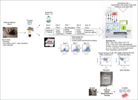
The spleen is the largest secondary lymphoid organ and plays a major role not only in immunologic functions but also in hematopoiesis and red blood cell clearance [16], [17]. Therefore, the spleen may play an important role in the progression or treatment of a WAS patients, and the study of the role of the spleen in autoimmunity in WAS-KO mice may help enhance our knowledge of the pathogenesis underlying autoimmune manifestations in patients with WAS. Single-cell RNA sequencing (scRNA-seq) has recently been widely adopted to identify the roles of immunity-related factors in a variety of fields because it can be used to detect changes in single cells and thus can provide insights into physiological and pathological processes.
In this study, we investigated differences in the spleens of 10-week-old WAS-KO and WT mice by performing scRNA-seq. These unprecedented data revealed the transcriptome in B cells, T cells, DCs, NK cells, macrophages, monocytes and granulocytes in the spleen and led to the identification of their gene expression signatures. To our knowledge, this is the first study to identify transcriptome sequences based on the cell types in the spleen of WAS-KO mice, which may help us understand the cellular and molecular mechanisms underlying WAS.


留言 (0)