
Remember me

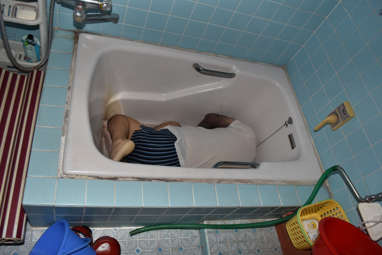
A 61-year-old man was found dead in an agricultural plot. The victim was occasionally there to pick olives for the owner of the agricultural plot. When officers came to investigate the crime scene, a dark dog that had been guarding the victim’s body wandered away and disappeared through a hole in a wire mesh (Fig. 1A). The victim’s upper clothes were torn, and pieces of his sweater and shirt were found around him (Fig. 1B). His trousers had been found pulled down to the ankles (Fig. 2A), further indicating that he had been dragged. The victim’s first on-site examination revealed face and abdomen injuries, as well as severe injuries of arms and left knee (Fig. 2B). During the “on-the-spot” investigation, six black Cane Corso dogs were found in the area surrounding the death scene. Four dogs belonged to one owner, and they were tagged as Dog_1, Dog_2, Dog_3, and Dog_4. Another person, living near the crime scene area, owned the other two dogs (Dog_5 and Dog_6). In Dog_1’s mouth, a mixed saliva-blood substance was found and collected for future comparisons. All the six dogs were taken to the kennel for further investigation. Due to the complexity of the injuries and the number of seized dogs, the prosecutor asked to determine the dogs’ involvement in the killing of the victim.
Fig. 1
A View of the hole in the wire mesh, where the dogs presumably walked through. “Dia” indicate the diameter of the hole (60 cm). “1 and 2” indicated two mesh broken extremities where dark tufts were found. B Detail of a shirt fragment collected in the second crime scene inspection (samples matched to the victim’s shirt)
Fig. 2
A Death scene. The corpse found close to the olive tree. Details of the trousers that had been found pulled down to the ankles. B Left knee extensive damage (the bite lesions were identified as postmortem due to no clinical evidence of tissue vitality)
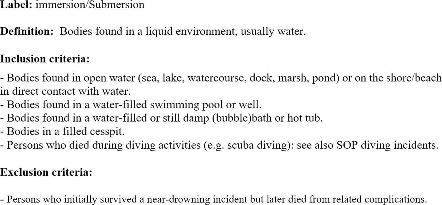
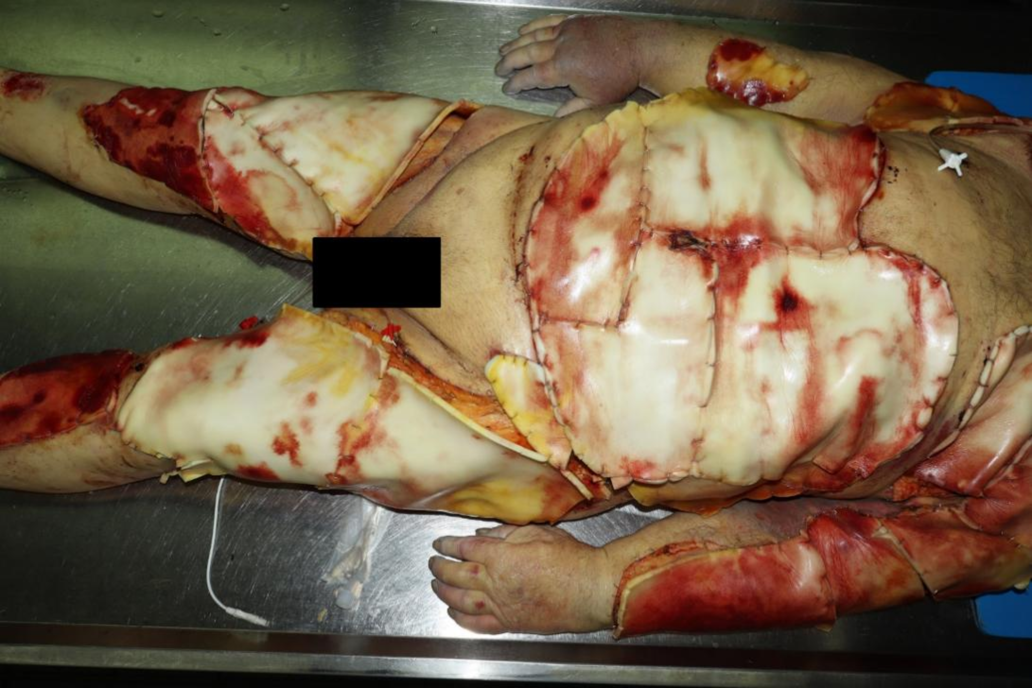
Autopsy findings and victim’s bite lesion analysisA general examination of the undressed body revealed traumatic wounds caused by several and deep dog bites. It was found that the deepest indentations, present in the upper right limb at the brachialis level, had caused losses of muscle-cutaneous substance at depths ranging from 2.5 to 4 cm. The right brachial artery was found slashed, suggesting heavy and massive blood loss, which could be the main cause of the victim’s death.
Before proceeding with autopsy, fourteen swabs were taken around the victim’s skin area near the dog’s bite marks in order to obtain dog saliva and DNA. The α-amylase test for the salivary enzyme presence could not be performed in victim’s wounds because salivary amylase is lacking or at very low abundance in mammalian carnivores such as cats and dogs [9, 10]. Regardless, the authors proceeded with swabbing the clearest bite-marks. Cardiac peripheral blood was taken to obtain the victim’s DNA reference sample. The biting victim’s clothes were packed and stored to be analyzed in a forensic genetic laboratory.
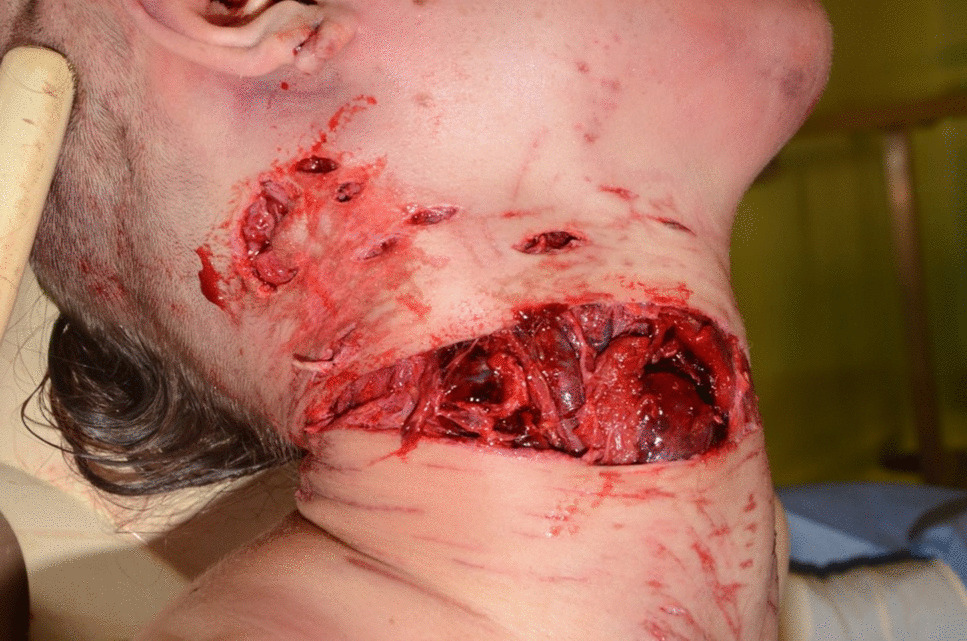
Subsequently, the “right Montgomery’s areola,” the “upper left hypochondriac area,” and the “left mid-tibial area” cutaneous bite injuries were selected for further specific analysis. The three anatomic areas were respectively numbered as AA1, AA2, and AA3 (Table 1), and their resulting bite imprints (B1, B2, and B3) were produced. A self-curing methacrylic resin ring was used to surround and border the damaged area. A high-viscosity addition silicone was added to completely fill the containment ring and remain in place until the polymerization was complete, and the bite silicone mark was obtained. After the silicone mark was removed (Fig. 3), it was sent to an odontological laboratory for a casting class IV hard plaster impression. Each anatomical area, AA1, AA2, and AA3, was again bordered with self-curing methacrylic resin, and the obtained ring was adhered to the skin with cyanoacrylate-based compound. Afterwards, a clean incision was made with a 22-blade scalpel from the skin to the muscle following the methacrylic resin ring border. The resulting muscle-cutaneous tissue containing the bite lesion was stabilized to the methacrylic resin ring by means of single, circular sutures for further comparisons. This activity was carried out in accordance with the guidelines of the American Board of Forensic Odontology (ABFO) [11], always taking care to avoid distortion of the tissue in order to photographically preserve the color and depth of the underlying bruises. Furthermore, cutaneous muscle samples were fixed in a solution of 5 mL 40% formaldehyde, 5 mL 99.8% glacial acetic acid, and 90 mL 7% ethanol. The samples were then stored for a period of 1 week after which they were removed from the formaldehyde bath and monitored for changes in dimension and stability, as well as their adherence or loss to the rings. The examined impressions of the dental arches on the skin were subjected to metric evaluations for subsequent comparative purposes.
Table 1 Skin lesion and corresponding Dog dental cast matches. Dog_2 was directly implicated in AA1 and AA2 injurie production present on the victim’s body. Its dental cast matched with these two anatomical lesions. Dog_1 was directly implicated in AA3 bite injury production. Its dental cast matched that anatomical lesionFig. 3
Silicone cast making and anatomic sample collection. A The methacrylate ring was applied around the bitten right Montgomery’s areola. B High-viscosity silicone was added to completely fill the containment ring, remained in place until polymerization was complete, and the bite silicone marks were obtained. C After the silicone was removed, the anatomical area was sutured to the methacrylate ring at several points, and then cut to the level of the muscular plane to stabilize the skin to the ring for further comparisons
Investigation of dogs’ bite marks analysisThe six Cane Corso dogs (Fig. 4) were taken to the kennel and subjected to judicial seizure. An initial analysis of the two dog groups showed that Dog_3 and Dog_4 were Dog_1 and Dog_2’s puppies, in adolescent stage (6–18 months). Whereas, the other two dogs were not related to the first four. After veterinary microchip recognitions and dog anesthetization, the oral mucosa cells were swabbed in order to obtain each dog’s reference sample, and the upper and lower dental impression were taken by modified steel dental tray. Before each dog was awakened, the dental formula was calculated (Table 2). Three dogs of the first owner (Dog_2, Dog_3, and Dog_4) had missing teeth. In addition, three of the six dogs (Dog_2, Dog_4, and Dog_6) exhibited a third-class malocclusion; this alteration is known as dysgnathia, where the lower jaw appears to be advanced compared to the upper jaw.
Fig. 4
Three of the six Cane Corso dogs subjected to judicial seizure before forensics veterinaries, odontological and genetics analysis were done at kennel. A Dog_1 was reported to be the alpha dog of his pack, a mixed saliva-blood substance was found in his mouth during the first on-site examination. B Dog_2 was the mother of the two puppies: Dog_3 and Dog_4. During the kennel investigations, a piece of fabric matching the victim’s shirt was found in her excrement. C Dog_3 was one of the juvenile dogs
Table 2 The dental formula of six Cane Corso dogs and related dental cast code. Dog_2, Dog_3, and Dog_4 presented lower jaw incisive anomalies (*). Dog_2, Dog_4, and Dog_6 were diagnosed with a class III dysgnathia. Dysgnathia was not detected (N.D.) in Dog_1, Dog_3, and Dog_5. Dental formula: I, incisors; C, canines; P, premolars; M, molars. Fraction slash (/) was used to divide left arch teeth from right arch teeth of each jawsSix sodium alginate canine dental impressions from Dog_1 to Dog_6 were obtained and numbered as follows: B1mg, B2mg, B3mg, B4mg, B5mg, and B6mg. A detailed photograph and analysis of each dog’s jaw was taken following ABFO recommendations [11]. Inter-canine distances and canine heights on each cast were also recorded using a digital caliper (Table 3). A piece of rose and green striped shirt was collected from Dog_2 excrement and preserved for further comparisons. Each dog dental cast was compared to the victim’s lesions by mechanical projection and by using DentalPrint© software [12].
Table 3 Dog dental anatomy measurements on dental casts. Upper inter-canine distance (U.I.C.D) is the distance measured in mm between canine in the upper cast. Lower inter-canine distance (L.I.C.D) is the distance measured in mm between canine in the lower cast. Upper canine height (U.C.H.) is the upper cast canine’s height expressed in mm. Lower canine height (L.C.H.) is the lower cast canine’s height expressed in mmDog DNA genotypingThe bitten victim’s clothes (e.g., blue jeans) together with dogs’ reference DNA were used to obtain the dogs’ DNA genetic profiles. DNA extraction was carried out using the QIAamp® DNA Mini Kit [13]. A preliminary amplification was performed on the extracts with universal primers for the canine mitochondrial cytochrome b gene [14,15,16,17]. This amplification provided information on the animal species that was eventually identified through sampling. The Dog DNA STR amplification was carried out using a ThermoFisher™ Canine STR panel 1.1 kit [18], composed of 18 autosomal loci and the amylogenic locus for sex determination. These regions are recommended by the International Society for Animal Genetics (ISAG) [19].
Human DNA genotypingThe victim’s clothes collected during the autopsy were initially observed by forensic lights. For presumptive trace detection of bloodstains [20], the Roche® tetramethylbenzidine (TMB) Combur3Test® was used. The samples that tested positive to the Combur3Test® reaction were subjected to the human blood detection by Bluestar® OBTI Immunochromatographic test [21]. All the samples that gave a positive result in the Bluestar® OBTI test, the blood collected from the Dog_1’s mouth and the victim’s control cardiac blood (taken during the autopsy), were used for the human DNA extraction using the QIAamp® DNA Mini Kit [13]. Human DNA quantification was conducted by the Quantifiler® Human kit [22]. Human STR amplification were conducted on a ThermoFisher GeneAmp® PCR System 9700 amplifier, and STR amplification was obtained using the GlobalFiler™ PCR Amplification Kit. The amplified products were separated into capillary electrophoresis with the 3500 Series Genetic Analyzer sequencer by Applied Biosystems™. Alleles were assigned by GeneMapper ID-X Software v1.1.2. C
Comments (0)