
記住我


Steady-state auditory evoked responses (SSAERs) are promising indicators of major auditory functions. Potential fields for the clinical application of SSAERs have been proposed in previous studies.1–7 However, the improvement and advancement of reliability and accessibility of SSAERs in clinical settings depends on simplification and minimalism in terms of standardization for the precise production and definition of the characteristics of SSAERs.
The interhemispheric asymmetry of SSAERs in normal controls has been inconclusive until now,8–10 although there might be insights with respect to changes in dominance for some clinical entities.1 The avant-garde results from published studies seem to be variable and controversial owing to factors including age, hearing condition, types/sides of stimulation, stimulus repetition rate, carrier frequency of stimuli, and recording methodology.8,10–16 Although previous studies revealed a tendency for the SSAERs to be right-side dominant under predetermined circumstances,10,17 the hemispheric asymmetry for the magnetic counterpart of SSAERs (ie, steady-state auditory evoked fields [SSAEFs]) induced by parameters reasonably accessible clinically has yet to be explored.
In the present study, the hemispheric dominance of SSAEFs was investigated in subjects with normal hearing by using magnetoencephalography (MEG). Acoustic sources ordinarily applied in an audiometric test, that is, binaural stimuli of pure tones modulated with amplitude at a rate of approximately 40 Hz for a carrier frequency of 1000 Hz were used as stimuli. We aimed to address repeatable precision and possible future standardization for the detection of hemispheric dominance in SSAEFs under clinically available circumstances.
2. METHODS 2.1. SubjectsTwelve healthy right-handed volunteers (seven males; aged, 21-42 years; mean age, 30 years) were recruited (Table 1). The inclusion criterion was normal pure-tone audiometry (PTA) results (see below). None of the subjects had implanted pacemakers, neurological deficits, or a history of trauma. This study conformed to the principles of the Declaration of Helsinki. Written informed consent was obtained from each subject, and the study protocol was approved by the Institutional Ethics and Research Committees of Cheng Hsin General Hospital and Taipei Veterans General Hospital.
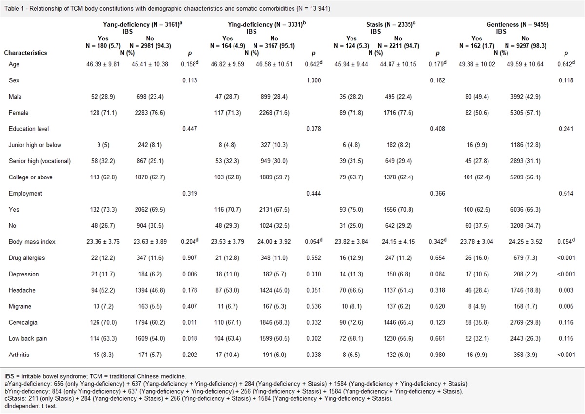
Table 1 - Amplitude of SSAEFs dipole moment and laterality index for subjects Hemisphere Left Right No Q(nAm) LI Sex Age Q(nAm) 1 12.3 1.8 M 23 22.7 2 10.0 2.0 F 35 20.3 3 11.3 2.3 M 31 25.9 4 14.4 1.9 F 42 26.8 5 7.8 2.2 M 40 17.2 6 10.3 1.8 M 29 18.5 7 11.9 1.1 F 22 12.6 8 9.2 1.3 M 24 11.9 9 9.5 2.3 M 27 21.5 10 14.6 1.7 M 35 24.1 11 18.5 1.5 F 21 28.1 12 14.6 1.7 F 35 24.1 m 12.0 1.8 30 21.1 SD 3.0 0.4 7.1 5.3 p <0.001Statistical significance using the t test was set at p < 0.05.
age = years; left = left hemisphere; LI = laterality index; m = mean; p, significance of difference for SSAEFs between responses of left vs right hemispheres; Q(nAm) = dipole moment strength of SSAEFs; right = right hemisphere; SSAEFs = steady-state auditory evoked fields.
All subjects underwent PTA to determine both air and bone conduction thresholds using test frequencies between 250 Hz and 8 kHz. All had normal PTA results (threshold ≤ 25 dB HL for all frequencies).
2.3. MEG paradigmMEG measurements were performed in a magnetically shielded room using a whole-head 306-channel neuromagnetometer (Vectorview 4-D Neuroimaging, Helsinki, Finland). The measurements were performed once during the study period. The subjects sat upright with their eyes open during the measurements. Auditory stimuli (1000 Hz, amplitude modulation [AM] frequency 43 Hz, modulation depth 100%, 180 seconds duration, and loudness matched individually to 50 dB SL at the exit end of the plastic tube) were delivered binaurally via molded earpieces using an analog-to-digital conversion card NI USB-6259 (National Instruments, Austin, TX) controlled by LabView (National Instruments). Triggers were given every second and reserved for subsequent signal processing. Trials with electro-oculographic amplitudes exceeding 150 μV were rejected. MEG signals were sampled at 400 Hz and band-pass filtered at 0.03 to 100 Hz. Two sessions (separated by 2 minutes of rest) were averaged before further analysis. An equivalent current dipole (ECD) model consisting of bilateral sources was used to explain the MEG signals.18 First, an initial guess of an independent source was made for both hemispheres, respectively. Each ECD was applied to a subset of 40 to 60 sensors around the maximum peak in one hemisphere, with a goodness-of-fit (g) >90% for acceptance. Because the accuracy of the dipole localization depends on the signal-to-noise ratio,19 we included a sensor only when the peak amplitude of the signal was stronger than two standard deviations above the baseline. After the ECD with the highest g value was identified, all channels were considered for further analysis to best explain the global recorded magnetic field.18 T1-weighted magnetic resonance imaging (MRI) of the subjects’ brains were acquired using a 3.0 T Bruker MedSpec S300 system (Bruker, Karlsruhe, Germany) for MEG-MRI co-registration. No obvious abnormalities (eg, vascular lesions or tumor growth) were found in any of the brain MRI scans.
2.4. Data analysisEach epoch was analyzed according to the concept of complementary ensemble empirical mode decomposition.20 The time window for each epoch was 1 second and was separated by the triggers provided in the recording. Because two sessions (each 180 seconds duration) were averaged before further analysis, a total of 360 epochs were used for the average and data normalization. The interhemispheric differences in the peak dipole strength of the SSAEFs observed in the left and right hemispheres for all subjects were evaluated using t tests. A laterality index (ratio of the SSAEF strength over the right hemisphere to that over the left hemisphere) was used to assess the degree of hemispheric asymmetry. Differences in the laterality index between sex were evaluated using the t test, too. Statistical significance was set as a p value <0.05.
3. RESULTSThe SSAEFs dipole was identifiable over each hemisphere in all subjects (Table 1). The SSAEFs source was localized bilaterally on the superior temporal plane, with an orientation centripetal to the auditory cortex. The laterality index (ie, the ratio of the SSAEF strength over the right hemisphere to that over the left hemisphere) ranged between 1.1 and 2.3 (m = 1.8, SD = 0.4) (Fig. 1). No differences were found between the laterality index of males and females (males: m = 1.9, SD = 0.4; females: m = 1.6, SD = 0.4; p = 0.226). In all subjects, the SSAEFs were significantly weaker in the left hemisphere (m = 12, SD = 3.0) than in the right hemisphere (m = 21.1, SD = 5.3, p = 0.014).
 Fig. 1:
 Neuromagnetic responses of SSAEFs to binaural stimulation in subjects with highest and lowest LI, respectively. A, Subject 3, male, demonstrated a pattern of right-side dominance for SSAEFs with the highest LI of 2.292. B, Subject 7, female, demonstrated a pattern of right-side dominance for SSAEFs with the lowest LI of 1.059. Dipole sources (red dots) were localized at the auditory cortices of bilateral temporal lobes in MRI scans. MRI views are displayed according to neurological convention, that is, the subject’s right hemisphere is on the right-side of the images. LI = laterality index; MRI = magnetic resonance imaging; SSAEFs = steady-state auditory evoked fields.
4. DISCUSSIONOne major and novel finding in the present study was the tendency for asymmetry of SSAEFs to the right brain with stimuli on both ears of AM pure tones in normal hearing. When SSAEFs from both hemispheres were collected and compared, the dipole moment of SSAEFs was consistently stronger in the right than in the left hemisphere. The laterality index, which was calculated as the ratio of SSAEFs strength over the right hemisphere to that over the left hemisphere, was therefore always >1.0 (with a range between 1.1 and 2.3). There were no differences in laterality index between sexes. To the best of our knowledge, our study is the first to report the right-side dominance of SSAEFs on binaural stimulation by a pure tone with the rate of AM at 43 Hz for a carrier frequency of 1000 Hz in subjects with normal hearing.
SSAERs can be potential probes for deficits in sound processing in the brain presented by medical conditions with or without auditory relevance. Alzheimer’s disease, for example, has been reported to enhance the strength of SSAEFs at its early stage, possibly suggesting an impaired optimization between the inhibition and excitation of central auditory processing.6 The dipole power of SSAEFs was also found to be increased in patients with tinnitus, which in turn could indicate that more neural fibers were entrained synchronously by the envelope of AM stimuli.21 However, the accessibility and application of SSAERs in clinical settings are yet to be improved. One of the major reasons resided in the lack of repeatable stability for the production of SSAERs due to parameters remaining to be simplified and standardized.
There are also some medical implications regarding the variation in interhemispheric asymmetry of SSAERs. For example, reduced hemispheric laterality of the SSAEFs has been observed in patients with schizophrenia.1 However, inconsistency persists for the hemispheric dominance of SSAERs in previous studies. One of the studies using intermittent stimuli, for example, revealed hemispheric asymmetry of SSAEFs to the right brain on both monaural and binaural stimulation.10 In contrast, other studies using monaural/binaural stimuli of AM continuous tones revealed a pattern of contralateral dominance for SSAEFs, regardless of the source strength or spectral amplitudes.8,9 Thus, it is necessary to confirm the tendency of the dominant side of SSAEFs in normal controls using reasonable parameters with clinical relevance.
However, in the present study, the interhemispheric dominance of SSAEFs upon binaural stimulation was constantly to the right brain. This is reflected by a laterality index ranging from 1.1 to 2.3 (Table 1 and Fig. 1). The laterality index had a different tendency from that of the transient fields. In a previous study, the auditory response to binaural stimulation was a nearly balanced condition.22
One of the main reasons for the repeatability in terms of hemispheric asymmetry for SSAEFs in this study was that it addressed factors associated with the optimization of the characteristics of SSAERs in subjects with normal hearing. The rate of AM was set at 43 Hz because the best frequency in terms of the interstimulus interval for the production of SSAERs with maximal strength has been shown to be approximately 40 Hz.23,24 Furthermore, 43 Hz instead of 40 Hz was selected to avoid possible interference by the frequency of the alternating current (60 Hz) in our country. In contrast to electroencephalography (EEG), MEG has the advantage of measuring cortical signals without the disturbance of noise from soft tissues including the scalp. Pure-tone stimuli were applied to both ears simultaneously, because binaural hearing is the most common mode of listening in the real world for human beings, and pure tones at a frequency of 1000 Hz are the style of stimulation most often encountered at the beginning of an audiometric test. It took approximately 3 minutes to complete the measurements in our study.
This study had some limitations. One is that MEG equipment is still difficult to access clinically. Owing to budget limitations, the sample size was small. However, the statistical strength was sufficient because there was a normal distribution inside. Because the primary goal of this study was to explore the possibility of establishing clinical standardization for the detection of hemispheric dominance in SSAEFs in the future, further studies with a similar setting and a larger sample size using EEG are warranted to investigate these issues.
In conclusion, the right-sided dominance of the SSAEFs was verified in subjects with normal hearing in the present study. Acoustic sources clinically available in audiometric tests were used as stimuli. Such simplification and minimalism of the parameters would be helpful for the standardization of precise production and definition of the characteristics of SSAERs, which in turn can improve the reliability and accessibility of the clinical application of SSAERs. Because MEG is still not easily accessible clinically, further studies using EEG with larger sample sizes are necessary to address these issues.
ACKNOWLEDGMENTSThis study was funded by the National Science Council of Taiwan (NSC-97-2314-B-075-025), Cheng Hsin General Hospital (99-43), National Yang Ming University (100F117CY14), and Taipei Veterans General Hospital (V98C1-150 and VGHUST97-P3-11).
We thank Mr. Chou-Ming Cheng and Mr. Chi-Cher Chou for their technical aid in MEG.
REFERENCES 1. Teale P, Carlson J, Rojas D, Reite M. Reduced laterality of the source locations for generators of the auditory steady-state field in schizophrenia. Biol Psychiatry. 2003;54:1149–53. 2. Poelmans H, Luts H, Vandermosten M, Boets B, Ghesquiere P, Wouters J. Auditory steady state cortical responses indicate deviant phonemic-rate processing in adults with dyslexia. Ear Hear. 2012;33:134–43. 3. Alaerts J, Luts H, Hofmann M, Wouters J. Cortical auditory steady-state responses to low modulation rates. Int J Audiol. 2009;48:582–93. 4. Hernandez-Perez H, Torres-Fortuny A. Auditory steady state response in sound field. Int J Audiol. 2013;52:139–43. 5. Pellegrino G, Arcara G, Cortese AM, Weis L, Di Tomasso S, Marioni G, et al. Cortical gamma-synchrony measured with magnetoencephalography is a marker of clinical status and predicts clinical outcome in stroke survivors. Neuroimage Clin. 2019;24:102092. 6. Osipova D, Pekkonen E, Ahveninen J. Enhanced magnetic auditory steady-state response in early Alzheimer’s disease. Clin Neurophysiol. 2006;117:1990–5. 7. Li LP, Shiao AS, Li CT, Lee PL, Cheng CM, Chou CC, et al. Steady-state auditory evoked fields reflect long-term effects of repetitive transcranial magnetic stimulation in tinnitus. Clin Neurophysiol. 2019;130:1665–72. 8. Tiihonen J, Hari R, Kaukoranta E, Kajola M. Interaural interaction in the human auditory cortex. Audiology. 1989;28:37–48. 9. Fujiki N, Jousmaki V, Hari R. Neuromagnetic responses to frequency-tagged sounds: a new method to follow inputs from each ear to the human auditory cortex during binaural hearing. J Neurosci. 2002;22:RC205. 10. Ross B, Herdman AT, Pantev C. Right hemispheric laterality of human 40 Hz auditory steady-state responses. Cereb Cortex. 2005;15:2029–39. 11. Kaneko K, Fujiki N, Hari R. Binaural interaction in the human auditory cortex revealed by neuromagnetic frequency tagging: no effect of stimulus intensity. Hear Res. 2003;183:1–6. 12. Hamalainen JA, Rupp A, Soltesz F, Szucs D, Goswami U. Reduced phase locking to slow amplitude modulation in adults with dyslexia: an MEG study. Neuroimage. 2012;59:2952–61. 13. Herdman AT, Lins O, Van Roon P, Stapells DR, Scherg M, Picton TW. Intracerebral sources of human auditory steady-state responses. Brain Topogr. 2002;15:69–86. 14. Schoonhoven R, Boden CJ, Verbunt JP, de Munck JC. A whole head MEG study of the amplitude-modulation-following response: phase coherence, group delay and dipole source analysis. Clin Neurophysiol. 2003;114:2096–106. 15. Yamasaki T, Goto Y, Taniwaki T, Kinukawa N, Kira J, Tobimatsu S. Left hemisphere specialization for rapid temporal processing: a study with auditory 40 Hz steady-state responses. Clin Neurophysiol. 2005;116:393–400. 16. Arutiunian V, Arcara G, Buyanova I, Gomozova M, Dragoy O. The age-related changes in 40 Hz auditory steady-state response and sustained event-related fields to the same amplitude-modulated tones in typically developing children: a magnetoencephalography study. Hum Brain Mapp. 2022;43:5370–83. 17. Poelmans H, Luts H, Vandermosten M, Ghesquiere P, Wouters J. Hemispheric asymmetry of auditory steady-state responses to monaural and diotic stimulation. J Assoc Res Otolaryngol. 2012;13:867–76. 18. Hari R, Makela JP. Modification of neuromagnetic responses of the human auditory cortex by masking sounds. Exp Brain Res. 1988;71:87–92. 19. Jacobson GP. Magnetoencephalographic studies of auditory system function. J Clin Neurophysiol. 1994;11:343–64. 20. Wang H, Li B, Feng Y, Cui B, Wu H, Shi H, et al. A pilot study of EEG source analysis based repetitive transcranial magnetic stimulation for the treatment of tinnitus. PLoS One. 2015;10:e0139622. 21. Wienbruch C, Paul I, Weisz N, Elbert T, Roberts LE. Frequency organization of the 40-Hz auditory steady-state response in normal hearing and in tinnitus. Neuroimage. 2006;33:180–94. 22. Scheffler K, Bilecen D, Schmid N, Tschopp K, Seelig J. Auditory cortical responses in hearing subjects and unilateral deaf patients as detected by functional magnetic resonance imaging. Cereb Cortex. 1998;8:156–63. 23. Pellegrino G, Arcara G, Di Pino G, Turco C, Maran M, Weis L, et al. Transcranial direct current stimulation over the sensory-motor regions inhibits gamma synchrony. Hum Brain Mapp. 2019;40:2736–46. 24. Galambos R, Makeig S, Talmachoff PJ. A 40-Hz auditory potential recorded from the human scalp. Proc Natl Acad Sci USA. 1981;78:2643–7.
留言 (0)