
記住我


The ulnar nerve function is crucial for performing intricate and dexterous tasks with our hands in daily life. Ulnar nerve injuries occurring at the wrist may cause loss of sensation at the little finger and the ulnar half of the ring finger, and weakness of the hypothenar muscles, the two medial lumbricals, interosseous muscles, adductor pollicis, and the deep head of the flexor pollicis brevis muscle.1,2 Impairment of the aforementioned functions results in substantial functional disability and decreased quality of life.
Past literature has concluded that some factors could lead to better surgical results for ulnar nerve repair, including younger patient age, injury at a lower level, shorter delayed time to surgery, and a clean transection injury.3–7 Furthermore, a direct end-to-end nerve repair is preferred over nerve grafting when a tension-free environment can be achieved.8,9 However, in cases of delayed repair or inadequate primary repair, a neuroma may develop, causing pain, and nerve conduction block. Neuroma excision results in a nerve gap, making end-to-end neurorrhaphy more challenging.3,10,11 Holding the wrist in a flexion position helps minimize the gap and, therefore, facilitates end-to-end neurorrhaphy.7 However, keeping the same position throughout the operation and until at least 4 weeks postoperatively can be very difficult to achieve.
Previously, we proposed our method of using a K wire to fix the wrist in flexion, which facilitates the nerve repair procedure and also protects the neurorrhaphy for 4 to 6 weeks before removal of the K wire.12 We now present our retrospective case series of five patients with delayed wrist-level ulnar nerve injuries who underwent nerve repair with this procedure, with a minimum of 12 months follow-up.
2. METHODS 2.1. Preoperative evaluationsThe basic profile of each patient was collected from medical charts, including age, gender, underlying diseases, hand dominance, time and mechanism of injury, and time and contents of the initial surgery. Specific neurological examinations for the ulnar nerve, including Froment sign, Wartenberg sign, and claw hand, were performed. The position of the Tinel sign was marked. General physical examination of the hand was performed to check for any associated injuries, such as tendon injuries, tendon adhesion, bony injuries, or ligament injuries, that should be addressed simultaneously. A baseline range of motion (ROM) of the wrist was recorded, and the tolerable flexion angle was tested.
Electrodiagnosis (EMG) was arranged before the operation to confirm that there was no reinnervation of the ulnar nerve and whether there was baseline median nerve neuropathy at the wrist, which could be exacerbated by the wrist flexion position.
The sensory and motor functions were graded according to the criteria of the Nerve Injuries Committee of the British Medical Research Council (the BMRC scale) for grading sensory and motor recovery after peripheral nerve injury.13–15 This retrospective study was approved by the institutional review board.
2.2. Surgical techniqueUnder general anesthesia, each patient was put in a supine position, with the arm extended over a hand table. A pneumatic tourniquet was applied to the upper arm. A zigzag incision was planned over the volar ulnar wrist, extending distally and proximally at the position of the Tinel sign. The zigzag design is very useful for wound closure later on when the wrist is fixed in flexion.
Due to the delayed treatment of our patients, severe scarring and adhesion were frequently noted, and careful dissection was performed to identify the neuroma. The neuroma was excised until healthy fascicles could be observed from both stump ends. Both stumps were further released to facilitate anastomosis and to minimize the gap distance. The wrist was then flexed to determine the position for which the two stumps could be anastomosed without tension. After the angle required for direct tension-free neurorrhaphy was determined, a 1.6-mm K wire was inserted antegrade from the dorsal cortex of the distal radius, spanning the radius-lunate joint, and the lunate-capitate joint to fix the wrist in flexion. Direct neurorrhaphy was then performed microscopically with 9-0 nylon epineural sutures. If concomitant tendon procedures were planned, they would be performed before the neurorrhaphy to avoid additional tension to the nerve anastomosis site once it had been completed.
2.3. Postoperative care and evaluationAfter the operation, a protective removable dorsal splint was given to each patient according to the flexion angle of the wrist. It was safe for the patient to remove the splint for wound care, hand hygiene, and finger exercise. The patients were regularly followed up at the outpatient clinic for wound condition and neurological examination. Between postoperative 4 and 6 weeks, the K wire was removed. Serial splinting was subsequently applied with the goal of gradually extending the wrist, advancing 20° to 30° per week. The ROM for the wrist usually returned to normal at postoperative 12 weeks. The patients were then followed up at postoperative 6 months, 12 months, and annually thereafter (mean follow-up: 17.5 months).
During the outpatient visits, the progression of the Tinel sign was recorded. The ROM of the wrist was measured by a goniometer. The grip strength and pinch strength for both hands were measured using a Jamar hand dynamometer and a Jamar pinch gauge (Baseline® Hydraulic Hand Dynamometers; Fabrication Enterprises, White Plains, NY). The sensory and motor functions were recorded according to the BMRC scale. Satisfactory motor recovery was defined as grade M4 or M5, and satisfactory sensory recovery was defined as grade S3+ or S4.13,15 We assessed subjective outcomes with patient-reported evaluations, including the quick disabilities arm, shoulder, and hand (DASH) score and modified Mayo wrist score (MMWS). Any intraoperative or postoperative complications were collected from the medical charts.
3. RESULTSThis was a retrospective case series. During October 2018 to July 2020, five patients (three men and two women) with delayed ulnar nerve injury at the wrist underwent this procedure. The mean patient age was 48.2 (range: 38-63) years. The mean delay from injury to index surgery was 84.6 (range: 63-111) days. Four patients were injured over the right wrist, all happened to be the dominant side, and one had the injury over the left wrist. Four patients underwent an initial surgery for ulnar nerve repair, and one patient only had treatment for a laceration wound. All patients were graded S0 in the ulnar half ring finger and little finger at presentation. Froment sign, Wartenberg sign, and claw hand were present in every patient. All preoperative EMGs showed complete ulnar nerve injury at the wrist with no reinnervation.
All patients underwent neuroma excision, pinning of the wrist with K wire and direct neurorrhaphy. Two patients had concurrent tenolysis of the flexor tendons, and one patient required flexor carpi ulnaris tendon repair. No intraoperative complications occurred. The mean flexion angle for the wrist (measured as the angle of the radial shaft and the third metacarpal shaft) was 52° (range 40°-60°). We did not identify any patient- or injury-related factors associated with the flexion angle required.
The mean follow-up time was 17.5 (range: 13-30.5) months. The mean grip strength was 76.8% (range: 64%-86%) of the contralateral hand and the mean pinch strength was 63.6% (range: 50%-82%) of the contralateral hand. The BMRC scale for sensory recovery was S3 for three patients and S3+ for two patients. The motor recovery was graded M4 for all five patients. The mean DASH score was 14.1 (range: 2.3-25), and the mean MMWS score was 83 (range: 70-95). All patients returned to normal wrist ROM within postoperative 12 weeks. There was no radiographic osteoarthritic change over the wrist, or wrist pain associated with cartilage penetration by the K wire. The details are shown in Table 1. We demonstrate a case in Fig. 1.
Table 1 - Details of the patients and their outcome Patient Age Gender Side of injury/side of hand dominance Time from injury to surgery, d Operative procedure Wrist fixation angle, °/K wire removal, wk Total follow-up time, mo Grasp (% of contralateral hand) Pinch (% of contralateral hand) BMRC (Sensory/motor) Quick DASH MMWS 1 63 F R/R 91 NE + DN 40/4.5 14 80 74 S3/M4 9.1 95 2 42 M R/R 63 NE + DN + FDP III-V, FDS IV-V tenolysis 60/6 13 75 52 S3+/M4 11.4 80 3 38 F L/R 76 NE + DN + FDP IV-V tenolysis 60/4 30.5 79 60 S3/M4 22.7 70 4 50 M R/R 111 NE + DN 50/6 15 64 50 S3+/M4 2.3 90 5 48 M R/R 82 NE + DN + FCU tendon repair 50/5 15 86 82 S3/M4 25 80 Mean 48.2 84.6 52/5.1 17.5 76.8 (SD 8.2) 63.6 (SD 14.0) 14.1 (SD 9.5) 8.3 (SD 9.7)Gender presented as F and M; side of injury and dominance presented as L and R.
BMRC = British Medical Research Council (the BMRC scale) for grading sensory and motor recovery after peripheral nerve injury; DN = direct neurorrhaphy; F = female; FCU = flexor carpi radialis; FDP = flexor digitorum profundus; FDS = flexor digitorum superficialis; L = left; M = male; MMWS = modified Mayo wrist score; NE = neuroma excision; Quick DASH = quick disabilities arm, shoulder, and hand score; R = right.
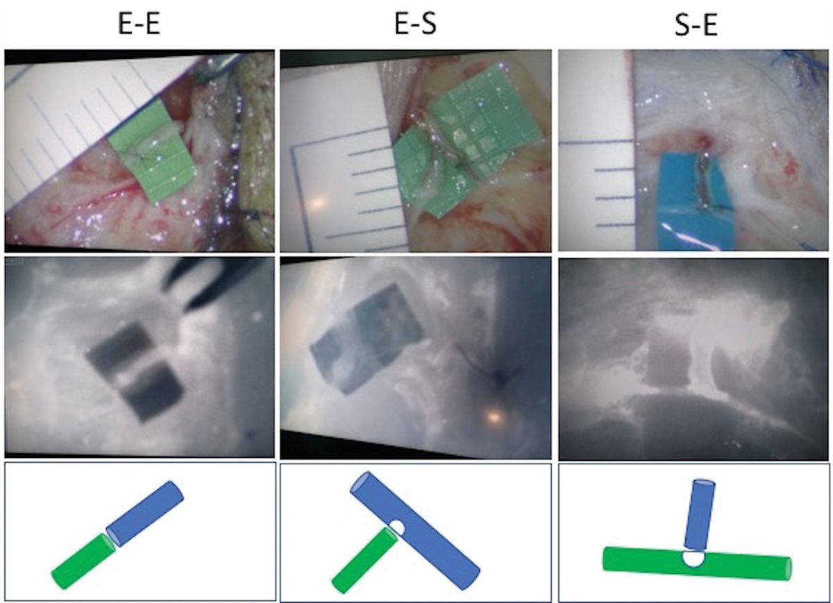
 Fig. 1: No. 5 patient is presented from Table 1. A, The blue vessel loops mark the ulnar nerve neuroma, and the red vessel loops mark the ulnar artery adhered to the neuroma. B, After excising the neuroma, wrist flexion, and pinning, direct ulnar nerve neurorrhaphy and artery repair with 9-0 nylon sutures were performed. C, The postoperative x-ray shows the 1.6-mm K wire spanning the radius, lunate, and capitate to fix the wrist in flexion by angle 50°. D, Right wrist range of motion was full and symmetric to the left wrist recorded at postoperative 15 months.4. DISCUSSION
We propose our method of temporary pinning of the wrist joint to facilitate end-to-end tension-free neurorrhaphy in delayed ulnar nerve injury at the wrist-level, and we present our case series. The results showed that direct neurorrhaphy was achieved with satisfactory motor recovery for all patients, and they also scored S3 or S3+ for sensory recovery. The mean DASH score was 14.1. No complications about wrist ROM were noted at a minimum of 1-year follow-up. An interesting finding was that two of our patients specifically reported difficulty in using chopsticks and thus had to use spoons and forks instead. This shows that using chopsticks requires more strength and control of the intrinsic muscles and is somewhat indicative of ulnar nerve functional recovery.
Past literature has shown that ulnar nerve repair is especially challenging, and the functional recovery is inferior compared to that of median and radial nerve repairs.5,16 Furthermore, it is even more difficult for satisfactory sensory recovery than satisfactory motor recovery.5,13 In their review article, Woo et al5 reported that approximately 55% of the patients who underwent primary ulnar nerve repair recovered to S2 sensation and 55% of the patients recovered to M3 in terms of motor function. No patient was able to recover to S4, and only 5% of the patients could achieve M5. The average DASH score was 22.5 Our results were comparable to the literature. Moreover, they mentioned that recovery can take up to 5 years,5,6 so further improvements could potentially be anticipated in our patients.
In our literature review, the factors that significantly affect the prognosis for ulnar nerve repair included age, injury level, gap of lesion, injury type, and delay time to surgical repair.3,4,6,7,15 In their meta-analysis of 260 patients, Lan et al6 showed that the rate of satisfactory motor recovery almost dropped to half for patients older than 40 years compared to patients aged between 30 and 40 years. They also noted that a delay time to surgery of more than 90 days was associated with worse motor function recovery.6 Although patient age, injury level, injury type, and delay time are nonmodifiable risk factors, there is room for management for the lesion gap.
The ideal scenario for nerve repair is to achieve tension-free end-to-end neurorrhaphy.8,9,17 However, this could be complicated by soft tissue loss, scarring, and neuroma formation, inevitably creating a nerve defect. The current strategies for managing nerve defects include positional adjustment, nerve transposition or rerouting, nerve grafting with various choices in graft selection, nerve transfer, and perhaps tendon transfer in more chronic cases.5,7,17,18 The common methods for positional adjustment for the ulnar nerve are elbow and wrist flexion by splinting. Anterior transposition of the ulnar nerve can help minimize nerve defects at the elbow level. Guyon canal and carpal tunnel release can help with nerve defects at the wrist level. In a cadaver study, Beldner et al9 proposed a creative method of removing the hamate hook that could shorten the distance of the motor branch by 21%. When a tension-free environment could not be achieved, nerve grafting with autograft is considered the gold standard treatment. However, the risk of donor site morbidity should be noted, including scarring, neuroma formation, and sensory loss. Nerve conduit and allograft spare the risks of donor site morbidity, but certain material properties should be taken into consideration with nerve conduit, and immune reaction should be considered in allograft use.17,18 Nerve transfer is also a strategy for ulnar nerve reconstruction. In more recent literature, supercharged end-to-side (SETS) anterior interosseous nerve to ulnar motor nerve transfer has been used in late reconstruction for intrinsic muscle function; this is particularly so in the early stages of nerve repair to protect and preserve the distal motor end plates and even adjunctively during the primary ulnar nerve repair for better and more rapid functional recovery in proximal ulnar nerve injuries.19–21 However, SETS is indicated for proximal ulnar nerve lesions and thus not as helpful in wrist-level lesions.
There are some contraindications for this method. There is a limitation to the length of gap between the nerve stumps. Lu et al12 showed that a maximal flexion angle of 60° to 75° was generally tolerated by patients, which could make up for about 3 cm of the gap distance. Thus, a backup plan with nerve graft should be prepared for a neuroma larger than 3 cm in length. If autograft is to be harvested, a preoperative explanation to the patient should be thorough and the risks and benefits clearly stated. Also, this method should be used cautiously in patients with preexisting carpal tunnel syndrome. Despite no patients in our series complaining of median nerve symptoms, preemptive carpal tunnel release could be considered in patients with baseline carpal tunnel syndrome. Finally, this was a small case series but nevertheless adds to the current literature of ulnar nerve repair.
As a short conclusion, we report our case series of patients with delayed ulnar nerve injury with a limited-sized neuroma formation. We performed radiocarpal pinning to fix the wrist in flexion, thereby facilitating neuroma excision and end-to-end neurorrhaphy. The minimum 1-year functional results were satisfactory. This method is simple, safe, and convenient for keeping the wrist in a flexion position during nerve repair intraoperatively and for protecting the neurorrhaphy postoperatively.
REFERENCES 1. Bertelli JA. Prior to repair functional deficits in above- and below-elbow ulnar nerve Injury. J Hand Surg Am. 2020;45:552.e1–e10. 2. Gottschalk HP, Bindra RR. Late reconstruction of ulnar nerve palsy. Orthop Clin North Am. 2012;43:495–507. 3. Grinsell D, Keating CP. Peripheral nerve reconstruction after injury: a review of clinical and experimental therapies. Biomed Res Int. 2014;2014:698256. 4. Basar H, Basar B, Erol B, Tetik C. Comparison of ulnar nerve repair according to injury level and type. Int Orthop. 2014;38:2123–8. 5. Woo A, Bakri K, Moran SL. Management of ulnar nerve injuries. J Hand Surg Am. 2015;40:173–81. 6. Lan CY, Tien HY, Lin YT, Hsu CC, Lin CH, Chen SH. Prognosis of traumatic ulnar nerve injuries: a systematic review. Ann Plast Surg. 2019;82:S45–52. 7. Pfaeffle HJ, Waitayawinyu T, Trumble TE. Ulnar nerve laceration and repair. Hand Clin. 2007;23:291–9, v. 8. Kim DH, Han K, Tiel RL, Murovic JA, Kline DG. Surgical outcomes of 654 ulnar nerve lesions. J Neurosurg. 2003;98:993–1004. 9. Beldner S, Rabinovich RV, Polatsch DB, Gonzalez DM. Ulnar nerve transposition in the hand: a cadaveric study. J Hand Surg Eur Vol. 2019;44:269–72. 10. Lans J, Baker DJ, Castelein RM, Sood RF, Chen NC, Eberlin KR. Patient-reported outcomes following surgical treatment of symptomatic digital neuromas. Plast Reconstr Surg. 2020;145:563e–73e. 11. Siemionow M, Brzezicki G. Chapter 8: current techniques and concepts in peripheral nerve repair. Int Rev Neurobiol. 2009;87:141–72. 12. Lu CC, Huang HK, Wang JP. Direct end-to-end neurorrhaphy for wrist-level long nerve defect with fixation of the wrist in flexion: technique note. J Wrist Surg. 2021; 11:362–6. 13. Ruijs AC, Jaquet JB, Kalmijn S, Giele H, Hovius SE. Median and ulnar nerve injuries: a meta-analysis of predictors of motor and sensory recovery after modern microsurgical nerve repair. Plast Reconstr Surg. 2005;116:484–94. 14. He B, Zhu Q, Chai Y, Ding X, Tang J, Gu L, et al. Safety and efficacy evaluation of a human acellular nerve graft as a digital nerve scaffold: a prospective, multicentre controlled clinical trial. J Tissue Eng Regen Med. 2015;9:286–95. 15. He B, Zhu Z, Zhu Q, Zhou X, Zheng C, Li P, et al. Factors predicting sensory and motor recovery after the repair of upper limb peripheral nerve injuries. Neural Regen Res. 2014;9:661–72. 16. Yang M, Rawson JL, Zhang EW, Arnold PB, Lineaweaver W, Zhang F. Comparisons of outcomes from repair of median nerve and ulnar nerve defect with nerve graft and tubulization: a meta-analysis. J Reconstr Microsurg. 2011;27:451–60. 17. Ray WZ, Mackinnon SE. Management of nerve gaps: autografts, allografts, nerve transfers, and end-to-side neurorrhaphy. Exp Neurol. 2010;223:77–85. 18. Beris A, Gkiatas I, Gelalis I, Papadopoulos D, Kostas-Agnantis I. Current concepts in peripheral nerve surgery. Eur J Orthop Surg Traumatol. 2019;29:263–9. 19. Barbour J, Yee A, Kahn LC, Mackinnon SE. Supercharged end-to-side anterior interosseous to ulnar motor nerve transfer for intrinsic musculature reinnervation. J Hand Surg Am. 2012;37:2150–9. 20. Chen SH, Mao SH, Lan CY, Huang RW, Lee CH, Hsu CC, et al. End-to-side anterior interosseous nerve transfer: a valuable alternative for traumatic high ulnar nerve palsy. Ann Plast Surg. 2021;86(2S Suppl 1):S102–7. 21. Koriem E, El-Mahy MM, Atiyya AN, Diab RA. Comparison between supercharged ulnar nerve repair by anterior interosseous nerve transfer and isolated ulnar nerve repair in proximal ulnar nerve injuries. J Hand Surg Am. 2020;45:104–10.
留言 (0)