
記住我


In 2016, the International Commission on Radiological Protection (ICRP) established Task Group 103 (TG 103) with the purpose of developing a new generation of the ICRP reference computational phantoms with the high-quality/fidelity mesh format, recognized as the most advanced format for phantom development at this point (Kainz et al. 2019). Since its establishment, TG 103 has been developing mesh-type reference computational phantoms (MRCPs) by converting the existing ICRP reference computational phantoms, the voxel-type reference computational phantoms (VRCPs) (ICRP 2009, 2020a), into the mesh format with anatomical and dosimetric improvements. In 2020, TG 103 completed the development of the adult male and female MRCPs (Nguyen et al. 2015; Yeom et al. 2016a–c; Kim et al. 2017), the mesh counterparts of the adult VRCPs of ICRP Publication 110 (ICRP 2009), and officially released them through ICRP Publication 145 (ICRP 2020b). The adult MRCPs include the complex and small organs, even all of the μm-thick target and source regions prescribed by the ICRP (ICRP 1994, 2006, 2010), thereby significantly improving the dosimetric accuracy relative to the adult VRCPs (Nguyen et al. 2015; Yeom et al. 2016a–c, 2019, 2020; Kim et al. 2017; ICRP 2020a). Following the completion of the adult MRCPs, TG 103 recently completed the development of the pediatric (newborn, 1-, 5-, 10-, and 15-y-old) male and female MRCPs (Choi et al. 2021a and b, 2022; Shin et al. 2021; Han et al. 2021; Yeom et al. 2022) and the mesh counterparts of the pediatric VRCPs of ICRP Publication 143 (ICRP 2020a), which share functionally the same level with that of the adult MRCPs.
Following up on our previous studies (Choi et al. 2021a and b, 2022; Shin et al. 2021; Han et al. 2021; Yeom et al. 2022), in the present study, we developed a new set of respiratory tract organs for the pediatric MRCPs, consisting of the following regions: extrathoracic (ET1 and ET2), bronchial (BB, airway generations 0–8), bronchiolar (bb, airway generations 9–15), and alveolar-interstitial (AI) regions. The ET1, ET2, tracheal (airway generation 0), and main BB (airway generation 1) regions and exterior lung boundaries were developed by converting those of the pediatric VRCPs into the mesh format with anatomical improvement, especially in the ET2 region. The remaining airways (airway generations 2–15) were produced inside the exterior lung boundaries using a dedicated C++ computer program (Kim et al. 2017). The AI region was assumed to be homogeneously present in the remaining regions within the lung boundaries. All of the μm-thick target and source regions prescribed in ICRP Publication 66 (ICRP 1994) were included in the developed respiratory tract organs. After development, the pediatric MRCPs with the respiratory tract organs were implemented in the Geant4 Monte Carlo code (Allison et al. 2016) to calculate the electron-specific absorbed fractions (SAFs). Finally, the resulting values were compared with those computed with the ICRP-66 data by ICRP Task Group 96 (TG 96) to investigate the potential dosimetric impact of the developed respiratory tract organs.
MATERIALS AND METHODS Respiratory tract organs of pediatric VRCPsFig. 1 shows an overall view of the respiratory tract organs of the pediatric VRCPs (ICRP 2020a), which consist of the ET1, ET2, tracheal, and main BB regions and exterior lung boundaries. The ET1, ET2, tracheal, and main BB regions are further subdivided into the wall and airway. Most of the organs and regions were constructed directly from the original CT images, but some organs (or some regions) were carefully designed using pipes based on the traces identified in CT images of insufficient image quality (Lee et al. 2007). The tracheal wall and exterior lung boundaries were aligned to the reference masses of ICRP Publication 89 (ICRP 2002). On the other hand, the ET1, ET2, and main BB walls’ masses were automatically determined according to their traces and thicknesses, due to the absence of ICRP-89 reference masses (Lee et al. 2007).
 Fig. 1: Respiratory tract organs of pediatric VRCPs (ICRP 2020a). This figure only shows male respiratory tract organs for 10 years and younger.
The major anatomical and dosimetric limitations of the respiratory tract organs of the pediatric VRCPs are that the airway generations other than the tracheal and main BB regions are not represented and that the μm-thick target and source regions prescribed in ICRP-66 (summarized in Tables 1 and 2) are also not included, due to insufficient image quality and finite voxel resolutions. Therefore, the SAFs to the respiratory tract organs for internal exposures to the charged particles (e.g., electrons) were computed with the ICRP-66 data by TG 96 (the results will be tabulated in an upcoming ICRP publication); more specifically, the SAFs were computed by dividing the absorbed fractions (AFs) calculated with the adult male stylized models by the age-dependent target-region masses (ICRP 1994). Note that the stylized models for other ages and sex are not provided and that the age-dependent target-region masses were obtained by scaling down the adult male masses from the adult male stylized models in proportion to the estimated airway surface areas corresponding to their ages (ICRP 1994). Besides, whereas the ET2 region should represent the regions connecting the posterior nasal passage, pharynx, and larynx (ICRP 1994), some of its regions are still missing (Fig. 1), and the larynx is included in the cartilage, not in the ET2 region. Thus, significant dosimetric errors in the SAFs to the ET2 may occur, especially for internal exposures from the adjacent organs (e.g., ET2 ← thyroid).
Table 1 - Summary of morphometric data of target regions in respiratory tract organs prescribed in ICRP Publication 66 (ICRP 1994). Organs Target region Target region deptha ET1 Basal cells 40 to 50 μm ET2 Basal cells 40 to 50 μm BB Basal cells 35 to 50 μm Secretory cells 10 to 40 μm bb Secretory cells 4 to 12 μmaIf there is no additional information, the airway surface is generally the basis of the depth. Positive and negative values indicate the depths outward and inward from the basis, respectively.
aIf there is no additional information, the airway surface is generally the basis of the depth. Positive and negative values indicate the depths outward and inward from the basis, respectively.
bAI as a source region is determined by radiation types and energies.
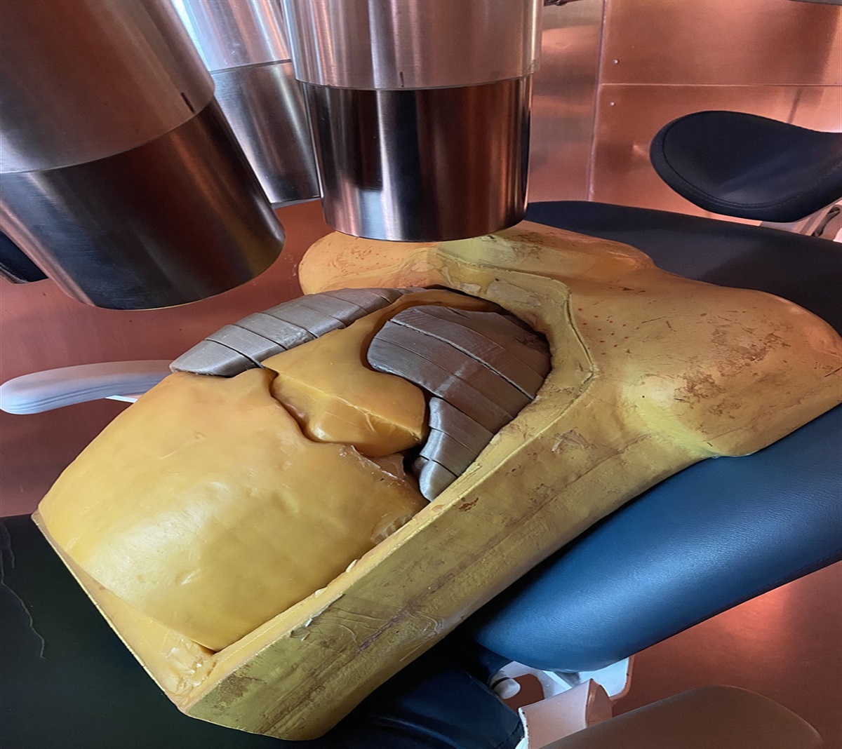
The respiratory tract organs of the pediatric MRCPs were produced first by converting the University of Florida and National Cancer Institute (UF and NCI) pediatric phantoms (Lee et al. 2010) into the polygonal mesh (PM) format and then by matching the mesh models to the pediatric VRCPs (ICRP 2020a), following the procedure shown in Fig. 2, which also had been used in previous studies (Choi et al. 2021a and b, 2022; Yeom et al. 2022). Note that the UF and NCI phantoms in the non-uniform rational B-spline (NURBS) and PM formats are the origins of the pediatric VRCPs (i.e., before voxelization and other adjustment processes), which strongly facilitated their conversion to the high-quality PM format. First, the respiratory tract organs of the UF and NCI phantoms (i.e., ET1, ET2, trachea, and main BB regions and exterior lung boundaries) were converted into primitive PM models, which were then refined into the high-quality PM format. Finally, the refined PM models were adjusted to match the masses and topologies of the respiratory tract organs of the pediatric VRCPs. Note that during the whole development process, the NURBS and PM models were visualized, handled, and refined using Rhinoceros 5.0 software (Robert McNeel & Associates, Seattle, WA) and Rapidform software (INUS Technology Inc, Seoul, Republic of [South] Korea), respectively.
 Fig. 2:
 Modeling method applied to respiratory tract organs of pediatric MRCPs for lung boundaries of 5-year-old male MRCP as example.
Then the ET2 region of the pediatric MRCPs was further improved to have a more realistic shape. First, the posterior nasal passage was manually reconstructed under the careful guidance of an anatomist based on the traces identified in the pediatric VRCPs. As shown in Fig. 1, the maxillary sinus in the posterior nasal passage was not defined in the newborn and 1-y-old VRCPs, whereas it was defined in the 5-, 10-, and 15-y-old VRCPs. According to Kozak et al. (2014), the maxillary sinus, although present from birth, starts to expand laterally beyond the infraorbital foramen from 5 y; we believe that it was not defined in the newborn and 1-y-old VRCPs because it was too small to be identified from the CT images or negligible, which was thus also reflected in the pediatric MRCPs. Then, the remaining regions (i.e., pharynx and larynx) were replaced with the new model, which was produced by scaling down those of the adult MRCPs (ICRP 2020b). For this, the mass ratio of the tracheal region between the adult and pediatric MRCPs was used as the scaling factor, considering the fact that the dimensions of the ET and tracheal regions are in a proportional relationship (ICRP 1994). Then the reconstructed (i.e., posterior nasal passage) and replaced (i.e., pharynx and larynx) regions were connected, which were finally divided into the ET2 wall and airway, matching their mass ratio in the adult MRCPs.
Next, the μm-thick target and source regions prescribed in ICRP 66 (Tables 1 and 2) were defined in the respiratory tract of the pediatric MRCPs. For the ET1, ET2, tracheal, and main BB regions, the target and source regions were defined simply by uniformly enlarging the airway surface following the method applied in previous studies (ICRP 2020b; Kim et al. 2017; Choi et al. 2021a, 2022). However, this method could not be applied to the other airway generations of the BB region (airway generations 2–8) or to any of the subsequent generations of the bb region (airway generations 9–15) because they are not represented in the pediatric VRCPs and, consequently, in the pediatric MRCPs. Therefore, the remaining airways (airway generations 2–15), including the target and source regions, were generated using a dedicated C++ computer program that had been used for the adult MRCPs (Kim et al. 2017).
Before generation of the remaining airways, the airway dimensions (i.e., lengths and diameters) were derived by using the scaling method described in ICRP 66. For the BB region except for the main BB region, the scaling factors were calculated by substituting the standing heights of the pediatric MRCPs in Table 4 of ICRP 66, and the airway lengths and diameters were then calculated by multiplying the scaling factors by those of the adult male. For the bb region, the airway lengths and diameters were calculated by hyperbolic and parabolic interpolation, respectively, between those of airway generations 8 and 16. Note that the lengths and diameters of airway generation 16 were calculated in advance by scaling those of the adult male by the cube root of the functional residual capacity (FRC) given in table 7 of ICRP 66, as described in Table 3 of ICRP 66. Note that the FRC for the newborn was newly calculated using the equation given in table B.4 of ICRP 66, because the newborn considered in ICRP 66 is a “3-month-old infant,” whereas the newborn considered in the present study was literally a “newborn (0-month-old) infant.”
Table 3 - Total branch length of each generation for remaining airways (airway generation 2–15) of pediatric MRCPs for male along with their ideal length. Generation Newborn male 1-year-old male 5-year-old male 10-year-old male 15-year-old male Ideal length (cm) Left lung length (cm) Right lung length (cm) Ideal length (cm) Left lung length (cm) Right lung length (cm) Ideal length (cm) Left lung length (cm) Right lung length (cm) Ideal length (cm) Left lung length (cm) Right lung length (cm) Ideal length (cm) Left lung length (cm) Right lung length (cm) 2 9.7 × 10⁻¹ 1.1 × 100 1.0 × 100 1.6 × 100 1.5 × 100 1.5 × 100 2.0 × 100 2.0 × 100 2.0 × 100 2.5 × 100 2.6 × 100 2.7 × 100 2.9 × 100 2.8 × 100 2.9 × 100 3 1.1 × 100 1.2 × 100 1.2 × 100 1.7 × 100 1.6 × 100 1.7 × 100 2.2 × 100 2.3 × 100 2.3 × 100 2.7 × 100 2.7 × 100 2.7 × 100 3.2 × 100 3.2 × 100 3.2 × 100 4 2.3 × 100 2.1 × 100 2.2 × 100 4.1 × 100 3.8 × 100 4.4 × 100 5.1 × 100 5.2 × 100 5.2 × 100 6.0 × 100 6.4 × 100 6.3 × 100 6.9 × 100 6.6 × 100 7.1 × 100 5 4.2 × 100 3.8 × 100 3.9 × 100 6.8 × 100 7.0 × 100 6.3 × 100 8.8 × 100 9.2 × 100 9.2 × 100 1.1 × 101 1.0 × 101 1.0 × 101 1.2 × 101 1.3 × 101 1.2 × 101 6 6.8 × 100 6.2 × 100 6.3 × 100 1.2 × 101 1.2 × 101 1.2 × 101 1.5 × 101 1.5 × 101 1.5 × 101 1.8 × 101 1.6 × 101 1.7 × 101 2.0 × 101 2.1 × 101 2.1 × 101 7 1.2 × 101 1.1 × 101 1.1 × 101 2.5 × 101 2.5 × 101 2.3 × 101 2.9 × 101 3.0 × 101 2.8 × 101 3.3 × 101 3.2 × 101 3.4 × 101 3.7 × 101 3.6 × 101 3.8 × 101 8 2.2 × 101 2.0 × 101 2.0 × 101 4.9 × 101 4.9 × 101 4.8 × 101 5.5 × 101 5.9 × 101 5.5 × 101 6.1 × 101 6.1 × 101 6.4 × 101 6.6 × 101 6.6 × 101 6.4 × 101 9 3.6 × 101 3.3 × 101 3.2 × 101 7.9 × 101 7.3 × 101 7.2 × 101 9.0 × 101 9.1 × 101 8.7 × 101 9.9 × 101 9.0 × 101 1.0 × 10² 1.1 × 10² 1.1 × 10² 1.1 × 10² 10 5.9 × 101 5.3 × 101 5.5 × 101 1.3 × 10² 1.2 × 10² 1.2 × 10² 1.5 × 10² 1.4 × 10² 1.5 × 10² 1.6 × 10² 1.5 × 10² 1.6 × 10² 1.8 × 10² 1.8 × 10² 1.7 × 10² 11 9.7 × 101 8.9 × 101 8.8 × 101 2.1 × 10² 1.9 × 10² 1.9 × 10² 2.4 × 10² 2.3 × 10² 2.3 × 10² 2.7 × 10² 2.5 × 10² 2.6 × 10² 3.0 × 10² 2.8 × 10² 3.0 × 10² 12 1.6 × 10² 1.5 × 10² 1.5 × 10² 3.3 × 10² 3.0 × 10² 3.0 × 10² 3.9 × 10² 3.6 × 10² 3.6 × 10² 4.4 × 10² 4.0 × 10² 4.1 × 10² 4.9 × 10² 5.0 × 10² 4.7 × 10² 13 2.6 × 10² 2.4 × 10² 2.4 × 10² 5.1 × 10² 4.7 × 10² 4.7 × 10² 6.2 × 10² 6.1 × 10² 6.0 × 10² 7.1 × 10² 6.5 × 10² 6.8 × 10² 8.1 × 10² 7.9 × 10² 8.1 × 10² 14 4.3 × 10² 3.9 × 10² 3.9 × 10² 7.8 × 10² 7.2 × 10² 7.2 × 10² 9.8 × 10² 9.5 × 10² 9.7 × 10² 1.1 × 10³ 1.1 × 10³ 1.0 × 10³ 1.3 × 10³ 1.3 × 10³ 1.4 × 10³ 15 6.8 × 10² 6.2 × 10² 6.3 × 10² 1.1 × 10³ 1.1 × 10³ 1.0 × 10³ 1.5 × 10³ 1.4 × 10³ 1.4 × 10³ 1.8 × 10³ 1.6 × 10³ 1.6 × 10³ 2.1 × 10³ 2.0 × 10³ 2.0 × 10³The remaining airways were then generated in the lung boundaries based on the derived airway dimensions using a computer program. The remaining airways for all of the pediatric MRCPs (with the exception of newborn MRCPs) were successfully generated. For the newborn MRCPs, however, it was not possible to construct the last bb airway generation (i.e., airway generation 15) due to the fact that the space in the lung boundaries was physically insufficient. To identify the problem, therefore, it was determined whether (1) the lung boundary volume of the newborn MRCPs and (2) the derived airway dimensions for the newborn were anatomically reasonable.
First, the lung boundary volume of the newborn MRCPs was compared with the conventional lung volume (CLV) calculated from the experimental data (Doershuk and Matthews 1969). The CLV considers mid-inhalation (or mid-exhalation) in the normal breathing cycle and can be calculated by the equation:
CLVcm3=MFRC+MTV2,
where the MFRC is the mean FRC (cm3) generally considered as the lung volume at end-exhalation, and the MTV is the “mean tidal volume” (cm3) defined as the normal volume of air displaced between normal inhalation and exhalation. Note that the MFRC and MTV were measured using a plethysmograph for 51 newborn subjects aged between 2 h and 11 d (Doershuk and Matthews 1969). Then, our comparison showed that the lung boundary volume of the newborn MRCPs (= 97.1 cm3) was slightly smaller than the calculated CLV (= 102.4 cm3) but remained within 1 standard deviation (= 15.6 cm3), thus validating that the lung boundary volume of the newborn MRCPs was within the normal lung volume range.
Secondly, the airway dimensions of the newborn derived from the ICRP 66 method were checked by examining the ratio of airway length (RAL) to the cube root of the lung volume, considering the statement of Hislop et al. (1972) that the pre-acinar airways of infants are regarded as a miniature version of those of adults and that they grow symmetrically. Fig. 3 shows the ratios of the RALs relative to the adult male. All of the ratios, except for that of the newborn, tended to be close to unity, with the maximum ratio of 1.13 (for 10 y), which means that the RALs were similar to that of the adult male. However, the ratio for the newborn was 1.42, a significant deviation from unity. This notable deviation for the newborn seems to have been due to the fact that the scaling method described in ICRP 66 was developed based on the anatomical data obtained from only 20 subjects aged between 11 d and 21 y (Phalen et al. 1985), thereby resulting in a relatively large discrepancy for the newborn, which is an extreme case.
 Fig. 3:
 Comparison of RAL of newborn, 1, 5, 10, and 15 years with RAL of adult male.
To address this discrepancy, in the present study, assuming that the airway dimensions are proportional to the cube root of the lung volume (Hislop et al. 1972; Hofmann 1982), the airway dimensions of the newborn were scaled down from the airway dimensions of the adult male by the cube-root ratio of the lung volume of the newborn MRCPs to that of the adult male MRCP. The derived airway dimensions were 3.1 times smaller than those of the adult male, which is consistent with the statement of ICRP 66, which is that the airway dimensions increase by around 3 times from birth to adulthood. Then, the remaining airways for the newborn MRCPs were again generated based on the newly derived airway dimensions for the newborn using the computer program.
The generated remaining airways are in the constructive solid geometry (CSG) format. They could have been converted to the PM or tetrahedral mesh (TM) format for easy incorporation into the pediatric MRCPs, but the resulting airways would require a very large number of facets and, so too, a large computer memory allocation (i.e., >50 GB) (Kim et al. 2017). In the present study, therefore, the remaining airways in the CSG format were not converted to the PM format but rather were directly incorporated into the pediatric MRCPs based on the overlaying approach used for the adult MRCPs (Kim et al. 2017; ICRP 2020b). Note that the overlaying approach makes it possible to perform the dose calculation for the remaining airways with only minimal added memory usage and computation time.
The target and source regions in the AI region were assumed to be homogeneously spread throughout the regions except for the BB and bb regions within the lung boundaries, on the bases that the interalveolar septa and the walls of blood of lymphatic capillaries are sufficiently thin to ensure this assumption (ICRP 1994).
Finally, the pediatric MRCPs incorporated with the respiratory tract organs (except for the remaining airways in the CSG format) were converted to the TM format through the tetrahedralization process, which fills the organs with tetrahedrons without any alteration to the original organ shapes, following the process described in Yeom et al. (2014). Note that TM models allow for computational speed hundreds of times faster in the Monte Carlo codes (Yeom et al. 2014) as well as better compatibility with the major Monte Carlo codes such as Geant4 (Allison et al. 2016), PHITS (Sato et al. 2013), and MCNP6 (Goorley et al. 2016), as compared with PM models.
Monte Carlo dose calculationsTo investigate the dosimetric impact of the developed respiratory tract organs, the SAFs were calculated using the pediatric MRCPs for some example cases of internal electron exposures and then compared with the TG 96 values. In the present study, the electrons were selected as the primary particles due to their ability to establish steep dose gradients within the organs, necessitating the use of thin target regions for dose calculation. Again, note that the pediatric VRCPs cannot be used for calculation of respiratory tract organs’ internal exposures to charged particles due to the absence of the thin target and source regions. Therefore, for this calculation, the pediatric MRCPs were implemented first in the Geant4 Monte Carlo code (version 10.06.p02, released in May 2020) (Allison et al. 2016); all organs in the TM format were implemented using the G4Tet class and were then overlaid with the remaining airways in the CSG format using the G4VUserParallelWorld class. They were considered to be in a vacuum. Then, the mono-energetic primary electrons with 25 energies ranging from 0.01 to 10 MeV were uniformly emitted from the source regions using the G4VUserPrimaryGeneratorAction class; the primary electrons were sampled using the barycentric-coordinate-system-based sampling method (Rocchini and Cignoni 2000) and the rejection method in the TM and CSG formats, respectively. The number of primary electrons varied from 104 to 108 depending on the example cases, keeping the statistical relative errors below 5%. They were then transported with a secondary production range cut value of 1 μm via the G4EmLivermorePhysics library. The variance reduction techniques were not used. Finally, the energies deposited in the target regions were calculated using the G4PSEnergyDeposit class and were divided by the primary electron energies and target-region masses to obtain the SAFs. The simulations were performed on a server computer equipped with the Intel® Xeon® Gold 6258R CPU [at 2.70 GHz with 56 cores (two hyperthreads per core) and 128 GB RAM] in the CentOS Linux release 7.9.2009 (Core). GNU compiler collection (GCC) 8.3.1 was used. Fig. 4 shows the 10-y-old female MRCP irradiated by 1 MeV electrons for the ET2 sequestered region and AI region as source regions as examples of simulations.
 Fig. 4:
 10-year-old female MRCP irradiated by 1 MeV electrons for the ET2 sequestered (left) and AI (right) regions as source regions.
RESULTS AND DISCUSSION Respiratory tract organs of pediatric MRCPsFig. 5 shows an overall view of the respiratory tract organs developed in the present study for the pediatric MRCPs. It can be seen that the developed respiratory tract organs closely preserve the original topologies of those of the pediatric VRCPs but boast improved anatomy, especially in the ET2 region, as compared with the pediatric VRCPs. It can also be seen that the developed airways are fully covered by the thin walls of the respiratory tract organs, unlike the pediatric VRCPs where they are partially exposed outside the walls. In addition, the developed respiratory tract organs include the remaining airways (airway generations 2–15) and the μm-thick target and source regions prescribed in ICRP 66. Fig. 6 shows the detailed structure of the developed respiratory tract organs, including the remaining airways and some target and source regions, for the 5-y-old male MRCP as an example.
留言 (0)