
Remember me

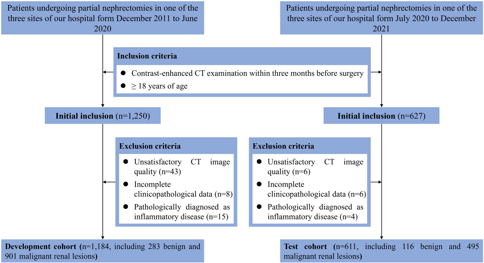


This two-center retrospective study’s ethical approval was provided by the two institutional review boards, and the requirement for informed consent was waived. Between June 2016 and July 2022, 160 patients with STS confirmed by pathology and met the inclusion criteria were retrospectively collected. Inclusion criteria:(1) Patients underwent surgical resection; (2)STS was diagnosed by histopathology; (3) Axial FS-T2WI MRI scans ≤ 2 weeks before surgery; Exclusion criteria: (1) Incomplete clinical or imaging data; (2) MRI image quality is poor, signal-to-noise ratio ≤ 1.0; (3) Development of other subsequent tumors; (4) The patient has received prior treatment, such as chemotherapy, radiation therapy, or needle biopsy.
In total, 160 STS patients were analyzed. Clinical-MRI characteristics included age, gender, location, and MRI morphological features. All images were independently reviewed by two radiologists with more than 5 years of skeletal muscle MRI experience while remaining blind to the clinical and histopathological data. Decisions on MRI findings were made through team negotiation. According to Zhao et al. [20], the following MRI morphological features were selected for comparison: (1) size (maximum diameter of tumor, < 5 cm or ≥ 5 cm); (2) margin (well- or poorly-defined); (3) signal intensity (homogeneous or heterogeneous, > 30% of the whole volume was considered heterogeneous); (4) peritumoral hyperintensity. All these MRI features were labeled as dichotomous variables and recorded using Yes or No.
The final histopathological results of the 160 STS patients were shown in Table 1. The FNCLCC system assigns a score to the tumor based on its mitotic index, differentiation, and amount of necrosis, and the tumor grade was calculated by adding these three scores. According to their FNCLCC tumor grade, the patients were divided into two groups: low-grade (N = 82) and high-grade (N = 78). The workflow was shown in Fig. 1.
Table 1 The pathologic data of the 160 STS patients Fig. 1
(Top) Flow chart of patient enrollment. (Bottom) Work flow of the radiomics implementation
MRI AcquisitionAll 160 patients underwent FS-T2WI with Siemens Verio3.0 T, Siemens Aera 1.5T (Siemens Medical AG, Erlangen, Germany), or Philips Achia1.5t, Philips Achieva3.0T (Philips Medical Systems, Best, The Netherlands), with adapted position and coils depending on tumor size and location. The scan parameters listed below were used: TR: 2640–5000 ms; TE:30-102ms; slice gap:1 mm; slice thickness:3-4 mm; matrix:320 × 320; The field of view ranges from 200 × 200mm2 to 400 × 400mm2.
Image segmentation and extractionFor image segmentation, all FS T2WI sequence images from patients were uploaded into 3D slicer (version 4.10.2, https://www.slicer.org/, Accessed 8 June 2023). In FS-T2WI images, tumor mass volume (TMV) VOIs were delineated within the margins of tumor masses, encompassing necrotic, cystic change, and hemorrhagic areas but omitting peritumoral edema. The TMV VOIs were then used as a template to construct the corresponding peritumoral tumor volume (PTV) VOIs. The PTV VOIs were generated automatically by uniformly dilating the tumor’s boundary by 10 mm in three dimensions, and adjacent air and bone were manually removed (Fig. 2). The segmentation process was independently performed by two readers (Reader1 and Reader 2) with more than five years of experience, blinded to clinical information and histopathological results. Reader 1 segmented 40 random cases to assess intra-observer reliability two weeks later. Additionally, Reader 2 completed the same 40 random cases to assess inter-observer reliability. Intra- and inter-class Dice coefficients were calculated to assess the stability of delineated VOIs. Features extracted from VOIs with ICCs greater than 0.75 were retained for subsequent investigation.
Fig. 2
Example of delineated ROI on FS-T2WI mapping. A 43-years-old woman with pleomorphic sarcoma. A The TMV region is marked in green. B The PTV region is marked in red, and the air region beyond the human tissue has been removed. 1037 feature values were extracted from each of the two disjoint regions through the Slicer-radiomics extension package of 3Dslicer (Feature types, and extraction methods are included in Supplementary Material 1–2)
Preprocessing procedures were used to reduce the bias of the features and to counteract the intensity inhomogeneity caused by different imaging protocols before radiomics feature extraction. All VOIs were normalized and resampled to the same resolution (1 mm×1 mm×1 mm) to eliminate data heterogeneity. The limitation of dynamics to µ ± 3σ (µ gray level mean, σ standard deviation) was used to minimize the influence of contrast and brightness variation [21].
Radiomics features were extracted via the Slicer-Radiomics extension in 3D Slicer which enables processing and extraction of radiomic features from medical image data using a large panel of engineered hard-coded feature algorithms by accessing PyRadiomics (https://github.com/AIM-Harvard/pyradiomics, Accessed 8 June 2023) [22]. The detailed operation of extracting features is shown in the Supplementary material (M1-2) 0.1037 radiomics features were extracted from each VOI of TMV and PTV, including first-order statistics(first-order), shape-based(3D) features, shape-based (2D) features, grey-level cooccurrence matrix (GLCM), grey-level run length matrix (GLRLM), grey-level size zone matrix (GLSZM), neighboring grey tone difference matrix (NGTDM), grey-level dependence matrix (GLDM), and wavelet decomposition features. Before further analysis, all the extracted radiomics features of TMV, and PTV were normalized by Z score transformation [23] and ComBat compensation [24] to eliminate the differences in the value scales of the data and remove the batch effects derived from multiple sources of variability caused by different scanners and protocols.
Feature selectionFeature selection was conducted using python software (version 3.8.8, https://www.python.org/, Accessed 8 June 2023), which is mainly implemented by calling the scikit-learn library, a widely used Python library for machine learning and data science [25]. In this step “levene,” “ttest” and “LassoCV” function will be used to select features. A two-step feature selection methodology was performed for the training - validation cohort. Firstly, A t-test was used to filter out the features that were significantly different between the low-grade and high-grade groups. Secondly, the least absolute shrinkage and selection operator (LASSO) method was applied to select the most powerful features in the training - validation cohort and selected non-zero coefficients based on 10 cross-validation. All codes and additional details can be found online (https://github.com/mystic1602/radiomics, Accessed 8 June 2023).
Model construction, rad-score buildingTo assess the feasibility and promise of the FS-T2WI-based peritumoral radiomics signature for detecting low- and high-grade STSs, the following 3 types of radiomic signatures were extracted: (1) radiomics signatures from TMV features; (2) radiomics signatures from PTV features; (3) radiomics signatures from the merged features of TMV and PTV (TM-PTV).TMV and PTV radiomics signatures were created utilizing the same approach described in the “Radiomics features extraction” and “Feature selection” subsections. TM-PTV features were created by combining TVM and PTV features, and the statistically significant features were chosen using the approach described in the “Feature selection” part.
Prediction models of 3 types of radiomics signatures were created using logistic regression, and three types of radiomics signatures were fed into the the GridSearchCV to establish an ideal parameter configuration [26]. In the external test cohort, their predictive performance was assessed utilizing the area under the curve (AUC) of receiver operating characteristic (ROC) curve analysis. The AUC of each model was evaluated first, and the best model was picked for further investigation. The Rad-score was then calculated using a LASSO logistic regression model based on the best type of radiomics signature.
Development and validation of NomogramUnivariate and multivariate logistic regression analyses were used to select clinical features and the Rad-score, and a nomogram was constructed based on the independent risk factors in the multivariate study. The model’s discriminative capacity was evaluated using Harrell’s concordance (C-index) with confidence intervals of 95% for both cohorts. The calibration curve was plotted to investigate the model’s predictive accuracy. To assess clinical usefulness, decision curve analysis (DCA) was used to calculate the net benefit of the nomogram model in training and validation groups.
Statistical analysisStatistical analyses were performed by GraphPad Prism (version 9.4, https://www.graphpad.com/, Accessed 8 June 2023), and R software (version 3.6.2, http://www.Rproject.org, Accessed 8 June2023). When comparing clinical data, The t-test or Mann-Whitney U test was used for continuous variables and Fisher’s exact test for categorical variables. The plot nomograms and calibration curves using the “RMS” software package and the DCA curve were drawn using the “RMDA” software package. Two-sided P < 0.05 was considered statistically significant for all tests.
Comments (0)