Characterization of breast changes in the early gestational period on automated breast ultrasound
Purpose
This study was conducted to determine the characteristics of milk duct development in early pregnancy on ultrasound images.
Methods
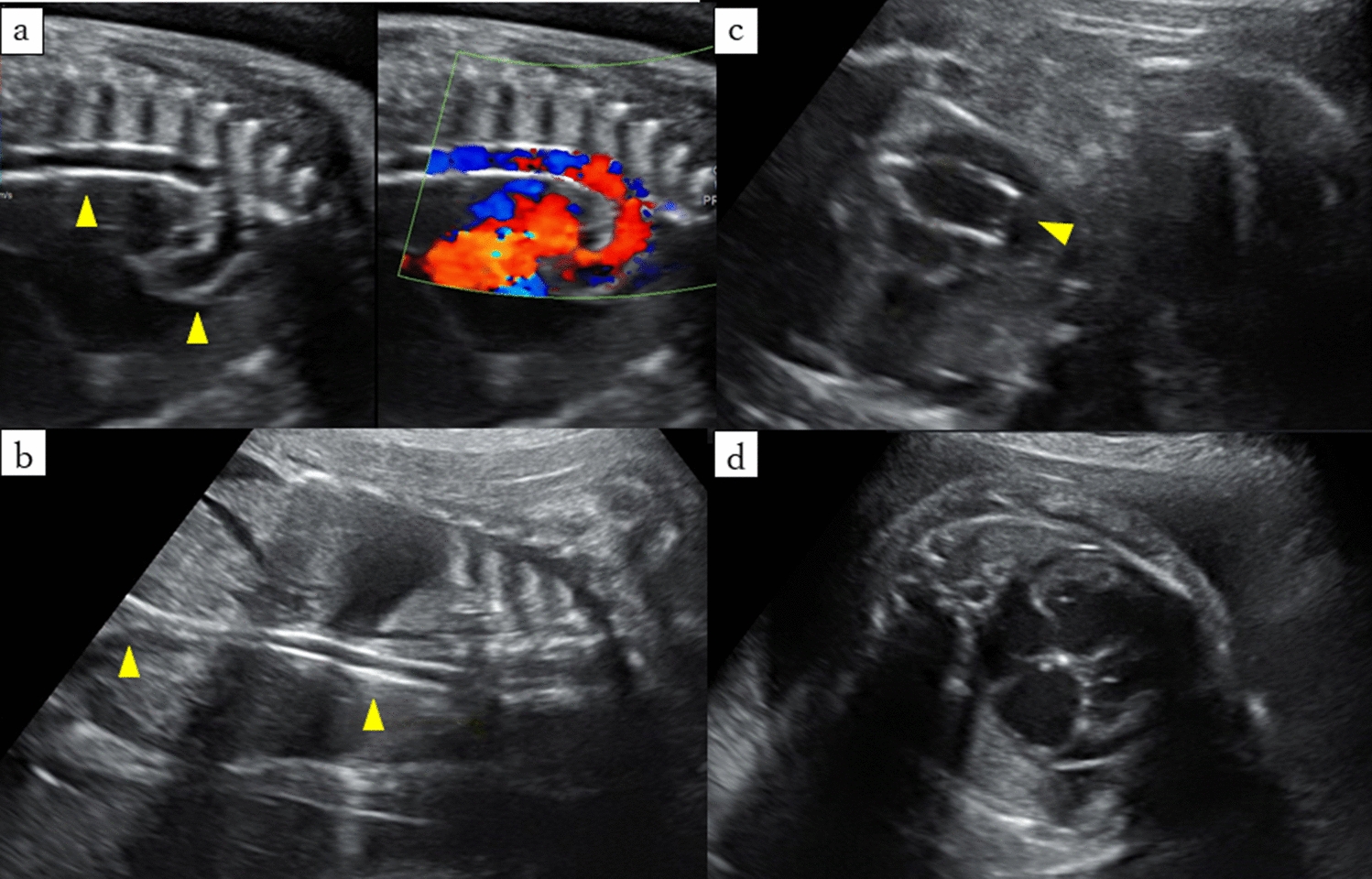
Automated breast ultrasound (ABUS) images used for breast cancer screening in 332 pregnant women were evaluated retrospectively to determine when and how ductal development becomes evident on ultrasonography. The diagnostic criteria used for mammary gland changes during the gestational period were extension of the ducts to the margins of the breast where little or no echogenic fibroglandular tissue is seen on sonograms and/or the appearance of ductal structures running along the ascending Cooper’s ligament tapering off or ending in a blind end at the superficial layer of the superficial fascia. The correlations between gestational stage and the prevalence of these criteria were verified by Spearman’s rank correlation coefficient (ρ). Assessments were performed by a single radiologist with experience reading ABUS images.
Results
With a few exceptions, the prevalence of the above findings increased sharply beginning at 10 weeks, and then increased with progression of gestation, reaching a plateau after 20 weeks (ρ = 0.766, P < 0.00001).
Conclusion
The findings in this study suggested that development of the milk ducts in early pregnancy can be observed using ABUS. These findings will be useful to gain a better understanding of breast ultrasound imaging characteristics during pregnancy.


留言 (0)