
記住我
We present a 63-year-old male with a background of congenital single kidney and end-stage renal failure on haemodialysis presumed secondary to hypertension and type 2 diabetes mellitus. He commenced haemodialysis 5 years ago. His background history included ischaemic heart disease with multiple coronary artery stents (most recent stents inserted 3 years ago), dyslipidaemia, right parietal stroke (at age 56 years old), gout, obstructive sleep apnoea and previous tuberculin test positive that was treated with standard three-drug treatment for 3 months.
A summary of his presentations is presented in Table 1.
Table 1 Summary of key events and results. MRI: magnetic resonance imagingTwo years following the commencement of his haemodialysis, he developed a non-blanching purpuric rash on his lower limbs with a skin punch biopsy demonstrating a small-vessel leukocytoclastic vasculitis that was successfully treated with a short course of oral prednisolone. Two years later, he subsequently experienced right-sided headaches, blurred vision and jaw claudication that was investigated with a right temporal artery biopsy revealing normal histopathology. However, his magnetic resonance imaging (MRI) brain scan (without contrast) showed cerebral vascular calibre irregularities, suggestive of cerebral vasculitis or intracranial atherosclerotic disease. He was treated as presumed giant cell arteritis with oral prednisolone and had resolution of his symptoms.
One year later, he had multiple presentations to hospital with generalised abdominal pain, fever and rigors without vomiting or diarrhoea. On the first presentation, his computed tomography (CT) abdomen scan showed soft-tissue stranding around the gallbladder and within the gallbladder fossa consistent with acute cholecystitis. He was medically managed with intravenous antibiotics. He also had an episode of headaches, conjunctival injection and blurred vision for which he received a single dose of intravenous methylprednisolone and symptoms promptly resolved. On his next presentation with abdominal pain, his CT abdomen scan showed mural thickening of the small bowel loops (in particular jejunum), with adjacent inflammatory stranding and mesenteric free fluid, suggestive of small bowel enteritis. He was treated with intravenous antibiotics with partial resolution of his symptoms. No pathogenic organisms grew on faeces culture. His blood test results at the time are summarised in Table 2.
Table 2 Summary of blood test results during the patient’s presentations with abdominal pain. RNP: ribonucleoprotein, PCNA: proliferating cell nuclear antigenHe had further hospitalisations for fever of unclear source, weight loss and ongoing abdominal pain. His inflammatory markers were raised: C-reactive protein > 200 mg/mL (ref < 5) and procalcitonin 2.75 ug/L (ref < 0.05). Abdominal imaging revealed new mild hepatosplenomegaly (liver 20 cm, spleen 14 cm). His serum immunoelectrophoresis demonstrated two trace kappa restrictions in the gamma region with no reactivity to IgG, IgA, IgM, IgD or IgE heavy-chain antisera. Peripheral blood flow cytometry revealed two B-cell populations: B cells which were polyclonal with a normal kappa:lambda (K:L) ratio (4% lymphocytes) and a light-chain restricted (kappa) B-cell population co-expressing CD45/11c/19/20/79b/81. This was suggestive of a CD11c + B-cell clonal lymphoproliferation likely non-Hodgkin’s lymphoma, either marginal zone or splenic lymphoma. Bone marrow biopsy showed moderate hypercellularity with trilineage hyperplasia, lymphocytes not increased (6% nucleated cells) and cytogenetics did not reveal any structural or numerical abnormalities.
Imaging at this time included a CT neck, chest, abdomen and pelvis scan that demonstrated discontinuous segments of bowel wall thickening involving the proximal-mid small bowel loops within the left side of the abdomen with free fluid and prominent mesenteric lymph nodes up to 11 mm. Whole-body gallium scan showed mild gallium accumulation in the small bowel loops in the left upper to mid-abdomen. Both transthoracic and transoesophageal echocardiograms did not reveal any valvular vegetations. A whole-body fluorodeoxyglucose (FDG) positron emission tomography (PET) scan was done to further investigate his ongoing fevers and showed non-specific sub-centimetre mesenteric and inguinal lymph nodes with increased uptake in the spleen and no evidence of vasculitis.
A diagnostic laparoscopy was performed with the aim to obtain a lymph-node biopsy. Laparoscopy revealed haemorrhagic ascitic fluid and haemorrhagic serositis mostly involving the mid to distal small bowel (Fig. 1). Lymphadenopathy was seen in retroperitoneum and deep mesenteric nodes adjacent to duodenojejunal flexure; however, biopsy was not attempted given the risk of vascular injury was considered too high.
Fig. 1
Diagnostic laparoscopy intra-operative images showing haemorrhagic serositis
Given a tissue biopsy was not obtained on diagnostic laparoscopy, gastrointestinal endoscopy was pursued. His upper gastrointestinal endoscopy and colonoscopy found patchy mild inflammation characterised by shallow ulcerations in the jejunum and non-specific inflammation in the right colon with loss of vascular pattern and mild friability. Biopsies of the gastrum, jejunum, ileum and left and right colon were obtained. The jejunal biopsy revealed focal vasculitis involving a small muscularised blood vessel and a separate isolated small blood vessel with Periodic Acid-Schiff (PAS)-positive intraluminal material. Cytomegalovirus (CMV) immunohistochemistry was negative and there were no granulomas. The terminal ileum biopsy revealed mild focal acute inflammation. The gastric biopsies showed minimal patchy chronic inflammation. The colon biopsies were normal.
In addition, during the admission, he developed a bilateral non-blanching rash on his lower limbs (Fig. 2a) and cyanotic discolouration of his fingertips particularly the right second, third and fourth digits (Fig. 2b). The skin biopsy of his lower limb rash revealed leukocytoclastic vasculitis with partial epidermal necrosis and PAS-positive laminal thrombi in small vessels. He also developed generalised weakness of his arms and legs requiring a lumbar puncture that was unremarkable.
Fig. 2
Lower limb skin rash (a) and digital ischaemia (b)
Testing for cryoglobulins revealed a type II cryoglobulinaemia consisting of an IgM kappa monoclonal band with polyclonal IgG (cryocrit 5%). His autoimmune blood tests at this time were as follows: C3 0.69 g/L (ref 0.69 to 1.85), C4 0.03 g/L (ref 0.15 to 0.53), rheumatoid factor 499 IU/mL (ref < 30), Hepatitis B and C antibodies negative, QuantiFERON gold negative, anti-cyclic citrullinated peptide (anti-CCP) antibodies < 1 U/mL (ref < 5), cardiolipin IgG < 3 U/mL (ref < 20) and cardiolipin IgM < 1 U/mL (ref < 20). He was also found to have a severe hypogammaglobulinaemia (IgG < 1.1 g/L [ref 7 to 16], IgA 0.72 g/L [ref 0.7 to 4], IgM 0.83 g/L [ref 0.4 to 2.3]) and was commenced on intravenous immunoglobulin replacement.
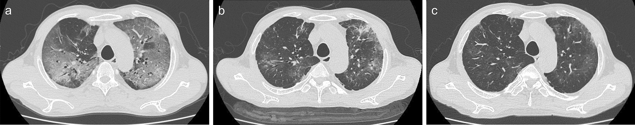
Given the above findings, he was diagnosed with a type II cryoglobulinaemia vasculitis with mainly cutaneous and gastrointestinal involvement. He was commenced on IV methylprednisolone 500 mg daily for 3 days (followed by oral prednisolone 75 mg daily on a weaning protocol) in addition to rituximab 375 mg/m2. His cryocrit reduced to 2% after treatment commencement. After three doses of rituximab, he developed haemoptysis and his CT chest showed extensive airspace consolidation in a perihilar distribution with sparing of the subpleural regions (Fig. 3). For further investigation, he underwent a bronchoscopy which showed bloodstained fluid that had negative Gram stain, scant polymorphs and was negative for acid fast bacilli, fungal infection, Mycobacterium tuberculosis and malignant cells. Respiratory virus polymerase chain reaction was negative for influenza A virus, influenza B virus, respiratory syncytial virus, parechovirus, human parainfluenza 1–4, human rhinovirus/enterovirus, metapneumovirus, adenovirus, bordetella and Mycoplasma pneumoniae. On blood tests, his cryocrit was 3%, anti-glomerular basement membrane antibodies (BIO-FLASH) was < 2.9 CU (ref < 20) and anti-neutrophil cytoplasmic antibodies (ANCA) were not detected at 1:20. Given the lack of another precipitating factor, the pulmonary haemorrhage was presumed to be cryoglobulinaemia vasculitis associated. This was successfully treated with five sessions of centrifugal plasma exchange separate to haemodialysis sessions and with substitution fluid warmed 4% albumin. After his final dose of rituximab therapy, his cryocrit improved to < 1% and C4 was 0.75 g/L (ref 0.15 to 0.53).
Fig. 3
Computed tomography (CT) chest coronial image consistent with vasculitis-associated pulmonary haemorrhage
He has had no relapses of cryoglobulinaemic vasculitis since treatment and his marginal zone lymphoma is being monitored as an outpatient.
留言 (0)