
記住我


The patients were treated from August 2011 to July 2021 in the Department of Oral and Maxillofacial Surgery, Seoul National University Dental Hospital (SNUDH), for a period of 10 years. The study included 333 patients with 1019 dental implants. The study protocol and access to patient records were approved by the Institutional Review Board of Seoul National University (IRB No. S-D20200007), Seoul, Korea. All implant placement surgical procedures were carried out by one surgeon at SNUDH. The minimal follow-up period was at least 3 months after prosthesis delivery.
Inclusion criteriaPatients who underwent dental implant (Luna®, Shinhung Co., Seoul, Korea) installation in the maxillofacial unit of SNUDH.
Patients with controlled systemic disease who received dental implant installation.
Patients with complete medical data, including the clinical and radiographic findings.
Exclusion criteriaPatients with uncontrolled systemic disease.
Previous history of trauma in the oral and maxillofacial area.
Incomplete data.
Patients who were lost during follow-up.
Treatment proceduresThe treatment involved the utilization of the submerged procedure. Under local anesthesia, a single oral and maxillofacial surgeon performed implant installation following the Luna® implant surgical protocol. All implants demonstrated good primary stability. The re-entry procedure was performed approximately 3 to 6 months after the initial implant installation. Once the soft tissue had adequately healed, the prosthesis was fabricated after 2 to 4 months.
Implant dataWe carried out vertical and horizontal bone augmenting procedures such as guided bone regeneration, block bone grafting, sinus lifting, and socket lifting based on each case’s remaining bone quality and the type of bone graft material. We installed the implants based on the third ITI Consensus Conference, based on the period between tooth extraction and implant placement [6].
Implant success criteriaImplant success criteria were based on the ICOI, Pisa, Consensus Conference 2007, which included the absence of pain and tenderness on function, absence of mobility, less than a 2-mm radiographic bone loss from initial surgery, and no exudates history [7].
Implant failure criteriaThe criteria are any of the following: radiographic bone loss exceeding half the length of the implant, pain on function, mobility, uncontrolled exudate, and no longer in mouth.
Implant survival criteriaEvaluation during the follow-up period for each patient included the clinical and radiographic situations such as implant stability, bone loss around the implants, signs of infection, and the level of bone around the implants.
Study variablesVarious clinical events were recorded, including the implant placement date, loading time, last follow-up period, and implant failure or removal date. The primary outcome variable for this study was implant failure. Survival time referred to the duration from implant installation to either implant removal or the last follow-up for surviving implants. The study variables were divided into two groups: healthy individuals without systemic disease and patients with medical conditions. Detailed information about the variables investigated can be found in Tables 1, 2, and 3. In our study, we did not include any uncontrolled variable such as previous history of trauma in the oral and maxillofacial area. Among 333 patients, only one patient had a history of car accident which did not affect the oral and maxillofacial area. Regardless of his trauma history, the patient had concomitant systemic conditions including hypertension and bronchial asthma, which met the inclusion criteria. In addition, we found successful treatment outcomes in this patient. Therefore, we believe that the previous trauma history in this case will not cause any bias in the result.
Table 1 Implant length distributionTable 2 Implant diameter distributionTable 3 Patient demographicsThe primary type of implant used in this study was the Luna® (Shinhung Co., Seoul, Korea) self-tapped bone-level implant, depicted in Fig. 1. The Luna® dental implants have a sand-blasted and acid-etched surface with a roughness of 2.5 μm or higher, resulting in a 20% improvement in bone healing period and cell response (Fig. 2). The optimal number of implants for each patient was determined based on the prosthesis design and the extent of edentulism.
Fig. 1
Macroscopic design of the Luna® implant system used in this study
Fig. 2
Scanning electron microscopy (SEM) view of the surface morphology of the self-tapped bone level, sand-blasted, and acid-etched surface. The SEM was operated at 20 kV. The secondary electron (SE) detection mode was used for the ultrastructural surface analysis. SEM × 60 magnification view (a), × 500 magnification view (b), × 1000 magnification view (c), and × 5000 magnification view (d)
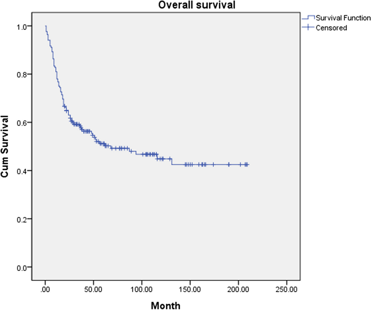
Statistical analysisStatistical analysis was performed using SPSS (version 23 IBM®, NY, USA), and implant-related data were calculated. The Kaplan–Meier analysis was used for the description of survival rates.
留言 (0)