In this predefined secondary analysis of the InventCOVID trial, we evaluated the correlation between four existing LUS scores and EVLWi in COVID-19 ARDS patients. The key findings of the study were: (1) the global LUS score, the LUS–ARDS score and the anterior–lateral score correlated with EVLWi, while the B-line score did not; (2) changes in the global LUS score and anterior–lateral score correlated with changes in EVLWi over time.
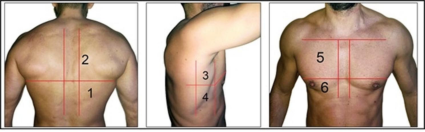
The 12- and 8-region scores examined in this study can quantify PiCCO-derived pulmonary edema measurements in COVID-19 ARDS. Combined with previous studies [24, 25, 32], our results further support the use of LUS for the assessment of pulmonary edema in patients with ARDS. The correlation of the shorter anterior-lateral score with EVLWi is in line with previous work that showed comparable performance of the 8-region score to more extensive protocols in assessing diagnostic accuracy and monitoring ARDS [33, 34]. The rationale of exempting the dorsal regions from examination is the prevalence of compression atelectasis and gravitational accumulation of pulmonary edema in the supine position [35]. Moreover, a score that requires less time to perform remains clinically attractive, as LUS is a bedside tool. Our data suggests that quantification of EVLW with the 8-region anterior–lateral score may be an alternative to the 12-region protocols to quantify pulmonary edema.
The performance of the LUS–ARDS score supports the score’s value in as an adjunct in the comprehensive assessment of patients with ARDS. Notably, this score was developed and validated for ARDS diagnosis [25] and not to predict pulmonary edema. Unlike other LUS aeration scores, the presence of pleural abnormalities contributes to the LUS–ARDS score. This choice was made to better capture the uncertain, non-binary nature of ARDS as a syndrome [36]. We hypothesize that taking into account pleural morphology in combination with the aeration score increases the likelihood of identifying severe pulmonary edema by functioning as an indicator of disease severity in the rest of the lung. Combined with the recently reported high accuracy for ARDS diagnosis [25], the score could be a useful adjunct to identify patients at risk of clinically relevant pulmonary edema. Validation in a non-COVID-19 ARDS cohort is needed to extrapolate our findings to the broader ARDS population.
To analyze the diagnostic accuracy of the LUS scores for detecting an EVLWi > 15 ml/kg, score cutoffs were chosen based on a sensitivity of > 90%. This comes at the expense of specificity—a choice which was made with clinical practice in mind. A clinician performing a LUS exam in a patient with ARDS can use a score below the determined cutoffs to rule out severe pulmonary edema at the moment of measurement. On the one hand, this may provide reassurance of the already implemented treatment. On the other hand, it can alert the clinician to monitor and/or to initiate proactive intervention in a patient who is clinically suspected to be at risk of deteriorating.
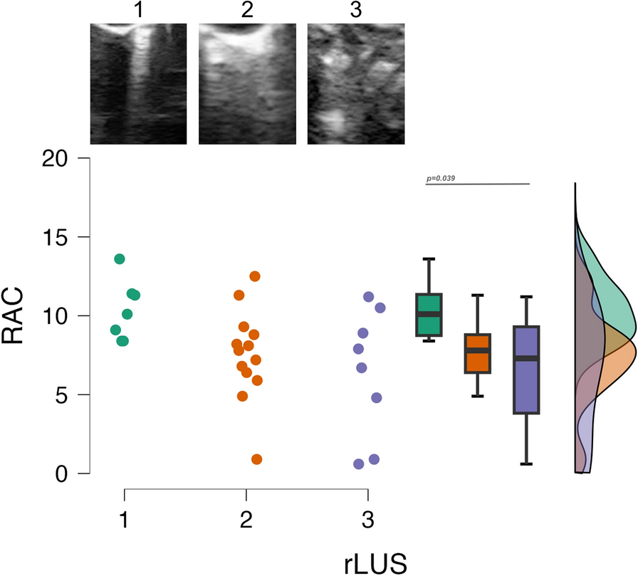
Considering the potential risk of over- or underestimation of pulmonary edema through the use of aeration patterns [33, 37,38,39], it follows that a score based solely on the number of B-lines may be more appropriate for focused quantification. Enghard et al. found an excellent correlation (r = 0.91) of a simplified 4-region B-line score with EVLWi in a mixed ICU population [22]. However, of the 50 patients, only 6 were classified as ARDS, considerably limiting the validity of their findings for the ARDS population. One study examined the same score in 26 ARDS patients and described a correlation (r = 0.66); however, it found that changes in B-line score could not predict variations in EVLWi [6]. In the current study, we found no significant correlation of the B-line score with EVLWi, nor with ∆EVLWi. Considering these discrepancies, it is questionable whether B-line counting is suitable for scoring pulmonary edema in ARDS patients. Reasons for the inconsistent performance of the score include that the choice of transducer and the interpretation of the sonographer significantly affect the reproducibility of this method [40].
Aside from assessing severity, monitoring changes in pulmonary edema and lung aeration is useful to evaluate treatment response. A change in global LUS score and the anterior–lateral LUS score between timepoints 1 and 2 was significantly associated with ∆EVLWi, and ∆LUS–ARDS score was positively associated with ∆EVLWi, despite not reaching statistical significance. Possibly, the global and anterior–lateral LUS scores are better suited to monitor pulmonary edema over time. A reason may be that the LUS–ARDS score considers pleural abnormalities, which may not be as sensitive to changes in EVLW as aeration patterns are. Based on the current findings and other studies [8, 41], LUS aeration scores seem useful to evaluate a change in EVLW in (COVID-19) ARDS. To validate this conclusion, a future study may include measurements at multiple timepoints.
The study has several strengths. First, the prospectively included population was exclusively comprised of patients with COVID-19 ARDS, making this a population with a single pulmonary etiology and thus providing a rare degree of relative homogeneity. Second, the availability of two timepoints of measurement allowed us to investigate the correlation of ∆LUS and ∆EVLWi, allowing for assessment of the value of LUS for monitoring pulmonary edema. Third, to our knowledge this is the first study to compare four previously proposed LUS scores that differ in terms of examined regions and/or means of score aggregation.
Some limitations should be acknowledged. The inclusion of COVID-19 ARDS patients with moderate-to-severe illness reduces external validity to ARDS populations with a different etiology or milder disease severity. However, we considered COVID-19 ARDS to be particularly suitable for this study, as it typically presents without the concomitant pathologies that challenge PiCCO measurement in other critically ill patients [42, 43]. Second, the study only included invasively ventilated patients, not patients receiving non-invasive modes, such as high-flow nasal oxygen. Therefore, we cannot draw conclusions about the use of LUS in a group that may particularly benefit from quantification of pulmonary edema [44, 45] and initiation of early intervention. Finally, the number of missing regions decreased the sample size and excluding patients with ≥ 4 missing regions may have induced a degree of selection bias.
This explorative study highlights that LUS can determine PiCCO-derived EVLWi, strengthening the rationale for its use to quantify pulmonary edema in patients with ARDS. Research into the application of LUS to quantify edema and use this information to guide adherence to a restrictive fluid balance is currently ongoing (ClinicalTrials.gov: NCT05188092). Yet, the results of this small study also underline the need for a larger sample, in which the different LUS techniques are systematically compared to the reference standard with the aim of drawing a definitive conclusion on the optimal score to be used in clinical practice.
In conclusion, both 12-region LUS scores and the 8-region anterior–lateral score correlated with PiCCO-derived pulmonary edema in invasively ventilated patients with COVID-19 ARDS. The anterior–lateral score seems to be as useful to quantify and monitor change in pulmonary edema as the 12-region scores. Combined with its recently reported high accuracy to diagnose ARDS, the LUS–ARDS score may be best-suited for a comprehensive assessment of ARDS diagnosis and pulmonary edema severity.


Comments (0)