Human samples
Blood samples (matched by sex and age) were collected from hospitalized VTE patients and physical examinations were conducted from January 2023 to April 2023. The patient’s blood samples were approved by the Ethics Committee of Run Run Shaw Hospital Affiliated with Zhejiang University School of Medicine. The serum specimens were packed according to the experimental requirements and transferred to a -80℃ refrigerator for preservation.
Mouse thrombus model
SPF grade, 8-week-old male C57 mice were reared in the Laboratory Animal Center of Run Run Shaw Hospital Affiliated with Zhejiang University. The feeding environment was kept at a constant temperature (25 ± 2) ℃ and constant humidity (50 ± 5) %. Artificial light was on 12 h each day. The animal experiments were approved by the Animal Ethics Committee of Run Run Shaw Hospital Affiliated with Zhejiang University School of Medicine. The process of establishing the mouse model of DVT was as follows. Mice were anaesthetized with 1% pentobarbital sodium (50 mg/kg), and the operation area was disinfected with iodophor. The abdomen of the mice was cut with line scissors along the midabdominal line, followed by the epidermis and muscle layer, and the abdominal cavity was exposed. The inferior vena cava was exposed after the intestinal tract was turned over, and the blood vessel was bluntly separated with sharp forceps. The main inferior vena cava below the left renal vein was carefully ligated, and the 5 − 0 suture was extracted. The mice were put back into the cage and recovered after half an hour to 1 h after successive sutures were used to close the intraperitoneal muscle layer and intermittent sutures to close the epidermis. The mice were again disinfected with iodophor. The mouse model samples were collected 1 week later. Twenty-one mice were randomly divided into the control group and the DVT group. There was no significant difference in body weight between the two groups. Decanoyl-RVKR-CMK treatment was administered by caudal vein injection (25 mg/kg) on the 1/3/5th day of modelling, and blank treatment was administered by an injection of the same amount of normal saline.
Immunostaining
Mouse vein tissue was fixed with 4% paraformaldehyde fixation solution for 24 h and cut into 0.2 cm tissue sections. After treatment, the slices were sliced at a thickness of 4–6 μm. HE staining, Masson staining, and Sirius red staining were performed. The primary antibody (Abmart, TD6006S) and secondary antibody (CST) against GDF15 were used for immunohistochemistry. Beyotime Biotechnology (AF5318) and fluorescent secondary antibodies (CST) were used for immunofluorescence.
HUVECs culture
HUCECs were purchased from Zhejiang Meisen Cell Technology Co., Ltd. ECM culture medium containing 5% foetal bovine serum + ECGs + 100 units/mL penicillin and 100 μg/mL streptomycin was used. Cells were stimulated with TGFβ1 (5 ng/ml) + TNFα (5 ng/ml) + IL1β (0.1 ng/ml) (Pepro Tech, USA) for 24 h after starving for 24 h.
Western blotting
Lysis buffer (Beyotime Biotechnology, China) and protease inhibitor (1%) and phosphatase inhibitor (5%) (Beyotime Biotechnology, China) were added to extract cell proteins. The protein concentration was detected by a BCA kit (Beyotime Biotechnology, China). Approximately 20 μg protein was added to each well during glue running. After electrophoresis and membrane transfer, the primary antibody was added overnight. The next day, after washing with PBS, the corresponding secondary antibodies (sheep anti-rabbit secondary antibody CST, sheep anti-mouse secondary antibody CST) were added, incubated at room temperature for 2 h, and exposed with an enhanced chemiluminescence kit HRP (FDbio science, China). Beta-actin was used as the internal control. The primary antibodies used in this study were: β-actin ((CST, #8457, 1:1000), SM22 (Beyotime, AF5318, 1:1000), GDF15 (Abcam, ab206414,1:1000), CD31 (CST, #77,699, 1:1000), TF (Beyotime, AF2455, 1:1000), Snail (CST, #3879, 1:1000), SMAD2 (CST, #5339, 1:1000), p-SMAD2 (CST, #7348, 1:1000), Smad2/3 (CST, #8685, 1:1000).
Cellular immunofluorescence
The cells were laid on 24-well plates, treated with drugs, removed from the medium and washed with PBS 3 times. After fixed and broken membrane treatment, the cells were blocked with 3% BSA for 1 h and incubated at 4 °C with primary antibody overnight. The cells were washed with PBS 3 times, and the secondary antibody was added and incubated for 1 h in the dark. The cells were washed with PBS 3 times, DAPI was added, and the cells were incubated for 15 min in the dark. After washing again, a fluorescence quencher was added and observed by fluorescence microscopy.
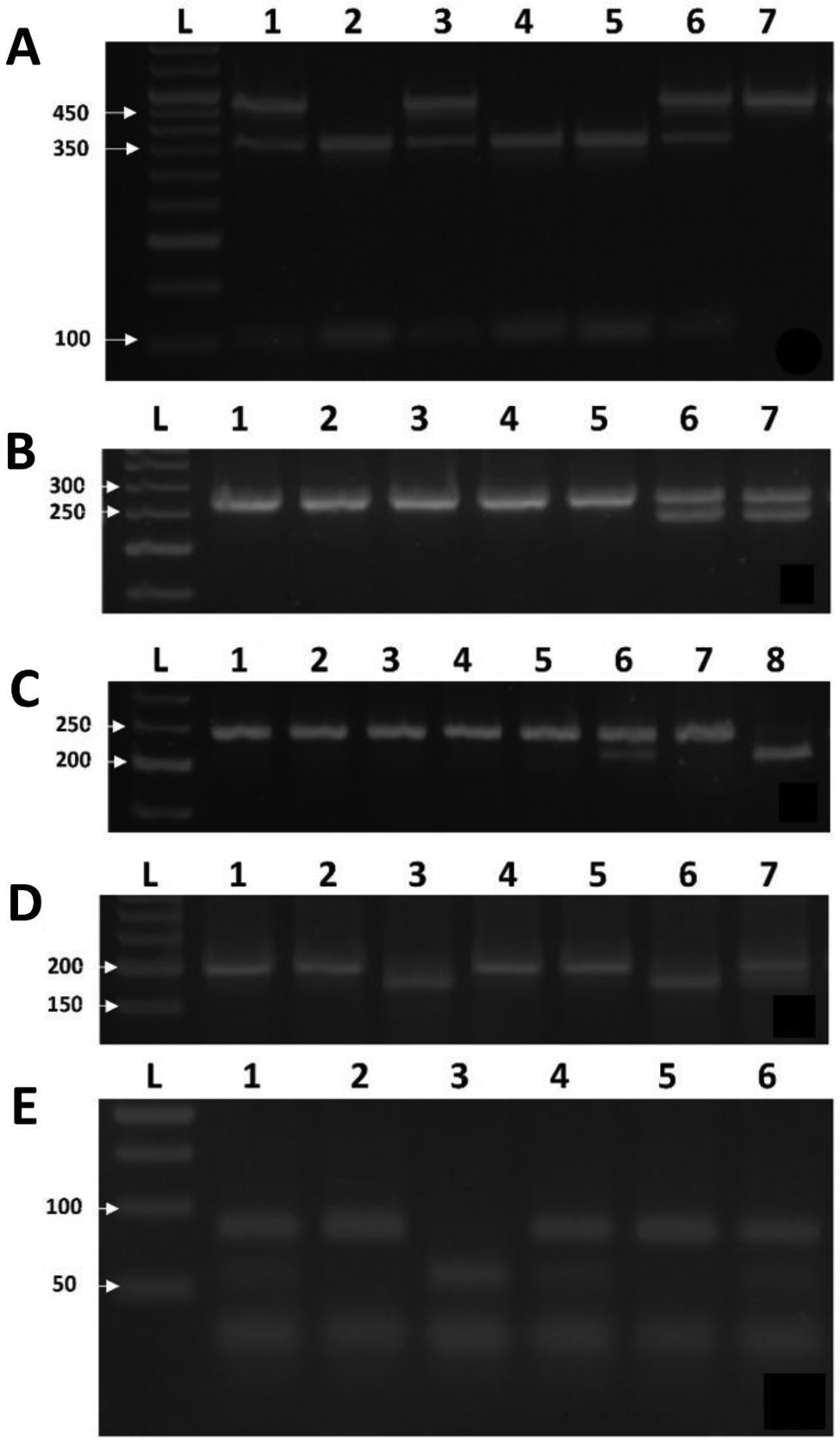
RNA extraction and RT‒qPCR
Cell RNA or mouse tissue RNA was extracted by the RNA-quick Purification kit. (Yishan Biotech, China) RNA concentration was measured on a NanoDrop2000 instrument. The Synthesiskit cDNA synthesis kit was transcribed by HiFiScript cDNA Synthesiskit after measurement. After that, TB Green® Premix Ex Taq™ II was added for RT‒qPCR in a LightCycler480 System. After the reaction, the corresponding cycle number (CT value) was recorded, and the relative gene expression was calculated by the 2-△ △ CT method. The primers used are shown in supplemental Table 1.
Overexpression and knockout
The siRNAs for GDF15 were synthesized by GenePharma (Shanghai, China). The sequences were as follows: siGDF15#1 (sense: 5′-CCAAACAGCUGUAUUUAUATT-3′, antisense: 5′- UAUAAAUACAGCUGUUUGGTT-3′). siGDF15#2 (sense: 5′- GACCUAUGAUGACUUGUUATT-3′, antisense: 5′- UAACAAGUCAUCAUAGGUCTT − 3′). The pcDNA3.1-GDF15 expression plasmid was synthesized by Genomeditech (Shanghai, China).
ELISA
Human or mouse serum GDF15 levels were detected by ELISA kits (purchased from Suzhou Sizhengbai Biotechnology Co., LTD.). Then, 45 μl human serum was added to 180 μl sample diluent or 110 μl mouse serum to 110 μl sample diluent and incubated with the diluted biotin antibody and enzyme conjugate at 37 °C. After that, the colour-developing agent was added and incubated for 15 min away from light. The OD450 value was measured after the termination solution was added.
Statistical analysis
SPSS 20 was used for analysis, and the chi-square (χ2) test was used for counting data. If the measurement data conformed to a normal distribution, the mean ± standard deviation was adopted; if they did not conform to a normal distribution, the median (front and back quartile) was adopted. For continuous data, the normality test was conducted first. If all groups met normality and the variance between the two groups was homogeneous, a t test was used for comparisons between groups. If the above conditions were not met, the nonparametric Mann‒Whitney U test was considered. The chi-square test was used for classification data and disordered classification data, and the nonparametric Mann‒Whitney U test was used for rank data. P < 0.05 was considered statistically significant.


Comments (0)