


 Figure 4.
Effect of the addition of a polycationic polymer, polyamidoamine dendrimer (5th generation-PAMAM), on the uptake of 60 nm nanoparticles compared to unmodified, negatively charged nanoparticles in the nasal olfactory mucosa (A) and the nasal respiratory mucosa (B) normalized per tissue surface area. Nanoparticle uptake was significantly decreased in both types of mucosae after incubation with PAMAM-modified nanoparticles. The two tailed t-test was applied to evaluate the statistical significance. Bars are mean ± SD, bars represent mean ± SD, ** ppn = 3 each. Data for untreated nanoparticles were previously reported [
Figure 4.
Effect of the addition of a polycationic polymer, polyamidoamine dendrimer (5th generation-PAMAM), on the uptake of 60 nm nanoparticles compared to unmodified, negatively charged nanoparticles in the nasal olfactory mucosa (A) and the nasal respiratory mucosa (B) normalized per tissue surface area. Nanoparticle uptake was significantly decreased in both types of mucosae after incubation with PAMAM-modified nanoparticles. The two tailed t-test was applied to evaluate the statistical significance. Bars are mean ± SD, bars represent mean ± SD, ** ppn = 3 each. Data for untreated nanoparticles were previously reported [
Remember me


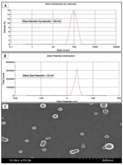


Figure 1. Characteristics of positively charged PAMAM-PLGA nanoparticles. (A) DLS measurements of nanoparticle diameter by scattering intensity showing unimodal distribution. (B) Zeta-potential (surface charge) shows a slightly positive surface charge. (C) SEM image shows spherical nanoparticles.
Figure 2. Amount of nanoparticles in the nasal mucosa after incubation with PAMAM-modified nanoparticles (cationic PLGA NP) for 30 and 60 min normalized per tissue surface area. The two tailed t-test was applied to evaluate the statistical significance. n = 3 each, bars represent mean ± SD.
Figure 3. Amount of nanoparticles quantified in each mucosal compartment and the receiver media in nasal olfactory mucosa (A), and respiratory mucosa (B) following incubation with the PAMAM-modified nanoparticles (cationic PLGA NP) for 30 and 60 min normalized per tissue surface area. The two tailed t-test was applied to evaluate the statistical significance. Bars are mean ± SD. ** p-value = 0.007, n = 3 each.
Figure 3. Amount of nanoparticles quantified in each mucosal compartment and the receiver media in nasal olfactory mucosa (A), and respiratory mucosa (B) following incubation with the PAMAM-modified nanoparticles (cationic PLGA NP) for 30 and 60 min normalized per tissue surface area. The two tailed t-test was applied to evaluate the statistical significance. Bars are mean ± SD. ** p-value = 0.007, n = 3 each.
Table 1. Size and surface charge of PLGA and PAMAM-modified nanoparticles.
Table 1. Size and surface charge of PLGA and PAMAM-modified nanoparticles.
FormulationAverage Diameter (by DLS) (nm)Polydispersity Index (PDI)Average Zeta Potential (mV)Nile Red Loading (µg/mg)PLGA nanoparticles600.15−280.60PAMAM Modified PLGA nanoparticles1030.1+31.40.59
Comments (0)