
Remember me


Motile cilia are present on the epithelial cell surface to generate a directional fluid flow that is crucial for various biological processes, such as the establishment of left–right symmetry in the ventral node, mucociliary clearance in airways, and cerebrospinal fluid circulation in the brain (Fliegauf et al, 2007; Brooks & Wallingford, 2014). The axonemal structure of most motile cilia has a “9 + 2” arrangement with nine peripheral outer doublet microtubules surrounding a central microtubule pair, however, motile nodal cilia have an either “9 + 0” or “9 + 4” structure (Feistel & Blum, 2006; Ishikawa & Marshall, 2011; Spassky & Meunier, 2017). The synchronous ciliary motility is governed by protein appendages attached to the axonemal microtubules, which include axonemal dyneins (inner dynein arm [IDA] and outer dynein arm [ODA] complexes), radial spokes, and nexin–dynein regulatory complexes (Teves et al, 2016). Radial spokes interact with IDA and the central pair of microtubules to coordinate the ciliary motility (Yang et al, 2006; Viswanadha et al, 2017). Defects in the assembly and motility of motile cilia can result in various human diseases such as sinopulmonary disease, hearing impairment, hydrocephalus, situs inversus, and male infertility (Nigg & Raff, 2009; Reiter & Leroux, 2017).
In Xenopus laevis epidermis and mammalian airways, the commitment of MCC precursors is transcriptionally controlled by GemC1 and Multicilin (Stubbs et al, 2012; Ma et al, 2014; Kyrousi et al, 2015; Arbi et al, 2016; Terre et al, 2016; Kim et al, 2018), which are necessary and sufficient to induce MCC differentiation. Upon activation of the transcriptional cascade, MCC precursors undergo massive centriole amplification mostly through a deuterosome-dependent pathway to produce hundreds of centrioles, which sequentially dissociate from deuterosomes, migrate and dock to the apical surface, and convert into basal bodies to initiate axoneme growth (Klos Dehring et al, 2013; Zhao et al, 2013; Brooks & Wallingford, 2014; Spassky & Meunier, 2017). In order for centrioles to dock with the apical membrane they must develop distal appendages following the deuterostome stage (Spassky & Meunier, 2017). MCCs also undergo remodeling of the subapical membrane cytoskeleton, in particular the actin network, to accommodate centriole membrane docking and motile cilium assembly (Antoniades et al, 2014; Sedzinski et al, 2016; Kulkarni et al, 2018). The establishment of the apical actin network involves focal adhesion proteins and components of the planar cell polarity (PCP) pathway including dishevelled (Dvl), Daam1, and the Daam1/Dvl regulator Drg1, which can stimulate the activation of RhoA, a Rho GTPase that regulates the actin polymerization (Park et al, 2008; Antoniades et al, 2014; Yasunaga et al, 2015; Lee et al, 2019).
Assembly and functional maintenance of the cilium require intraflagellar transport (IFT) particles (Davenport et al, 2007; Lechtreck, 2015; Zhu et al, 2017). IFT particles regulate the bidirectional trafficking of ciliary proteins along the axonemal microtubules, and can assemble into linear IFT train structures of varying length (Pigino et al, 2009; Stepanek & Pigino, 2016; Yang & Huang, 2019). The IFT particle is composed of two multiunit subcomplexes, IFT-A and IFT-B. The IFT-A complex functions in retrograde trafficking from the ciliary tip to the base powered by the dynein-2 motor complex, whereas the IFT-B complex mediates anterograde protein trafficking from the cell to the ciliary tip through kinesin-2 motor proteins (Rosenbaum & Witman, 2002; Ishikawa & Marshall, 2011; Sung & Leroux, 2013; Liang et al, 2014). Defects in anterograde trafficking mediated by the IFT-B complex cause shortened or an absence of cilia (Pazour et al, 2000; Davenport et al, 2007; Lechtreck, 2015; Kubo et al, 2016). IFT-B subunits IFT20, IFT25, IFT27, and IFT74 are also required for sperm flagella formation in mice (Zhang et al, 2016, 2017; Shi et al, 2019). In the primary cilium, IFT-B complex proteins accumulate at the mother centriole following removal of the CP110/CEP97 cap that blocks axoneme formation (Goetz et al, 2012; Lu et al, 2015; Kanie et al, 2017). In spite of the important role of IFT proteins in cilium assembly, little is known about how IFT-B proteins are recruited to centrioles/basal bodies during early ciliogenesis and how this process is scaled/amplified in multiciliogenesis.
CCDC108 (also known as CFAP65) is the ortholog of Akap240, identified as an axonemal protein in Chlamydomonas reinhardtii (Gaillard et al, 2001; Zhao et al, 2019b; Dai et al, 2020). Interestingly, CCDC108 mutations cause abnormal sperm flagellum in patients (Wang et al, 2019; Zhang et al, 2019; Li et al, 2020), and its orthologs in mouse and chicken are essential for sperm motility (Imsland et al, 2012; Li et al, 2020; Wang et al, 2021). These studies suggest that CCDC108 is involved in regulating motile ciliation and/or movement. Here, we investigated the function of Ccdc108 in motile multiciliogenesis. We showed that Ccdc108 is required for ciliation in frog, fish, and mouse multiciliated cells. In Xenopus MCCs, Ccdc108 is needed for basal body migration/docking to the plasma membrane and apical enrichment of F-actin during multiciliogenesis. We demonstrated that Ccdc108 localizes to centrioles as they migrate to the apical cell surface and in the cilium in Xenopus MCCs. Loss-of-function and replacement experiments in vivo demonstrated that the Ccdc108 interacts with the IFT machinery during ciliogenesis and in the mature cilium, and this association is essential for the migration/docking of centrioles to the apical membrane during cilium assembly in MCCs. Finally, we showed that Ccdc108 and its interaction with IFT machinery are crucial for the centriolar distribution of planar cell polarity-associated actin cytoskeleton regulators Drg1 and RhoA, which contribute to the apical actin polymerization during multiciliation. Together, our findings demonstrate that Ccdc108 and IFT-B complex components function together in multiciliogenesis.
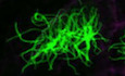
Results Ccdc108 is essential for ciliation in multiciliated epidermis of the Xenopus embryoPrevious studies indicate that Ccdc108 is important for proper sperm motility in chicken, mouse, and human (Imsland et al, 2012; Wang et al, 2019, 2021; Zhang et al, 2019; Li et al, 2020). To further investigate Ccdc108 association with motile cilia function, we injected Xenopus laevis embryos with morpholino (MO) oligonucleotides against the ccdc108 mRNA sequence to block the translation of the protein. Morpholino efficiency was confirmed using a GFP reporter RNA containing the MO target site of ccdc108 at the 5’UTR (Fig EV1A) (Romaker et al, 2014). Since the motile cilia of MCCs on the skin of Xenopus embryos generate a directed fluid flow along the posterior ventral axis of the embryo epidermis (Mitchell et al, 2007), fluorescent microbeads were used to visualize the fluid flow. ccdc108 morphants displayed significantly affected bead motility over the epidermis (Fig 1A and B; Movies EV1–3). Importantly, this defect was rescued by co-injection of a hemagglutinin (HA)-tagged ccdc108 mRNA, validating the specificity of the MO (Fig 1A and B; Movies EV1–3). Defects in fluid flow may be caused by either reduction in cilia number or abnormal cilia motility. To evaluate Ccdc108 requirements in ciliary bead flow in MCCs, we performed immunostaining to examine cilia of epidermal MCCs by confocal and super-resolution structured illumination microscopy (SIM) and surprisingly found a > 4-fold reduction in cilia levels upon depletion (Fig 1C and D). We further confirmed that Ccdc108 depletion resulted in fewer and slightly shorter cilia in MCCs using scanning electron microscopy (SEM) (Figs 1E and EV1B). Moreover, we confirmed ciliation requirements for Ccdc108 using CRISPR/Cas9 technology to deplete the protein (Cong et al, 2013; Wang et al, 2015). Consistent with our MO studies, ccdc108 CRISPR mutants displayed a significant reduction in MCC cilia (Figs 1F and EV1C). Because the Chlamydomonas reinhardtii ortholog of Ccdc108, Akap240, is associated with the axoneme microtubule central pair which is important for cilia motility (Rao et al, 2016; Teves et al, 2016), we examined the ciliary beat pattern (CBP) and ciliary beat frequency (CBF) of MCCs by high-speed video microscopy. MCCs of Ccdc108 morphants displayed an altered CBP and an increased CBF (Fig EV1D; Movies EV4–6). Importantly, ciliary defects were restored by the expression of wild-type (WT) Ccdc108 (Figs 1C–E and EV1D). This indicates that cilia present in Ccdc108 morphants have impaired motility. Examination of Ccdc108 expression in Xenopus laevis embryos by in situ hybridization chain reaction (HCR) combined with immunostaining for cilia confirmed that ccdc108 is highly expressed in MCCs but not in the neighboring non-MCCs (Fig 1G). To further test if Ccdc108 is required for other types of cilia formation, primary cilia of neural progenitors in the neural tube and motile monocilia in the gastrocoel roof plate (GRP) were examined (Schweickert et al, 2007; Toriyama et al, 2017). Notably, neither of these monocilia was affected by Ccdc108 depletion (Fig EV1E and F). Thus, our results indicate that Ccdc108 plays a crucial role in regulating multiciliation in epidermal MCCs.
Click here to expand this figure.
Figure EV1. Ccdc108 is essential for multiciliogenesis in epidermis of the Xenopus embryo (related to Fig )
In vivo validation of morpholino efficiency. ccdc108 morpholino inhibits the expression of the GFP reporter containing the MO target site of ccdc108 at the 5’UTR. Scanning electron microscope images and graphical plot show slightly shorter cilia in Ccdc108-depleted MCCs. Experiment was performed once and 50 cilia from 10 MCCs in different microscopic fields were scored. Mean ± s.d. values are presented. Representative confocal images and graphical plot display reduced ciliary Ac-tub levels in ccdc108 CRISPR mutants. A total of 20 images of 20 embryos for each condition. Cell membranes (mGFP, purple) and cilia (Ac-tub, green) were labeled with indicated antibodies. Quantitative data from three independent experiments were scored. Unpaired two-tailed t-test was performed (***P < 0.001). Mean ± s.d. values are also presented. Graphical plot and cartoons show that epidermal MCC cilia beat frequency (CBF) and cilia beat pattern (CBP) are both affected in ccdc108 morpholino-treated Xenopus embryos from stage 27. The cartoons were generated based on the results of imaging live MCCs by high-speed video microscopy (Movies EV4–6). Two biologically independent experiments were performed. Greater than 15 MCCs from four embryos for each condition. Transverse section views show that Ccdc108 is dispensable for primary ciliogenesis in the Xenopus neural tube. Embryos at stage 30 were fixed, and stained with the acetylated tubulin antibody (red) and DAPI (blue). mRNA of a membrane-bound form of GFP (mGFP; green) was co-injected with each morpholino to indicate targeted cells. Arrows mark primary cilia. Three biologically independent experiments were performed and images from the same experiment were presented. Effects of Ccdc108 depletion on motile monocilia formation and length in the gastrocoel roof plate (GRP). Embryos were fixed and stained with the acetylated tubulin antibody (red). mGFP (green) was co-injected with each morpholino to indicate targeted cells. GRP explants were prepared from embryos at stage 18. Greater than 6 embryos from three biologically independent experiments for each condition were images and 10 cilia from each embryo were scored. Unpaired two-tailed t-test was performed. Mean ± s.d. values are also presented.Source data are available online for this figure.

Figure 1. Ccdc108 is essential for ciliogenesis in multiciliated epidermis of the Xenopus embryo
A, B. Depletion of Ccdc108 markedly affects bead movement by decreasing velocity (Movies EV1–3). Fluorescent beads were applied to the dorsal surface of Xenopus embryos. Bead tracks every 500 ms over 2 s are shown (A). Plot of average bead velocity shows significantly reduced bead flow in ccdc108 morphants, which can be fully rescued by co-injecting with Xenopus ccdc108 mRNA (B). A total of 150 beads from 15 embryos for each condition. Arrowheads (A) indicate beads that showed no movement on the embryo surface through the recording. C, D. Defective cilia formation in ccdc108 morphants. Embryos at stage 27 were fixed and stained with the acetylated tubulin (Ac-tub) antibody and/or Cep164 antibody. mRNA of a membrane-bound form of GFP (mGFP) was co-injected with each morpholino to indicate targeted cells. Representative confocal images (C) and 3D-SIM images (D) of the epidermis for each condition and plots show significantly reduced cilia number in Ccdc108-depleted embryos. (C) Twenty images of 20 embryos and (D) greater than 80 MCCs from six embryos for each condition. E. Scanning electron microscope images of Xenopus embryos display defective cilia formation in Ccdc108-depleted MCCs. White arrowheads mark MCCs with reduced ciliation. Boxed areas are magnified to show the details. F. Defective cilia formation occurs in ccdc108 CRISPR mutants. Embryos at one-cell stage were injected with Cas9 protein with or without the sgRNA against ccdc108 (108sg) and fixed at stage 27. Representative 3D-SIM images and plot show significantly reduced cilia number in ccdc108 CRISPR mutants. Greater than 70 MCCs from six embryos for each condition. Cell membranes (mGFP, purple), cilia (Ac-tub, green), and basal bodies (Cep164, yellow) were labeled with indicated antibodies. G. In situ hybridization chain reaction (HCR) reveals that ccdc108 mRNA is specially detected in MCCs. Embryos at stage 35 were fixed and subjected to in situ HCR (red). Embryos were then incubated with the Ac-tub antibody (cyan) and DAPI (gray).Data information: Quantification data were collected from three independent experiments. Unpaired two-tailed t-test was performed (***P < 0.001). Mean ± s.d. values are presented.
Source data are available online for this figure.
Ccdc108 ciliation requirement is evolutionarily conserved in fish and mouse cellsSince Ccdc108 plays an important role in multiciliation in Xenopus, we asked whether this requirement is evolutionarily conserved. We first examined Ccdc108 function in zebrafish. In situ HCR analysis in zebrafish embryos at the long-pec stage (48 h post-fertilization) showed that the ccdc108 mRNA was detected in the multiciliated olfactory placode cells and neuromast hair cells, but not in the surrounding cells (Fig 2A), suggesting a potential role of Ccdc108 in these two cell types with 9 + 2 structure cilia (Song et al, 2016). To investigate Ccdc108 ciliary requirements, we injected embryos with a morpholino targeting the translation start site of zebrafish ccdc108. ccdc108 morphants displayed hydrocephalus, a phenotype associated with ciliary motility dysfunction and excess cerebrospinal fluid (CSF) in the brain, which could be rescued by Xenopus Ccdc108 (Fig 2B). Additionally, ccdc108 morphants displayed a reduced number of acetylated tubulin-positive cilia in both olfactory placode cells and neuromasts, which could be significantly restored by the expression of Xenopus Ccdc108 protein (Fig 2C and D). Using SEM, we confirmed ciliation was affected in olfactory placodes of ccdc108 morphants (Fig 2E). Collectively, these findings demonstrate that Ccdc108 is required for the formation of MCC cilia and the neuromast kinocilia and causes ciliopathy-like phenotypes upon depletion in zebrafish.

Figure 2. The function of Ccdc108 in regulating multiciliogenesis is evolutionarily conserved across species
A. In situ HCR reveals that ccdc108 mRNA is specially detected in ciliated organs in zebrafish. Zebrafish embryos at the long-pec stage (48 h post-fertilization) were fixed and subjected to in situ HCR (purple). Embryos were then incubated with the Ac-tub antibody (green). B. Representative images and plots show that ccdc108 morphants display severe hydrocephalus (white arrowhead) in zebrafish, which was rescued by expression of Xenopus Ccdc108. C, D. Representative confocal images and plots show that ccdc108 morphants displayed ciliogenesis defects in neuromasts (C) and olfactory placodes (D). Zebrafish embryos were fixed and stained with the Ac-tub antibody (green), phalloidin (purple), and Hoechst (blue). E. Scanning electron microscope images of zebrafish embryos display a failure in cilia formation in Ccdc108-depleted olfactory placodes. F. CCDC108 shows similar expression pattern to that of indicated cilia-related proteins in mEPCs. Cells were collected at the indicated day after serum starvation and used for immunoblotting. IFT81 is an IFT complex protein, and RSPH1 is a component of the ciliary radial spoke structure. GAPDH was used as loading control. G. Four independent shRNAs (108-i1, -i2, -i3, and -i5) effectively depleted CCDC108 without affecting the indicated cilia-related proteins. GAPDH served as loading control. Ctrl-i, a control shRNA. H. Representative 3D-SIM images and plot reveal that depletion of CCDC108 leads to a significant reduction in multiciliogenesis in mEPCs. mEPCs that were infected with lentivirus expressing indicated shRNA were serum starved for 5 days and subjected to immunostaining. GFP-CETN1 (blue) marked mEPCs infected with lentivirus. Basal bodies (CEP164, purple) and cilia (Ac-tub, green) were labeled with indicated antibodies. Greater than 60 MCCs from three independent repeats for each condition were counted.Data information: Quantitative data were from three independent repeats. Unpaired two-tailed t-test was performed (***P < 0.001; *P < 0.05). Mean ± s.e.m. (B, C, and D) and mean ± s.d. (H) values are presented.
Source data are available online for this figure.
Next, we evaluated CCDC108 function in motile multiciliated mouse ependymal cells (mEPCs). During the course of ependymal cell differentiation, MCC precursors can be serum starved to induce differentiation and progressively undergo centriole amplification, centriole migration and docking to the plasma membrane, and axoneme extension similar to Xenopus MCCs (Brooks & Wallingford, 2014; Spassky & Meunier, 2017; Zhao et al, 2019a). Proteins functioning in ciliation are upregulated following serum starvation in mEPCs such as the IFT-B protein IFT81 and the radial spoke protein RSPH1 (Fig 2F) (Pigino et al, 2011; Bhogaraju et al, 2013; Kubo et al, 2016). Thus, we examined the expression pattern of CCDC108 in the differentiating mEPCs. Immunoblotting showed that CCDC108 was gradually upregulated as mEPCs differentiated (Fig 2F), suggesting a potential role in regulating ciliation. To investigate CCDC108 requirements in multiciliation, lentivirus-mediated stable expression of specific short hairpin RNAs (shRNAs) that decreased CCDC108 protein levels was utilized to deplete CCDC108 in mEPCs (Fig 2G). Strikingly, multiciliation was significantly inhibited by all four CCDC108-depleting shRNAs without affecting levels of expression of other ciliary proteins (Fig 2G and H). Taken together, our functional analysis in Xenopus, zebrafish, and mouse cells indicates that Ccdc108 has an evolutionarily conserved requirement for multiciliation across species.
Ccdc108 is required for apical trafficking of centrioles and actin assembly in MCCs of the Xenopus epidermisSince fewer multicilia were observed in frog, fish, and mouse cells following Ccdc108 depletion, we next investigated at what stage Ccdc108 is required during multiciliogenesis. We first examined whether ccdc108 morphants affected centriole amplification and maturation in Xenopus MCCs (Balestra & Gonczy, 2014; Ma et al, 2014; Zhang & Mitchell, 2015; Yan et al, 2016; Spassky & Meunier, 2017; Loncarek & Bettencourt-Dias, 2018). Although our results revealed a slight decrease in centriole numbers in ccdc108 morphants (162 ± 17 in control morphants vs. 153 ± 17 in Ccdc108 morphants), this reduction does not explain the dramatic reduction in motile cilia in ccdc108 morphants (Figs 1D and 3A). Moreover, the majority of centrioles in Xenopus ccdc108 morphants and CRISPR mutants displayed Cep164 accumulation (Figs 1D and F, and 3A), suggesting that distal appendages form and therefore centriole maturation is unlikely to be affected by Ccdc108 depletion. Likewise, Cep164-positive structures were observed on the centrioles of mEPCs depleted of Ccdc108 (Fig 2H). Together, these results indicate that Ccdc108 is dispensable for centriole amplification and maturation during ciliogenesis.

Figure 3. Ccdc108 is required for apical trafficking of basal bodies and F-actin enrichment in MCCs of the Xenopus epidermis
A. Depletion of Ccdc108 causes disorganized basal body distribution in MCCs. Representative 3D-SIM images (x–y) and 3D reconstructions (x–z) of MCCs of embryos show a failure of apical trafficking of basal bodies in Ccdc108-depleted cells. Cell membranes (mGFP, blue), basal bodies (Cetn1, green), and distal appendages (Cep164, purple) were labeled with indicated antibodies. Select area of Cep164 channel was zoomed to show centriolar Cep164 accumulations. Orange arrowheads and numbers mark individual centriole. Greater than 70 MCCs from six embryos for each condition were counted. B. Transmission electron microscopy images of Xenopus embryos displaying aberrant apical trafficking and docking of centrioles in Ccdc108-depleted MCCs. Orange arrowheads mark centrioles. Percentage of centrioles docked to plasma membrane in each field was scored. At least seven cells for each sample were counted. C. Representative 3D-SIM images reveal that depletion of Ccdc108 has no effect on apical expansion in MCCs. Embryos treated as in Fig 1C were fixed and stained with phalloidin (purple). Cilia (Ac-tub, gray) were also presented. Apical area of MCCs in each condition was measured as described in Methods and plotted. Greater than 80 MCCs from six embryos for each condition. D. Representative confocal images show a significant reduction in apical actin following depletion of Ccdc108. Cell boundaries and apical actin network were marked with mGFP (green) and phalloidin (purple) and phalloidin intensity levels were measured and plotted. Greater than 80 MCCs from six embryos for each condition.Data information: Quantitative data in (A, C, and D) were from three independent experiments. Unpaired two-tailed t-test was performed (***P < 0.001; **P < 0.01; *P < 0.05). Mean ± s.d. values are presented.
Source data are available online for this figure.
Examination of centriole positioning in Xenopus ccdc108 morphants revealed defects in centriole migration and/or docking to the apical membrane (Fig 3A and B). In comparison to controls, ccdc108 morphants displayed centriole clustering in the cytoplasm by fluorescence microscopy (Fig 3A). This finding was further confirmed by transmission electron microscopy (TEM) (Fig 3B). Only 4 centrioles of 30 from 13 different cells of control morphants were observed in the cytosol, whereas most of the centrioles (71 centrioles of 75 from 12 cells) in ccdc108 morphants remained in the cytoplasm (Fig 3B). As expected, expression of WT Ccdc108 rescued this apical trafficking defect (Fig 3A and B). Together, these results demonstrate that Ccdc108 is important for the apical trafficking and/or docking of basal bodies.
MCC progenitors originate from the basal layer ectoderm and migrate apically before differentiating into MCCs. Once protruded out of the superficial epithelium, the apical surface has to expand to accommodate hundreds of motile cilia (Stubbs et al, 2006; Sedzinski et al, 2016; Kulkarni et al, 2018). Thus, we next examined whether Ccdc108 function in multiciliogenesis is associated with expansion of the apical surface in MCCs. Compared to control MCCs, the area of the apical surface was not affected in Ccdc108-depleted MCCs at ciliated stage 27 (Fig 3C), indicating that Ccdc108 is dispensable for the apical expansion at this stage in MCCs. However, we observed that the apical enrichment of F-actin was significantly reduced in MCCs of ccdc108 morphants, while re-expression of exogenous Ccdc108 rescued the F-actin levels (Fig 3D). Since there was a noticeable reduction in apical F-actin but no obvious effect on the apical surface area at this stage, we performed the same analysis with embryos at a later stage (stage 32), and we observed that in MCCs of ccdc108 morphants, centrioles still failed to completely migrate/dock to the apical surface (Fig EV2A). However, unlike MCCs of embryos at stage 27, stage 32 MCCs displayed a significantly reduced apical size with less F-actin enrichment (Fig 3C vs. Fig EV2B), suggesting that ccdc108 morphants fail to maintain the apical surface due to the reduction in the F-actin network. Given these results, we conclude that Ccdc108 is required for the apical migration and/or docking of centrioles and apical F-actin enrichment in multiciliated Xenopus epidermis.
Click here to expand this figure.
Figure EV2. Ccdc108 is required for apical trafficking of basal bodies and F-actin enrichment in MCCs of the Xenopus epidermis (related to Fig )
Ccdc108 depletion affects centriole migration/docking to the apical surface in MCCs. Representative 3D-SIM images (x–y) and 3D reconstructions (x–z) of MCCs of embryos at stage 32 show a failure of apical trafficking of basal bodies in Ccdc108-depleted cells. Cell membranes (mGFP, blue), basal bodies (Cetn1, green), and distal appendages (Cep164, purple) were labeled with indicated antibodies. Greater than 60 MCCs from six embryos for each condition were counted. Representative 3D-SIM images show a significant reduction in apical expansion in MCCs of embryos at stage 32. Embryos treated as in Fig 1C were fixed at stage 32 and stained with phalloidin (purple). Greater than 60 MCCs from six embryos for each condition.Data information: Quantitative data were from three independent experiments. Unpaired two-tailed t-test was performed (***P < 0.001; **P < 0.01). Mean ± s.d. values are presented.
Source data are available online for this figure.
Ccdc108 localizes to the ciliary axoneme and basal body in fully ciliated MCCsTo better understand Ccdc108 ciliary function, we investigated its subcellular localization in Xenopus epidermal MCCs. GFP-Ccdc108 localized to the basal body and along the cilium in fully ciliated MCCs of embryos at stage 27 (Fig 4A). Notably, in MCCs showing higher expression levels, ciliary Ccdc108 was prominently detected in a single punctum in most cilia and could be found along the axoneme anywhere from the ciliary base to the tip, and cytosolic Ccdc108 formed globular structures (Fig EV3A). To further examine the punctate ciliary localization of Ccdc108, we performed immunogold electron microscopy on the Xenopus epidermis using embryos expressing GFP-Ccdc108, and found that gold particles labeled GFP-Ccdc108 in electron-dense regions at the tip of the cilia and at bulges along the axoneme (Fig EV3B), the latter resembling an IFT train-like structure (Stepanek & Pigino, 2016; Vannuccini et al, 2016). Given that singular Ccdc108 puncta are observed in different areas of cilia, this suggested that Ccdc108 may be trafficked along the axoneme via these IFT train-like structures. To examine Ccdc108 ciliary trafficking, we performed live cell imaging and found that Ccdc108 puncta display bidirectional movement along the axoneme (Fig EV3C; Movie EV7). Together, these results indicate that Ccdc108 localizes to the basal body and the cilium; localizations consistent with Ccdc108 requirements in multiciliogenesis as well as motile cilia structure and/or function.

Figure 4. Ccdc108 localizes to the ciliary axoneme and basal body in ciliated MCCs, and its ciliary accumulation is dependent on the Ccdc108–IFT interaction
A. Live imaging of MCCs shows the distribution of Ccdc108 in MCCs cilia and basal bodies. Embryos expressing GFP-Ccdc108 (green) and Cetn4-RFP (purple) were imaged at stage 27. To better visualize ciliary structures, maximum intensity projections of images stacks for regions containing cilia and basal bodies are shown separately. B, C. Ccdc108 interacts with IFT proteins. IFT candidate proteins were identified by Shotgun mass spectrometric analysis (B). Interactions were further determined by co-immunoprecipitation of endogenous IFT proteins (C) from HEK293T cells with GFP-Trap agarose beads. The table lists unique peptide numbers of indicated IFT proteins in the GFP and GFP-Ccdc108 samples. D. Live imaging of MCCs shows both Ccdc108 and Ift74 proteins localize to the ciliary axoneme and the basal bodies in MCCs. Embryos expressing mCherry-Ccdc108 (green) and GFP-Ift74 (purple) were imaged at stage 27. E. Identification of an evolutionarily conserved seven amino acid motif (dashed box) is essential for the Ccdc108 interaction with IFT-B proteins. Multiple protein sequences were analyzed with Clustal Omega program (‘*’ indicates fully conserved residue positions; and ‘:’ indicate strongly and weakly conserved residue positions, respectively). Interactions were determined by co-immunoprecipitation of endogenous IFT74 and IFT81 from HEK 293T cells expressing GFP-luciferase, GFP-Ccdc108, and mutant proteins. F. Live imaging of MCCs shows the inability of Ccdc108 mutant proteins lacking the seven amino acid IFT interaction domain to localize to cilia. Embryos expressing GFP-tagged Ccdc108 mutant proteins (green) and Cetn4-RFP (purple) were imaged at stage 27.Source data are available online for this figure.
Click here to expand this figure.
Figure EV3. Overexpression of Ccdc108 forms cytoplasmic granules and ciliary puncta along the axoneme in Xenopus epidermal MCCs (related to Fig )
A. Representative confocal images show the distribution of Ccdc108 in MCCs cilia and cytoplasm. Embryos were injected with 500 pg mRNA of GFP-Ccdc108 (purple) at four-cell stage, fixed at stage 27 and labeled with the Ac-tub antibody (cyan). B. Immuno-EM demonstrates that Ccdc108 localizes to the IFT train-like structures along the axoneme. Embryos expressing GFP-tagged Ccdc108 were immune labeled with 10 nm gold particles. C. Live cell imaging shows that Ccdc108 displays a bidirectional movement along the axoneme (Movie EV7). A live embryo expressing GFP-Ccdc108 (purple) and membrane-bound RFP (blue) was imaged at 2 s intervals using a spinning disk confocal microscope. D, E. Representative confocal images show Ccdc108 puncta co-localize with IFT proteins at the ciliary axoneme in MCCs. Embryos expressing mCherry-Ccdc108 (purple) and Ift74-GFP (D), or GFP-Ift80 (E) were fixed and labeled with related epitope tag antibodies. F. Representative confocal images show the inability of Ccdc108 mutant proteins lacking the seven amino acid IFT interaction domain to localize to cilia. Embryos expressing GFP-Ccdc108 or mutant proteins (purple) were fixed and labeled with the acetylated tubulin antibody (Ac-tub, cyan). The ciliary localization of Ccdc108 relies on the Ccdc108–IFT interaction in MCCsGiven the similarity between IFTs and Ccdc108 in ciliary-associated localization, we investigated whether overexpressed Xenopus Ccdc108 may interact with IFTs using the non-motile ciliated mammalian HEK293T cell line. Following co-immunoprecipitation and subsequent Shotgun mass spectrometry, we identified seven of the sixteen IFT-B complex components (Fig 4B; Dataset EV1) (Nakayama & Katoh, 2018). To validate these potential IFT-B complex interactions, we performed co-immunoprecipitation in HEK293T cells expressing GFP-Luciferase (Luci) and GFP-Ccdc108 (108F) and confirmed that IFT-B complex proteins IFT74, IFT81, and IFT56 interact with Ccdc108 (Fig 4B). We also examined interactions with other IFT-B complex subunits not identified by mass spectrometry and validated interactions between GFP-Ccdc108 and IFT80, and IFT57 (Fig 4C). We next examined mCherry–Ccdc108 association with the IFT-B complex in the cilium of MCCs. Consistent with our biochemical observations, both Ccdc108 and Ift74 were located to the basal body and ciliary axoneme (Fig 4D). In embryos injected with higher levels of ccdc108 mRNA, where single prominent mCherry–Ccdc108 puncta were observed in cilia, the IFT-B complex proteins consistently co-localized with these structures (Fig EV3D and E).
To identify the regions of Ccdc108 that are required for IFT-B complex interactions, Ccdc108 truncation and deletion mutants were generated (Fig 4E). Biochemical pull-down studies identified a region between Ccdc108 amino acids 1,408–1,459 needed for IFT-B interactions. Analysis of Ccdc108 ortholog protein sequences in this region identified a conserved seven amino acids motif (IFT-binding motif—K/RYKTLPP) necessary for IFT binding (Fig 4E). To further confirm the requirement for IFT association, the seven amino acids of this motif were mutated to glycine. The Ccdc108–IFT interactions were dramatically similarly weakened upon deletion or glycine mutation of these seven amino acids (Fig 4E). Shotgun mass spectrometry analysis further confirmed that glycine mutant Ccdc108 protein failed to pull d
Comments (0)