
Remember me

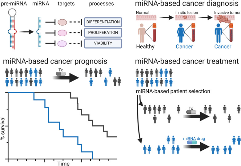
The hematopoietic system plays life-essential roles thanks to its various blood elements: oxygen transport and delivery to all tissues of the body is performed by erythrocytes, wound healing by platelets, host defense and tumor surveillance by white blood cells. Most mature blood elements are short-lived and must be promptly replenished to ensure proper functionality; thus, a pool of immature blood HSPCs must be constantly maintained, in the correct proportions between HSCs and MPPs. Hematopoiesis proceeds through well-defined steps of differentiation, in a hierarchical organization with a small pool of multipotent HSCs at the apex. HSCs generate progenitors with higher proliferation capacity but progressively more restricted lineage potential, intermediate precursors, and finally mature cells. To constantly support the hematopoietic hierarchy, HSCs are endowed with self-renewal as well as multipotent differentiation capacity throughout the life of the individual; balancing these two opposite fates is crucial to maintain the homeostasis of the hematopoietic system and avoid BM failure, immunodeficiency, onset of autoimmune diseases, or hematological disorders including malignancies. Different mechanisms must be tightly regulated to achieve this balance: to maintain the HSC pool, most HSCs are dormant or quiescent at any given time, with only a few in active state of proliferation, to be protected from DNA damage that can occur during cell cycle. At the same time, the capacity to exit the dormant state, proliferate, and differentiate according to peripheral requests without exhaustion must be preserved. All these mechanisms are primarily orchestrated by transcription factors and chromatin remodeling molecules in response to intrinsic or extrinsic signals, like cytokines, chemokines, or hormones, produced either by the surrounding microenvironment or by distant sites. However, posttranscriptional mechanisms, including the action of miRNAs, represent further checkpoints for fast and fine-tuning the quantity of intracellular proteins essential for HSC function. The role of specific miRNAs in several differentiation steps along the hematopoietic hierarchy beyond the HSC stage has been reviewed elsewhere; we here focus on their role in maintaining long-term HSC identity by affecting all mechanisms described above. In addition, miRNAs whose function has been described in HSPCs (murine LSK and/or human CD34+ cells) but their specific role in HSCs has not been dissected out, will not be considered here.
The majority of what is currently known on the role of miRNAs in HSCs is derived from mouse studies, where the expression of specific miRNAs has been altered with different methods (Table 1). Among the first evidences that miRNAs play a role in HSCs there is the observation that in the absence of Dicer, HSPCs display altered function and a higher rate of apoptosis (S. Guo et al., 2010). While the role of some miRNAs has been linked to HSC biology for several years now (miR-125, miR-126, miR-29a, and miR-193), the association of other miRNAs to HSC is quite recent (miR-143/145, miR-127-3p, miR-34a, and miR-21) and will be integrated in the context of HSC maintenance mechanisms. A few examples of miRNAs associated to HSC-related hematological disorders and involved in their pathogenesis will be also discussed.
2.1 Maintenance of HSC identity through cell-intrinsic mechanismsMost biological mechanisms that are in place to maintain HSC identity are a peculiarity of the adult hematopoietic system. In vertebrates, HSCs develop in the aorta–gonad–mesonephron (AGM) area of the dorsal aorta of day 10.5–11.5 or day 27–40 of mouse and human embryos, respectively (Dzierzak & Medvinsky, 2008; Julien et al., 2016). At later developmental stages they are found in the fetal liver, where they expand, and then finally migrate and lodge in the BM. The rate of proliferation and metabolic activity is different during the embryonal, fetal, and perinatal life compared with the adult, when HSCs are mostly quiescent. This review will mainly focus on the role of miRNAs in HSCs of adult individuals, for which more information is available, with only a brief reference to few miRNAs involved in HSC specification in Section 2.3.
2.1.1 Prevention of cell cycle entry: miR-126a, miR-193b, and miR-21Different miRNAs contribute to prevent cell cycle entry and to restrain proliferation, thus avoiding HSC expansion when unnecessary. Unlike other cell types, in HSCs proliferation is a prerequisite for differentiation toward the subsequent steps in the hematopoietic hierarchy. In HSCs the fate of proliferation depends on the capacity to undertake symmetric and asymmetric cell divisions. With asymmetric cell division, one daughter cell remains HSC, while the other becomes a progenitor, thus the proportion between HSCs and differentiating cells is maintained. On the other hand, symmetric cell division can result in increased self-renewal when both daughter cells are still HSCs, or to increased differentiation when both daughter cells become progenitors (Morrison & Kimble, 2006). Therefore, an excess of HSC proliferation can paradoxically result in two opposite outcomes: expansion (in some instances at the expenses of differentiation), or exhaustion and/or loss of functionality. For example, HSC expansion without exhaustion occurs when miR-126a is experimentally downregulated. miR-126a is differentially expressed in HSCs compared with lineage-committed progenitors, but still highly expressed at the MPP stage. It prevents cell cycle entry by affecting PI3K/AKT signaling. Attenuation of its activity in mouse and human HSPCs results in expansion of phenotypically and functionally defined HSCs, without inducing exhaustion or malignancies, while overexpression prevents HSC cell cycle entry and reduces hematopoietic output in vivo (Lechman et al., 2012). Interestingly, as opposed to normal HSCs, prevention of cell-cycle entry due to high miR-126a expression is instead advantageous for leukemic stem cells (LSC), since their quiescent state confers them resistance to chemotherapy (Lechman et al., 2016).
Another example of miRNA whose function is linked to cell cycle is miR-193b, identified as a regulatory feedback molecule restricting excessive HSC self-renewal upon the activation of the TPO-MPL-STAT5 signaling. Expansion of HSCs in miR-193b−/− mice, as well as in primary and secondary recipients of miR-193b−/− cells, is not transient and does not ultimately result in HSC exhaustion, but rather in increased self-renewal (Haetscher et al., 2015). This is justified not only by a higher proportion of HSCs that exit from their quiescent state, but also by a shorter time from first division at the single cell level. Moreover, an in vitro delay in differentiation was observed in HSCs from miR-193b−/− mice. miR-193b is one of the miRNAs that we found to be expressed in HSCs and downregulated in the very first maturation step that from HSCs brings to Flt3−MPPs (Crisafulli et al., 2019) when loss of self-renewal occurs.
Like miR-126a and miR-193b, the absence of miR-21 causes HSC exit from quiescence and results in the expansion of phenotypically defined HSCs; nevertheless, these expanded HSCs are myeloid-biased and less functional as demonstrated by the severely reduced reconstitution ability of BM cells from mice with miR-21 conditional deletion. In detail, wild-type (WT) recipients transplanted with BM cells from miR-21-conditional knock-out (KO) mice died earlier than those transplanted with miR-21-positive BM cells in secondary non-competitive BM transplantation (M. Hu et al., 2021). In competitive BM transplantation experiments, miR-21 deficiency resulted in a lower proportion of donor-derived cells in recipients' peripheral blood and LSK cells after primary and secondary transplants. miR-21 role in maintaining HSC homeostasis is mediated by targeting Programmed cell death 4 (Pdcd4), thus supporting the nuclear factor kappa B (NF-kB) pathway, already implicated in preserving HSC function (M. Hu et al., 2021). Thus, increased proliferation results in increased HSC self-renewal when miR-126 or miR-193 are absent, or in HSC exhaustion without miR-21.
2.1.2 Induction of proliferation: miR-125, miR-143/145, and miR-22An excess of proliferation may be deleterious for HSCs, nevertheless the capacity to proliferate is a pre-requisite to preserve both self-renewal and differentiation. Overexpression of miR-125a, mammalian ortholog of lin-4, the first discovered miRNA in Caenorhabditis elegans, was shown to affect HSC expansion at the single cell level. This led to increased clone longevity, size, contribution to multilineage engraftment, and trafficking from the BM to the circulation after transplantation (Wojtowicz et al., 2019). Continuous miR-125a expression appears to be necessary to induce HSC proliferation and self-renewal (Luinenburg et al., 2021). Other miR-125 family members (miR-125b1 and miR-125b2), which share with miR-125a the same seed sequence, exert similar effects on HSCs when overexpressed through retroviral vectors (Wojtowicz et al., 2014). Contradictory data regarding the function of miR-125b in lymphoid versus myeloid skewing have been reported (Ooi et al., 2010). However, properly regulated expression of miR-125 in HSCs is crucial to maintain their correct overall pool size and blood cell numbers (S. Guo et al., 2010) thus reducing the likelihood of expansion of malignant clones. Indeed, miR-125 overexpression in murine HSCs has been associated to myeloid skewing and to myeloproliferative neoplasm (MPN) development (S. Guo et al., 2012; Wojtowicz et al., 2014). Of note, also the absence of miR-125 in KO mice results in myeloproliferative disorders (Tatsumi et al., 2016), and this is in line with the reported role for miR-125 as tumor suppressor in vivo, both in mouse models and in patients (Berg et al., 2021). On the other hand, miR-125a was found to be overexpressed in CD34+ cells from myelodysplastic syndrome (MDS) patients and to play a role in the disease (see below). The opposing roles for miR-125a and for other family members described in the literature lead to the conclusion that fine tuning of miR-125 expression must be achieved and regulated at any given time and differentiation step in the hematopoietic hierarchy. In this way, a correct balance of proliferation and quiescence, as well as of myeloid and lymphoid differentiation, prevents the development of hematopoietic malignancies. Given that the miR-125a locus is highly conserved between human and mouse, the knowledge of how miR-125a expression is controlled in HSCs would be of crucial interest.
Other miRNAs positively regulating HSC proliferation are miR-143 and miR-145, which are transcribed as a single primary transcript. In a mouse model with miR-143/145 inactivation, depletion of HSCs was documented both in targeted mice and in mice transplanted with cells overexpressing the miR-145 mRNA target DAB2, a positive regulator of TGFβ signaling, which dampens HSC proliferation. Decreased activity of miR-143/145−/− HSC was demonstrated by secondary colony assays (Lam et al., 2018). Since mice lacked both miRNAs, whether both exert this function or if one of the two is prominent it remains to be investigated.
Another example of miRNA inducing HSC proliferation is miR-22, which emerged as being markedly upregulated in MDS patients (Song et al., 2013). Mice conditionally expressing miR-22 in the hematopoietic compartment displayed increased HSC self-renewal accompanied by myeloid skewing, ultimately leading to an MDS-like disease. This is due to concomitant silencing of ten-eleven-translocation gene 2 (Tet2) and of phosphatase and tensin homolog (Pten).
2.1.3 Restraining differentiation: miR-29a, miR99, and miR-127Another crucial biological mechanism to preserve HSC maintenance is the inhibition of excessive differentiation, which may or may not be linked to increased HSC proliferation. Like miR-126a, miR-29a is differentially expressed in HSCs compared with lineage-committed progenitors, but still highly expressed at the MPP stage. It limits HSC maturation in part through regulation of epigenetic changes by targeting DNA methyltransferase 3A (Dmnt3a) (W. Hu et al., 2015). Deletion of miR29a results in reduced number of both HSCs and MPPs, which display cell-intrinsic self-renewal defects in vivo. These defects are due to loss of quiescence and premature differentiation, with phenotypically defined HSCs from miR-29a KO mice harboring a transcriptional profile resembling that of committed progenitors.
All three members of the miR-99 family were found to be expressed at higher levels in mouse HSCs compared with more differentiated populations (Khalaj et al., 2017). miR-99 inhibits both differentiation and cell cycle entry by targeting HOXA, thus maintaining HSC long-term reconstitution ability. Functional inactivation of miR-99 leads to HSPC depletion due to accelerated differentiation.
A further example of miRNA inhibiting differentiation is miR-127, which is almost absent beyond the HSC developmental stage in steady state BM (Crisafulli et al., 2019). Inhibition of miR-127-3p function in HSPCs through a lentiviral-sponge vector led to severe stem cell depletion, as assessed with serial transplantation assays. The observed HSC reduction was accounted for by their accelerated differentiation, in the absence of increased proliferation (Crisafulli et al., 2019), as opposed to what reported upon miR-99 and miR-29a inactivation. A peculiarity of miR-127 is its strong HSC specificity; moreover, it emerged as the highest downregulated miRNA in HSCs from the Pbx1-conditional KO mouse model, which are characterized by a profound self-renewal defect and a tendency to differentiate prematurely (Ficara et al., 2008; Ficara et al., 2013). Despite its crucial function in maintaining HSC self-renewal, miR-127 is not expressed at high levels in HSCs; this is probably the reason why it has not been identified earlier among miRNAs regulating HSC biology. Even when purified with the best available combination of markers, phenotypically defined HSCs are still heterogeneous, and include cells with different degrees of differentiation potential and cell cycle state, as shown with experiments of single cell sequencing (Wilson et al., 2015). A selective expression of miR-127 in a stem cell subpopulation might theoretically justify its apparently low abundance in the sorted HSCs.
2.1.4 Protection from DNA damage: miR-34a and miR-21Various mechanisms are in place to protect HSCs from different harms, including DNA damage that can occur physiologically during replication, or that can be induced by external insults such as radiation. One example of miRNA affecting this mechanism is miR-34a, shown to promote irradiation-induced DNA damage repair in HSCs. In the absence of miR-34a, increased radiosensitivity in HSCs has been reported (Zeng et al., 2019), despite miR-34a is not essential for steady-state hematopoiesis. miR-34a involvement in DNA damage repair is likely linked to promotion of homologous recombination (HR) and nonhomologous end-joining (NHEJ) DNA repair processes. DNA damage in miR-34a−/− mice induces long-term functional consequences: their HSCs displayed reduced quiescence up to at least 3 months after total body irradiation, and loss of long-term reconstituting ability. DNA damage has been shown to induce aging of HSCs (Beerman et al., 2014; Flach et al., 2014), therefore miR-34a activity might also be key to prevent premature aging (see also Section 2.2.1). A low but detectable level of expression of the mature form of miR-34a in HSCs was observed also by our group (GEO Series accession number GSE113062; Crisafulli et al., 2019). However, only upon radiation or other DNA damage-inducing agents, miR-34a is likely induced as needed.
Another miRNA contributing to protect HSCs from radiation-induced DNA damage (such as double-strand breaks) is miR-21, already mentioned in Section 2.1.1; mediators of this function might be the NF-κB pathway, thrombopoietin-mediated NHEJ in HSCs, or other not yet identified molecular pathways (M. Hu et al., 2021).
2.1.5 Protection from apoptosis: miR-23a/miR-23b, and miR-125miR-23a has been recently included among the HSC-maintaining miRNAs due to its function in inhibiting apoptosis. miR-23a gene codes for a miRNA cluster including miR-23a itself, miR-24-2, and miR-27a; a paralogous miRNA cluster gives rise to three mature miRNAs (miR-23b, miR-24-1, and miR-27b) with identical seed sequences to miR-23a itself, miR-24-2 and miR-27a, respectively. Deletion of miR-23a alone did not result in HSPC defects, while miR-23a−/−miR-23b−/− conditional double-KO mice displayed decreased BM cellularity including fewer hematopoietic progenitors and HSCs, which also had a competitive disadvantage compared with WT or miR-23a−/− cells in a transplantation setting. Increased apoptosis was suggested as the mechanism at the basis of decreased HSPC populations (Kurkewich et al., 2018).
Decreased apoptosis had been also observed upon miR-125a over-expression (S. Guo et al., 2010). miR-125b, which belong to the same family of miR-125a as mentioned, is produced from a single miR-99b/100-125b polycistronic message. The combined activity of all three mature miRNAs converges to block the TGFβ pathway and amplify Wnt signaling thus escaping TGFβ-induced apoptosis (Emmrich et al., 2014).
2.1.6 Prevention of lineage skewing: miR-23a/miR-23b and miR-21To preserve multipotency, skewing to specific lineages must be prevented. In addition to affecting apoptosis as seen in Section 2.1.5, compound loss of miR23a/miR23b miRNAs gives rise to augmented CLP production at the expense of myeloid progenitors (Kurkewich et al., 2018). miR-21 has the opposite effect compared with miR-23a/miR-23b, since its absence in HSCs leads to myeloid-skewed differentiation at the expenses of the B-lymphoid lineage (M. Hu et al., 2021). The described effects of miR-21 deletion in HSCs underline how avoidance of excessive cell cycle, prevention of apoptosis, and preservation of multipotency are linked and all crucial to maintain HSC properties.
2.1.7 Resistance to inflammatory stress: miR-146aAlthough miR-146a is expressed in HSCs, it is more abundant along with maturation and it does not seem to be essential for hematopoietic development nor for steady-state hematopoiesis; its function appear evident with aging or upon the occurrence of inflammatory stress. In general, miR-146 expression level is modest, but it is upregulated by different inflammatory stimuli including LPS and proinflammatory cytokines, through NF-kB direct regulation. It represses IRAK1 and TRAF6 through direct mRNA targeting, thus acting as feedback inhibitor for NF-kB activation, since IRAK1 and TRAF6 are upstream of NF-kB, and help resolving the inflammation-induced immune reaction by reducing myelopoiesis.
2.1.8 Other miRNAs maintaining self-renewalmiR-10a has recently been included among miRNAs positively affecting self-renewal, although the biological mechanism through which miR-10a exerts its function has not been clarified yet, nor its mRNA target(s). Importantly, it was found preferentially expressed in human cells with long-term engraftment capabilities. Functionally defined miR-10-expressing HSCs were identified using gamma-retroviral vector integration sites in gene therapy patients 6 years after autologous HSC transduction and transplantation (Wünsche et al., 2018). Overexpression of miR-10 in murine HSCs resulted in increased replating ability in CFC assays and in augmented proportion of HSCs and MPPs upon transplantation, leading to increased BM cellularity and T cell lymphopoiesis.
The miR-212 and miR-132 cluster (or Mirc19) is enriched in HSCs and is upregulated during aging (Mehta et al., 2015). Mirc19 regulates HSC cycling, function, and survival through autophagy thus balancing over time HSC maintenance and normal immune cell production.
2.2 Maintenance of HSC identity through the microenvironmentIn post-natal life, hematopoiesis occurs mainly within the BM, where HSCs are in close contact with cells belonging to the microenvironment. Niche cells include osteoblast bone-lining cells, mesenchymal stromal cells (MSCs), macrophages, megakaryocytes (MKs) and endothelial cells. In one view of the BM microenvironment, “dormant” HSCs are located in the endosteum in association with arterioles, while “active” HSCs reside in vascular niches and are located adjacent to sinusoids. High resolution image studies revealed that indeed HSCs reside mainly close to sinusoids, to Cxcl12+ stromal cells, and to MKs (Kokkaliaris et al., 2020), although the picture is evolving continuously. Cells belonging to the BM microenvironment provide HSCs with a tridimensional niche that nurtures them and maintains their stem cell characteristics by releasing growth factors and/or other molecules, by inducing intracellular signals through cell–cell interaction, and by retaining them in the BM or releasing them when needed. miRNAs are emerging as additional players in this complex network, either by affecting the function of niche cells, or by being released and up taken by HSCs.
2.2.1 Vesicles produced by cells belonging to the HSC nicheExtracellular vesicles (EVs) are membranous nanoparticles (30–10,000 nm) secreted by all cell types, carrying bioactive proteins, lipids and nucleic acids including miRNAs. They can be transferred to other cells to mediate both local and distant intercellular communication (Valadi et al., 2007). EVs include microvesicles (MVs, 50–1000 nm), generated by calcium mediated budding and cleavage of the plasma-membrane and remodeling of the cortical cytoskeleton, as well as exosomes (30–150 nm), which mature from endosomes and then fuse to the inner layer of the plasma membrane for release (Butler et al., 2018). MSCs produce both MVs and exosomes. BM-MSC-derived EVs were shown to contain several miRNAs and to alter gene expression profile of cord-blood CD34+ cells exposed to them ex-vivo (De Luca et al., 2016). Among BM-MSC-EV contained miRNAs, miR-21, and miR-22 are known to play a role in HSCs (Tables 1 and 2); some of their predicted target mRNAs were indeed downregulated in CD34+ cells upon exposure to EVs (De Luca et al., 2016).
TABLE 2. HSC related miRNAs expressed in other adult tissue-specific stem cells miRNA Stem cell Function References miR-21 MSCa Promotes osteoblast, osteocyte, and adipocyte differentiation; limits cartilage differentiation Sekar et al. (2015); Vail et al. (2021) Periodontal ligament stem cells (PDLSCs) Promotes osteogenic differentiation following orthodontic force H. Huang et al. (2018) miR-22 BM-MSCa Inhibits osteoblast differentiation Yin et al. (2020) PDLSCs Promotes osteoblast differentiation Yan et al. (2017) Adipose tissue derived MSC (ADMSCs) Inhibits adipogenesis, promotes osteogenesis S. Huang et al. (2012) Fibro/adipogenic progenitors (FAPs) Prevents adipogenesis Lin et al. (2020) Hair follicle stem cells (HFSCs) Inhibits proliferation and differentiation, regulates hair cycle Cai et al. (2020); Yuan et al. (2015) miR-23 BM-MSC Suppresses osteogenic differentiation K. Jiang et al. (2020) Myoblasts Induces skeletal muscle differentiation Mercatelli et al. (2017) miR-29a BM-MSC Promotes osteogenic differentiation, inhibits cartilage formation, inhibits proliferation, prevents senescence Guérit et al. (2014); Jung et al. (2020); Kapinas et al. (2010); Tan et al. (2018); Y. Zhang and Zhou (2015) Multipotent adipose stem cells (MADS) Inhibits adipogenesis Glantschnig et al. (2019) Myoblasts Improves differentiation into myotubes X. H. Wang et al. (2011) Neural stem/progenitor cells (NSPCs) Promotes neural differentiation, neurite outgrowth and complexity, still maintaining stemness Y. Gao et al. (2020); Ma et al. (2020) miR-34a BM-MSC Inhibits/promotes osteogenic differentiation; proapoptotic and prosenescence roles Chen et al. (2014); Liu et al. (2019); Pi et al. (2021); F. Zhang et al. (2015) Adipose-derived stem cells (ADSCs) Promotes senescence rather than differentiation Park et al. (2015) Dental pulp stem cells (DPSC) Induces senescence S. Zhang et al. (2021) Neural stem cells (NSCs) Induces/inhibits differentiation Aranha et al. (2011); Morgado et al. (2015) miR-125 BM-MSC Inhibits osteoblast differentiation Tu et al. (2016) Glial progenitor cell (GPC) Primes astrogliogenesis Shenoy et al. (2015) miR-126a BM-MSC Promotes proliferation and endothelial differentiation while inhibits apoptosis and osteogenic differentiation Kong et al. (2020) DPSC Induces apoptosis Ge et al. (2021) Endothelial progenitor cells (EPCs) Maintains stemness Pei et al. (2020) miR-127-3p Myoblasts Inhibits proliferation, enhances differentiation Li et al. (2020); Zhai et al. (2017) miR-212/132 BM-MSC Suppresses osteogenic and promotes chondrogenic differentiation Y. Zhang, Jiang, et al. (2020); Zhou et al. (2018) PDLSCs Inhibits osteogenic differentiation Xu et al. (2019) Precartilaginous stem cells (PCSCs) Promotes proliferation and inhibits apoptosis F. Y. Zhang, Zhen, et al. (2020) NSCs Induces hippocampal neurogenesis Walgrave et al. (2021) miR-143/145 BM-MSC Negative regulator of osteogenesis and of EC differentiation Cha et al. (2016); Feng et al. (2018) ADSCs Negative regulator of osteogenesis Hao et al. (2018) miR-146a BM-MSC Inhibits proliferation, promotes immune-modulatory properties, and osteoblast differentiation/suppresses osteoblastogenesis and bone formation Cui et al. (2020); Kuang et al. (2017); Zheng et al. (2021); Zhou et al. (2016) ADSCs Inhibits osteogenic differentiation S. Wan et al. (2020) miR-193b BM-MSC Promotes chondrogenesis/inhibits early chondrogenesis, promotes proliferation Hou et al. (2015); Meng et al. (2018); J. Wang et al. (2012) ADSCs Promotes adipogenesis Mazzu et al. (2017) a MSC is the acronym of “mesenchymal stromal cells” in some studies, or of “mesenchymal stem cells” in others. MSCs are multipotent clonogenic progenitors that give rise to several cell types including osteoblasts, osteocytes, cartilage, and adipocytes. The definition of MSC as “stem cells” is sometimes controversial, mainly because it is not clear if their multipotency is exerted in vivo at the single cell level. All studies mentioned in Table 2 consider MSC as stem cells. MSC can be isolated from different tissues; BM-MSC are BM-derived.The role of miRNA-containing exosomes secreted by MSCs has been also associated to aging. In detail, MSC exosomes from young mice contribute to rejuvenate HSCs upon ex-vivo exposure; they were shown to contain significant lower levels of miR-34a compared with exosomes from aged MSCs (Kulkarni et al., 2018). The role of miR-34a contained in MSC-secreted exosomes has been linked to its ability to target the autophagy-related protein Sirtuin, thus reducing autophagy. This in turn accelerates aging, since autophagy protects HSCs from metabolic stress (Ho et al., 2017). Fine tuning of miR-34a is likely crucial: in steady state hematopoiesis under physiological conditions miR-34a levels must be kept low both in HSCs (see Section 2.1.4) and in MSCs serving as HSC niche.
As mentioned previously and reviewed elsewhere (Ghosh et al., 2021), MKs are also part of the HSC niche. In the BM, MKs localize in close proximity to HSCs, promoting their quiescence and contributing to their retention within the BM. MVs produced by MKs have been shown to be internalized by HSPCs, to contain miRNAs, and to induce differentiation into MKs (J. Jiang et al., 2017). Whether miRNAs contained in MK MVs also participate to the maintenance of HSC quiescence it remains to be investigated.
2.2.2 HSC-supporting ability by niche cells: miR-23aThe chemokine CXCL12 regulates the interaction between HSCs and BM stromal cells (BMSC). An intact CXCL12-CXCR4 signaling is crucial to maintain the quiescent HSC pool and its retention within the BM (Sugiyama et al., 2006). CXCL12 produced specifically by BM MSC (but not by other cells belonging to the BM microenvironment) has been shown to be essential for HSC self-renewal and reconstitution ability (Agarwal et al., 2019). However, temporary HSC release from the BM is physiologically needed in response to peripheral stimuli and must be properly regulated at any given time. miR-23a (see also Section 2.1.6) was shown to be expressed in primary BMSC, to be upregulated upon TGFβ1 exposure, and to inhibit CLCX12 production in BMSC by direct binding to its 3′-UTR (Arabanian et al., 2014). Additional functional studies are needed to further clarify the role of stromal miR-23a in vivo; however, it is tempting to speculate that a TGFβ1-mediated mechanism may be responsible for miR-23a-mediated reduction of CXCL12, leading to HSC release from the BM.
Comments (0)