
Remember me


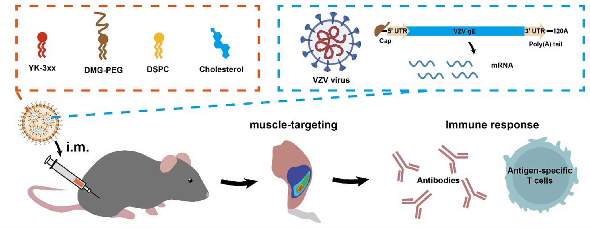
We sought to better understand how the multi-step process of incorporating mRNA-LNPs into MNPs may impact mRNA-LNP stability (Fig. 1). mRNA-LNPs were formulated via microfluidic mixing [36], followed by formulation with excipients containing a water-soluble polymer (to form the MN matrix) and, in some cases, a stabilizer (to protect mRNA-LNP integrity) to form the MN cast solution. This solution was cast onto a PDMS mold under vacuum. After drying, a backing cast solution was prepared and cast onto the mold. This solution contained excipients without mRNA-LNPs and was used to connect the MNs and form a patch backing. After the entire MNP was dried, it was demolded for analysis (Supplementary Fig. 3). The casting solution formulation, casting process, drying process, and other components of MNP manufacturing can destabilize and damage mRNA-LNPs.
Fig. 1
Schematic diagram of the mRNA-LNP MNP manufacturing process. A MN cast solution is cast onto a PDMS mold to fill the MN cavities of the mold with formulated mRNA-LNPs. A backing cast solution is then cast onto the mold to form the patch backing. After drying, the resulting MNP is demolded
Concentration of LNPsThe limited volume of MNPs constrains the mRNA-LNP dose that can be incorporated. While intramuscular injection can deliver up to 50 µL in mice and 2 mL in humans [39], MNPs are typically made with a MN cast solution of up to ~ 10 µL, which is about three orders of magnitude less than the typical human IM injection volume. To achieve effective mRNA dosing, mRNA-LNPs generally have to be significantly concentrated compared to conventional formulations to be incorporated into a MNP.
We therefore tested three methods to concentrate mRNA-LNPs: centrifugal filtration, TFF, and reverse dialysis. For initial experiments, polyA was used as a model poly-nucleic acid as a substitute for mRNA. Though we were able to reduce liquid volume by up to 20-fold, the recovered polyA concentration in the LNP formulation was increased to a much lesser extent, indicating substantial polyA loss (Fig. 2a). Of the tested methods, TFF demonstrated least polyA loss and highest final polyA concentrations. Therefore, we chose TFF to test for in vivo application.
In control groups, intradermal (ID) injection of naked mRNA in BALB/c mice produced measurable reporter protein expression, as measured by bioluminescence measurements (Fig. 2b). ID injection of mRNA-LNPs yielded an order of magnitude greater expression, consistent with the enhanced transfection expected for LNP delivery. However, no significant luciferase expression was seen after mRNA-LNP delivery by MNP, neither for mRNA-LNPs concentrated by TFF nor for unconcentrated mRNA-LNPs. This indicates that concentrating mRNA-LNPs by TFF was not effective to enable mRNA-LNP delivery by MNP.
Fig. 2
Effect of concentrating mRNA-LNPs after their fabrication. (a) PolyA-loaded LNPs were concentrated by TFF, centrifugal filtration, or reverse dialysis. Intended polyA concentration was determined by the change of volume of the polyA-LNP solution, while measured polyA concentration was determined by UV spectroscopy. (b) In vivo luminescence after delivery of luciferase-encoding mRNA-LNPs as: 1 µg naked mRNA delivered intradermally (ID), 1 µg mRNA loaded in LNPs delivered ID, 0.9 µg mRNA in unconcentrated LNPs delivered via MNP, and 1.4 µg mRNA in LNPs concentrated by TFF (7-fold by volume) and delivered by MNP. MNPs were fabricated with PVA and sucrose. The dashed line represents background luminescence levels. (n = 3–4, *p < 0.05, **p < 0.01, ANOVA with multiple comparisons to the ID mRNA-LNP control group)
Instead of concentrating mRNA-LNPs after their fabrication, we increased the concentration of lipid and mRNA during fabrication to attain a more-concentrated mRNA-LNPs suspension. Using this approach, we increased mRNA-LNP concentration roughly three-fold (Fig. 3a), and correspondingly increased mRNA-LNP content in MNPs roughly three-fold compared to MNPs made with conventional lipids and mRNA concentrations (Fig. 3b). These higher-concentration MNPs led to significant expression in vivo (Fig. 3d-e). Notably, the three-fold increase in mRNA-LNP delivery by MNP resulted in a 93-fold increase in luciferase bioluminescence (Fig. 3c), suggesting that the increased dose exceeded the threshold for detectable protein expression [40]. We also note that these concentrated mRNA-LNPs did not undergo a buffer exchange and therefore remained at pH 3 during MNP manufacturing, a factor which is investigated in the next section.
Fig. 3
Effect of concentrating mRNA-LNPs during their fabrication. (a) Intended mRNA concentration (determined by change of volume of mRNA-LNP solution) compared to measured concentration (determined by UV spectroscopy). (b) mRNA loading in MNPs. (c) In vivo expression after administration of MNPs made from low-concentration (0.9 µg per patch) or high-concentration (3.0 µg per patch) luciferase-encoding mRNA-LNPs. Representative IVIS images of mice with (d) low-concentration mRNA-LNP MNPs or (e) high-concentration mRNA-LNP MNPs. (n = 3–4, *p < 0.05, ****p < 0.0001, Student’s t-test)
We wanted to further increase mRNA-LNP loading in the MNP, which we achieved by casting MN cast solution twice before adding the backing cast solution. This second cast increased the amount of mRNA-LNPs incorporated into the MNPs and filled in voids in the MNs created during drying of the first cast (Fig. 4a). This two-cast approach increased mRNA-LNP loading by roughly two-fold (Fig. 4b) and increased reporter protein expression by roughly six-fold in vitro (Fig. 4c). However, this approach did not significantly increase protein expression in vivo (Fig. 4d). Adding a third cast did not further increase mRNA-LNP loading (Supplementary Fig. 4), suggesting saturation of MN capacity. We concluded that increasing the number of sample casts was not a useful strategy for increasing mRNA expression in vivo.
Fig. 4
Effect of repeated casting of MN cast solution. (a) Schematic diagram showing the two-cast MN manufacturing approach. (b) mRNA loading in MNPs. (c) In vitro expression of luciferase-encoding mRNA-LNPs after reconstitution from MNPs. (d) In vivo expression of luciferase-encoding mRNA-LNPs in BALB/c mice after delivery by MNPs. The mRNA doses were 3.6 µg for one-cast and 8.4 µg for two-cast MNPs. Representative IVIS images of mice after administration of mRNA-LNP MNPs prepared with (e) one cast or (f) two casts MN cast solution. (n = 2, ns = no significance, **p < 0.01, ****p < 0.0001, Student’s t-test)
Effect of pHWhile mRNA-LNPs are generally formulated at pH 3 to facilitate mRNA incorporation into LNPs by ionic bonding between negatively charged mRNA and positively charged ionizable lipids, they typically undergo subsequent buffer exchange to bring them to a physiological pH of 7.4 [11]. However, this pH change may not be needed for MNPs, since MNPs are administered in a dry state and not as a liquid injection. We hypothesized that acidic pH may be beneficial to mRNA stability by maintaining positive charge on the ionizable lipids for binding to mRNA.
We screened the effects of formulation pH between 3.0 and 7.4 after casting and drying and found that lower pH produced smaller mRNA-LNPs (Supplementary Fig. 5a), which is generally believed to improve transfection ability due to reduced aggregation and fusion [41]. pH had no significant effect on mRNA encapsulation efficiency (Supplementary Fig. 5b), which generally needs to remain high for efficient transfection [42]. We found that in vitro expression after reconstitution from a MNP was greater at low pH, while in vitro expression of non-dried LNPs appeared to be reduced at low pH (Supplementary Fig. 5c). Additionally, we found that there was no significant difference between using a phosphate or a citrate buffer when concentrating mRNA-LNPs (Supplementary Fig. 6).
Fig. 5
Effect of pH on mRNA-LNP stability during MNP manufacturing. (a) mRNA-LNP hydrodynamic radius. (b) mRNA encapsulation efficiency in LNPs. (c) In vitro cellular expression of luciferase-encoding mRNA-LNPs normalized to initial LNPs at pH 7.4. mRNA-LNPs were formulated, dialyzed in phosphate buffer at pH 3.0 or 7.4, cast onto MNPs, and reconstituted. (d) In vivo expression of luciferase-encoding mRNA-LNP MNPs in BALB/c mice after delivery by MNPs. Representative IVIS images of mice after application of MNPs with luciferase-encoding mRNA-LNPs at (e) pH 3.0 or (f) pH 7.4. (n = 4, ns = no significance, **p < 0.01, ANOVA with multiple comparisons relative to the pH 7.4 control group, Student’s t-test)
Follow-up experiments performed by keeping the mRNA-LNPs at either pH 3.0 or 7.4 throughout the MNP manufacturing process showed no significant effect of pH on mRNA-LNP size (Fig. 5a) or encapsulation efficiency (Fig. 5b), significantly better in vitro expression at pH 3.0 (Fig. 5c) and no significant difference in in vivo expression (Fig. 5d). Mice observed for several days after application of MNPs showed no signs of reactogenicity or ulceration at the MNP application site at either pH. We concluded that there was no clear advantage to either low or high pH to mRNA-LNP effectiveness but favored use of low pH since it simplified manufacturing by eliminating the buffer-exchange step.
Effect of MNP formulation excipientsFig. 6
Effect of polymeric MNP formulation excipients. (a) mRNA-LNP hydrodynamic radius. (b) mRNA encapsulation efficiency in LNPs. (c) In vitro cellular expression of luciferase-encoding mRNA-LNPs normalized to initial LNPs. mRNA-LNPs were evaluated after reconstitution from MNPs formulated with polyacrylic acid (PAA), poly(vinyl methyl ether-co-maleic acid) (PVME/MA), polyvinyl alcohol (PVA), or carboxymethyl-cellulose (CMC). (n = 6, ns = no significance, ****p < 0.0001, ANOVA with multiple comparisons relative to the PVA control group)
Water-soluble polymers are used to stabilize excipients and provide mechanical strength to MNPs [43]. Our initial comparison of MN cast solutions containing PVA and PVP demonstrated that PVA preserved mRNA-LNP size after MNP manufacturing (Supplementary Fig. 7). Further screening against polyacrylic acid (PAA), poly(vinyl methyl ether-co-maleic acid) (PVME/MA), and carboxymethyl-cellulose (CMC) across multiple measures of LNP stability demonstrated PVA’s superiority. MNPs manufactured with PVA maintained a small size, greater encapsulation, and greater in vitro cellular expression than the other formulations (Fig. 6). We therefore used the PVA-based MN cast solution formulation in all other experiments.
We also studied the effects of PVA concentration and observed that higher PVA content increased in vitro cellular expression. Low PVA content (1% or 5%) maintained smaller mRNA-LNP size after MNP manufacturing (Supplementary Fig. 8a) and any amount of PVA decreased mRNA encapsulation in LNPs (Supplementary Fig. 8b). However, the in vitro expression post MNP manufacturing exponentially increased with increasing PVA content (Supplementary Fig. 8c). We tested the effect of PVA on mRNA-LNPs before MNP manufacturing and found that PVA content increased in vitro expression only in the presence of sucrose (Supplementary Fig. 8d). From these data, we concluded that PVA has a positive effect on mRNA-LNP transfection beyond its protective effects during the MNP manufacturing process, but further research is needed to determine whether this offers translational potential.
In addition to water-soluble polymers, other excipients may be added to a MNP to increase stability, often including sugars [19]. We screened two disaccharides (sucrose and trehalose) and a monosaccharide (glucose) to assess their effects of mRNA-LNP stability [17, 44] We found that addition of sucrose to the MN cast solution over a range of concentrations did not significantly affect mRNA-LNP size (Fig. 7a) or encapsulation (Fig. 7b) after MNP manufacturing, but did generally decrease in vitro expression (Fig. 7c). Formulation with trehalose, glucose, and combinations of two or three sugars indicated that trehalose helped maintain smaller mRNA-LNP size (Fig. 7d). Combination of sucrose and glucose maintained smaller mRNA-LNP size (Fig. 7d), greater encapsulation efficiency (Fig. 7e) and increased in vitro cellular expression (Fig. 7f). However, all of these improvements were modest, indicating that additional sugars provided minimal benefit beyond the stabilization already conferred by PVA. However, additional exploration into other stabilizers may further improve mRNA-LNP MNPs.
Fig. 7
Effect of sugar-based MNP formulation excipients. (a, d) mRNA-LNP hydrodynamic radius. (b, e) mRNA encapsulation efficiency in LNPs. (c, f) In vitro cellular expression of luciferase-encoding mRNA-LNPs normalized to initial LNPs. mRNA-LNPs were evaluated after reconstitution from MNPs formulated with 5% PVA and (a-c) varying concentrations of sucrose, or (d-f) 10% sucrose (Suc), trehalose (Tre), glucose (Glu), sucrose/glucose, or sucrose/glucose/trehalose. (n = 6, ns = no significance, *p < 0.05, **p < 0.01, ***p < 0.001, ****p < 0.0001, ANOVA with multiple comparisons relative to the 0% control group and sucrose control group)
Effect of MNP drying conditionsDrying process represents a critical stress factor for lipid-based nanoparticles, and previous studies on air-drying of liposomes indicate that slowing the drying process can increase stability [45]. Additionally, lower temperatures generally slow degradation processes for LNPs and mRNA [46, 47]. Therefore, we hypothesized that maintaining a lower temperature (i.e., which would also slow the drying process) during the MNP manufacturing process would improve stability. We manufactured MNPs by casting the MN casting solution and backing cast solution at 25 °C, and then storing the MNPs with desiccant for two days at 5 °C, 25 °C, or 40 °C.
Fig. 8
Effect of drying temperature during MNP manufacturing. (a) mRNA-LNP hydrodynamic radius, (b) mRNA encapsulation efficiency in LNPs, and (c) in vitro cellular expression of luciferase-encoding mRNA normalized to initial LNPs as a function of MNP drying temperature. mRNA-LNPs were evaluated after reconstitution from an MNP formulated with PVA-sucrose, backed with epoxy, and stored for two days with desiccant in a controlled-temperature stability chamber. (n = 6, ns = no significance, *p < 0.05, **p < 0.01, ****p < 0.0001, ANOVA with multiple comparisons between all groups after MNP manufacturing)
The drying temperature had no significant effect on mRNA-LNP size (Fig. 8a). There was a slight reduction in encapsulation efficiency at 40 °C (Fig. 8b). In contrast, the in vitro expression showed a significant correlation between lower drying temperature and greater mRNA expression (Fig. 8c). We concluded that drying at a low temperature favored mRNA-LNP stability. This discrepancy between physical characteristics and functional activity points to a factor other than LNP integrity, such as mRNA integrity, that might be compromised by higher temperatures.
We also investigated the effects of drying time on mRNA-LNP stability. First, we measured the effects of adding the backing cast solution on the same day as the sample cast solution (before complete drying of the sample cast occurred) and compared with more-fully drying the sample cast overnight before adding the backing cast solution. When the sample cast was dried overnight, the mRNA-LNPs had a smaller size (Fig. 9a), a similar encapsulation efficiency (Fig. 9b), and a lower in vitro expression (Fig. 9c). We concluded that it was beneficial to add the backing cast solution on the same day rather than more-fully drying the sample cast overnight. We hypothesized that MNPs in the mold dried slowly and, to understand this, we studied the impact of the time the MNPs remained in the PDMS mold before demolding. When varying the time of MNP demolding between one and six days, we found that the demolding time had no significant effect on mRNA-LNP size (Fig. 9d). The encapsulation efficiency was higher in MNPs that were demolded after more than one day (Fig. 9e). The cellular expression was highest from the MNPs demolded after one day, but the effect was minimal (Fig. 9f). We concluded that there was no clear advantage to waiting more than one day to demold the MNPs.
Fig. 9
Effect of drying conditions on mRNA-LNP stability in an MNP. (a-c) Effect of adding the backing cast solution on the same day (Day 0) or after drying the sample cast overnight (Day 1). (d-f) Effect of the length of the drying process time. Effects of drying conditions were assessed by measuring (a, d) hydrodynamic radius of mRNA-LNPs, (b, e) mRNA encapsulation in an MNP, and (c, f) in vitro cellular expression of luciferase-encoding mRNA-LNPs normalized to initial LNPs. (n = 6, *p < 0.05, **p < 0.01, ***p < 0.001, ****p < 0.0001, ANOVA with multiple comparisons between all groups, Student’s t-test)
Effect of MNP backing cast formulationAfter loading the MNs with the sample cast, we added a backing cast solution to form the rest of the MNP and provide mechanical strength for insertion. We tested the effects of the backing cast solution on the mRNA-LNPs during MNP manufacturing. We screened four different backings solutions: a self-setting epoxy, ultraviolet (UV)-curable glue, an aqueous solution of PVA and sucrose, and an aqueous solution of PVP. We also tested epoxy backing with exposure to UV light to evaluate the effect of UV light exposure independent of its use to cure the UV-curable glue.
The choice of backing cast solution had a significant effect on mRNA-LNP size, with PVA-sucrose and PVP casting solution resulting in the smallest mRNA-LNPs (Fig. 10a), and no significant effect on mRNA encapsulation (Fig. 10b). The epoxy backing resulted in the highest in vitro expression with and without UV light, but exposure to UV light roughly halved the expression (Fig. 10c). An additional factor to consider was that the backings required different drying/curing times, which affected the overall manufacturing time of the MNP. Aqueous backings, such as PVA-sucrose and PVP, took up to seven days to dry at refrigeration temperatures. The epoxy backing took less than one day to fully cure. The UV-curable glue cured within 30 s during exposure to UV light. Because of the stability benefit and relatively short manufacturing time, we concluded that epoxy was the best backing cast solution.
Fig. 10
Effect of MNP backing cast solutions. (a) mRNA-LNP hydrodynamic radius, (b) mRNA encapsulation efficiency, and (c) in vitro cellular expression of luciferase-encoding mRNA-LNPs normalized to initial LNPs shown as a function of backing cast solution. mRNA-LNPs were evaluated after reconstitution from a MNP formulated with PVA-sucrose and backed with epoxy, epoxy with exposure to UV light, UV-curable glue and exposure to UV light, an aqueous solution 18% w/v PVA and 18% w/v sucrose, or 20% w/v PVP. (n = 4, ns = no significance, *p < 0.05, **p < 0.01, ****p < 0.0001, ANOVA with multiple comparisons between all groups after MNP manufacturing)
Thermostability of mRNA-LNPs in MNPsIn addition to assessing mRNA-LNP stability during MNP manufacturing, we also tested the thermostability at 25 °C for 2 months and 40 °C for one month. At both temperatures, mRNA-LNP size (Fig. 11a and d) and encapsulation (Fig. 11b and e) did not change significantly between day 0 and the end of the study. At 25 °C, in vivo expression dropped by 59% in the first week and then remained just above background levels without significantly decreasing after day 7 for up to 60 days (p = 0.50, Fig. 11c). At 40 °C, in vivo expression dropped by 87% in the first two days and then continued to decrease out to 28 days, ending with a cumulative drop of 97% (p = 0.02, Fig. 11f). We concluded that mRNA-LNP MNPs lost activity during storage, and the most significant loss in mRNA expression occurred within the first week.
Fig. 11
Thermostability of mRNA-LNP MNPs. (a-c) Thermostability of mRNA-LNP MNPs at 25 °C over 60 days, as measured by (a) mRNA-LNP hydrodynamic radius, (b) mRNA encapsulation efficiency in LNPs, and (c) in vivo expression of luciferase-encoding mRNA-LNP MNPs in mice. (d-f) Thermostability of luciferase-encoding mRNA-LNP MNPs at 40 °C over 28 days, as measured by (d) mRNA-LNP hydrodynamic radius, (e) mRNA encapsulation efficiency in LNPs, and (f) in vivo expression of luciferase-encoding mRNA-LNP MNPs in mice. The dashed lines represent background levels of luminescence. (n = 2–6, *p < 0.05, **p < 0.01, ***p < 0.001, ****p < 0.0001, ANOVA with multiple comparisons to control group at Day 0)
Comments (0)