
Remember me


The findings of this study provide critical insights into the relationships between zygomatic implant configurations and the anatomical structures surrounding the zygomatic bone and orbital floor. By introducing the zygomatic orbital floor (ZOF) classification system, we aimed to offer a novel tool for assessing the risk of orbital damage during free-hand zygomatic implant placement. The results of our study show statistically significant differences between implant configurations and their proximity to key anatomical landmarks, particularly in relation to the ZOF classification, which is consistent with the findings in prior studies. This section discusses how our results compare to existing knowledge and the clinical significance of these findings.
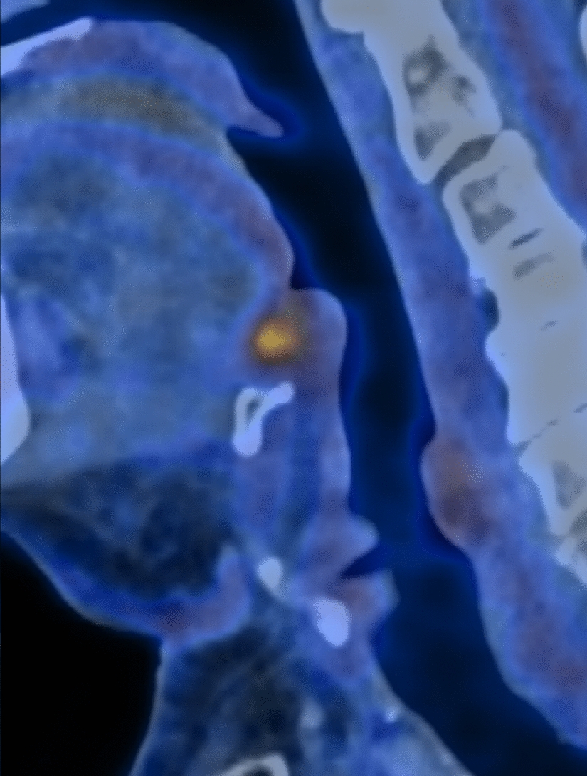
Implant Configurations and Orbital Floor ProximityOur results show that in patients with higher ZOF classifications (Class III and IV), implants were placed lower in the zygomatic bone to avoid the orbital floor, with a significant difference observed between the left and right sides (p < 0.05). Specifically, implants placed in patients with deeper ZOF classifications tended to be positioned at greater distances from the orbital rim, thus reducing the risk of orbital damage. This confirms that ZOF classification is an effective method for planning implant placement.
Implant Length and Sinus PenetrationThe study found that the average implant length across all groups was 42.8 mm (range: 32.5–52.5 mm). Implant length varied significantly between groups, with longer implants more frequently used in patients with more severe maxillary atrophy (p < 0.05). Interestingly, intramaxillary placement of zygomatic implants was associated with a reduced incidence of Schneiderian membrane rupture (p < 0.05), confirming that this technique is safer for avoiding sinus-related complications. Notably, implants placed intramaxillary reduced the risk of Schneiderian membrane rupture (p < 0.05), a finding consistent with the work of Grecchi E., et al. [10], who reported that intramaxillary placement results in fewer sinus complications compared to extrasinus placement. While Grecchi et al. used a guided surgical approach, our results show that free-hand surgery, when combined with proper anatomical assessments like ZOF classification, can minimize sinus-related complications as well. However, our study further expands on this by quantifying the specific relationship between implant length and anatomical safety, showing that longer implants, when placed correctly, do not increase the risk of sinus complications.
ZOF Classification as a Novel ToolThe ZOF classification system introduced in this study provides a practical and effective method for assessing the depth of the orbital floor and its impact on zygomatic implant placement. Our results indicate that the deeper the ZOF classification, the greater the risk of complications if not properly accounted for. Specifically, patients with Class III or IV ZOF classifications benefited from lower implant placement in the zygomatic bone, reducing the likelihood of orbital penetration (p < 0.05). This supports the utility of the ZOF classification in clinical practice, particularly in cases where advanced guided surgery tools are not available. Previous research has largely focused on the use of CAD and guided systems to mitigate the risks of orbital damage [11]. While those studies advocate for guided surgery, which can reduce complications, the ZOF classification offers a practical alternative for surgeons who may not have access to advanced digital tools. Our findings demonstrate that by categorizing patients based on their orbital floor anatomy, clinicians can plan the angle and depth of osteotomies to minimize the risk of orbital complications.
Orbital penetration is a well-documented complication of zygomatic implant surgery, with studies by Mertens et al. [1] and Wu et al. [12] reporting cases of orbital damage in free-hand surgeries. Our study had no cases of orbital perforation, which can be attributed to the operator’s consistent use of preoperative CBCT planning and the avoidance of high-risk ZOF classifications. This contrasts with Krauthammer et al. [13], who reported extraocular muscle damage in a case of orbital penetration during free-hand surgery. Our findings underscore the importance of understanding the patient’s specific orbital anatomy through detailed imaging, even in the absence of digital guides, thereby reinforcing the clinical utility of ZOF classification as a protective measure. Furthermore, we have found that surgery duration correlated with both implant configuration and patient-specific anatomical challenges (p < 0.05). This reinforces the importance of individualized surgical planning based on precise anatomical measurements.
Success Criteria of Zygomatic Implants—ORISThose who work with zygomatic implants concur that the success criteria for traditional implants do not directly apply to zygomatic implants. Thus, Aparicio et al. outlined four distinct criteria to be considered: offset of the definitive prostheses, rhinosinus condition, soft tissue infection, and stability. [9] Evaluating these criteria allows a practitioner to categorize a patient into one of the following five conditions:
• Success condition I: The optimal stage.
• Success condition II: A deviation from the norm without clinical impact.
• Success condition III: A borderline situation with clinically evident alterations, yet still treatable.
• Condition IV: Reflects a surviving implant supporting the prosthesis but not evaluated by the proposed criteria.
• Condition V: Represents implant failure.
Offset of the Prostheses [O]The emergence of zygomatic implants in the palate may lead to prostheses with "piping". Occasionally, a substantial dental bridge on the palatal side can cause discomfort, impacting speech, and limiting hygiene access (Fig. 10). Conversely, placing the implant too buccally can result in zygomatic implant failure (Fig. 11).
Fig. 10
The emergence of zygomatic implants in the palate may lead to prostheses with "piping"
Fig. 11
Implant screwed in region 26 screwed too buccally (a) resulted in implant removal after 2 years of surgical procedure (b). Lack of keritanized mucosa caused implant dehiscence
Rhinosinus Status [R]The health and status of the sinuses are crucial and should be assessed both clinically and radiographically, as detailed in the Lanza and Kennedy survey (Table 4) (L-K survey) and the Lund-Mackay system. We suggest using the Lund-Mackay.
Table 4 Lanza and Kennedy table shows rhinosinusitis criteria necessary for assessment for ORISstaging system, a validated scoring system recommended by the task force on rhinosinusitis for research outcome. The radiological test includes six regions: anterior ethmoid; posterior ethmoid; maxillary; frontal; sphenoid; and osteomeatal complex. Each region is given a score of 0, 1 or 2—0: no abnormality; 1: partial opacification; and 2: complete opacification. The ostiomeatal complex (OMC) is scored as either 0 (not obstructed) or 2 (obstructed). Any scan with a score of > 0 would be considered an abnormal or ‘positive’ scan. [14, 15]
Periimplant Soft Tissue Condition [I]Implant exposure resulting from soft tissue dehiscence can cause partial visibility of the implant. This also reveals the delicate bone layer situated between the neck of the implant and the sinus cavity. Over time, this exposed bone tends to undergo more resorption, generally without inducing pain. If this condition remains unchecked, especially in critical areas with minimal bone thickness, it could eventually form a pathway to the sinus. The accumulation of bacterial films, particularly pronounced on implants with threads or rough surfaces, can lead to sustained inflammation of the soft tissue surrounding the implant. Such inflammation can further complicate matters by hastening bone remodeling, which may then result in a range of issues. These include potential communication with the sinus, esthetic problems, mucositis, and in severe cases, cellulitis.
Stability [S]Confirming osseointegration via the clinical mobility test is a widely accepted method, and this has been extended to zygomatic implants as well. Nevertheless, when examining a zygomatic implant, variations in implant stability can be evident. For instance, when non-axial forces are applied to an externally positioned zygomatic implant anchored in suboptimal zygomatic bone quality, slight movement might be detected. It is essential to understand that this movement doesn't always align with clinical symptoms or pathological signs. Such movement is attributed to the bone’s elasticity counteracting the lateral force applied externally on the protruding head of the implant, inducing bending stress. This mobility should not be of concern if other symptoms are absent, and it usually diminishes after fastening the superstructure using screws. In essence, minor, pain-free mobility can be attributed to the elastic nature of the anchoring zygomatic bone when the implant faces lateral force externally. A Grade I success indicates an absence of detectable movement, Grade II suggests minimal discernible movement, while Grade III denotes noticeable movement without evidence of loss of osseointegration. On the other hand, failure is characterized by apparent movement combined with rotation or pain. Any observed rotational movement of the implant should be unequivocally considered a sign of implant failure, regardless of accompanying discomfort. The ORIS scale emerges as the most pertinent tool for evaluating the success of zygomatic implants.
A Surgical TemplateOnly highly experienced and skilled surgeons should undertake the placement of zygomatic implants to minimize the occurrence of postoperative complications, particularly those affecting the adjacent anatomical structures, such as the orbit [16, 17]. Traditional implants differ from zygomatic implants in many aspects [18,19,20,21,22].
Even though all of the implants in the study were inserted free-hand, with only the help of a three-dimensional (3D)-printed skull, inferior alveolar nerve and orbit damage did not occur. However, some articles describe the advantages of fully guided surgical procedures for zygomatic implants. Thanks to surgical templates, the risk of dysesthesia or orbit penetration should be low, even in less experienced surgeons. By superimposing preoperative digitally planned zygomatic implants and postoperative CBCT.stl files, the accuracy of using the template was examined. Many authors state that surgical templates minimize the risk during implant placement, and make implant positioning more precise [23,24,25].
Flügge et al. claim that preoperative radiological assessment can be imprecise and endanger the patient’s health. However, based on our numerous patient groups, we must disagree with the statement when the osteotomy channel is visible during surgery, with the implant probe palpable on the skin in the zygomatic region.
The main drawbacks of surgical templates for zygomatic implants are the price, the necessity for specially designed software, delivery time, and the time required to design and melt them. All articles on surgical templates for zygomatic implants contain information on the price of the device. Even though the results of discrepancies between the planned and final position of zygomatic implants when using templates are very promising, further investigations should be performed. Indeed, operators must remember that a template is just a tool that might also be faulty. Gao et al. compared the final zygomatic implant position between free-hand and guided-off surgery with the planned position and found big differences, especially in angular positions (p < 0.05). Another study showed the difference between plan and post-surgery angulation ranging from 0.5 to 14°, with an average of 5°. [26] Regarding zygomatic implants, some authors describe up to 2.5 mm linear discrepancies and 3 degrees in angulation [27]. In the current study, the distance between the zygomatic implant and the orbit was about 1 mm in some cases, and given that a 2-mm deviation could have occurred, orbit perforation was possible. This conclusion shows that surgical experience and knowledge of patient’s anatomy, especially the orbital wall localization and ZB height and width, are of paramount importance.
Precise pre-surgical planning, with or without a template, and a strict drilling protocol are essential for the zygomatic implant procedure. Previous studies highlighted the risk of orbital damage, and some reported specific incidents. Duarte et al. conducted the implantation of quad-zygomatic implants in 12 patients, resulting in two cases of orbital cavity penetration [28]. Similarly, Davo et al. reported one instance of orbital cavity penetration out of 17 cases, but no permanent ocular damage was observed in any of these incidents [29]. Krauthammer et al. reported permanent extraocular muscle damage [7].
To address this risk, Wu et al. [12] proposed a new technique involving the pre-exposure of the inferiolateral orbital rim by an ophthalmologist. However, this method demands an experienced ophthalmologist on the surgical team and makes the procedure more aggressive. [30] We decided not to use surgical templates in order not to dull the vigilance of the operating doctor and to avoid subjecting the patient to additional costs.
Stella’s MethodAnother risk of zygomatic implants is maxillary sinus inflammation. To prevent or at least reduce this risk, zygomatic implants should be placed extrasinusally or extramaxillary. Such an approach to osteotomy preserves the Schneiderian membrane. In the extramaxillary approach, multi-units attached to the implant nest are positioned at or near the top of the alveolar crest in the most convenient location. [31, 32] The main disadvantage of this method is the high risk of dehiscence of the zygomatic implant within the maxillary sinus as a result of Schneiderian membrane’s rupture during surgical procedure. However, its primary advantage is the preservation of alveolar bone. As a result, the risk of dehiscence is much lower because the gingiva lies directly on the bone and not on the implant. With this method, however, there is a risk that the position of the zygomatic implant head may not be ideal. The authors determined that the risk of palatal placement of the zygomatic implant head poses less danger in the long run than the dehiscence of the zygomatic implant. Thus, to prevent or at least mitigate this risk, the authors of the article opted to place implants using Stella's method (Fig. 12).
Fig. 12
a Intrasinusal and extramaxillary and b intrasinusal and intramaxillary implant placement according to the Stella technique
Zygomatic Orbital Floor (ZOF)Instead of this invasive method, Zielinski et al. suggested improved orbital anatomy investigation, especially for the aspects of ZOF classification, as described in their article. We demonstrated that extramaxillary insertion of zygomatic implants was an appropriate method for prosthodontic reasons, as the mounting screws on the bridge are located on the occlusal surface of the tooth; however, the more external from maxilla the higher risk of dehiscence of an implant. Moreover, the intra/extrasinusal or intra/extramaxillary placing of zygomatic implants in majority cases did not influence the ORIS scale. The distance between two zygomatic implants in the zygomatic bone (ZB) was statistically different on the left side (p < 0.05) but not on the right side (p > 0.05). The lowest risk of orbit’s damage in ZOF classification is class I: 0–3 mm—flat. In class I, the orbit’s undercut is minimal so during osteotomy between zygomatic bone and alveolar crest, there is straight line without orbit in the drill’s path.
A possible reason was the position of the operator, as they were always on the right side of the patient’s head and did not use a template.
OncologyZygomatic implants are also helpful in patients following a hemi-maxillectomy. 27 Smaller defects in the oral cavity can be managed using dental obturators. However, larger defects, especially those that need bone support, are best treated with microvascular grafts. The donor soft tissue flaps are the fasciocutaneous radial forearm flap or the anterolateral thigh flap. For more extensive defects, alternatives such as the fibula graft [33, 34], the iliac crest flap [35], and the scapular flap [36] have been proposed.
In patients with unstable health status, the method of choice is a dental obturator. However, this approach leaves much to be desired, as much depends on retention, sealing, and the area of remaining teeth. Xerostomia induced by radiation or edentulism also influences poor retention. Using zygomatic implants after hemi-maxillectomy and conventional implants if there is adequate bone on the healthy side enhances obturator stability.
Strengths and Limitations and Clinical SignificanceStrengths- The use of a large cohort of 81 patients over a long follow-up period (up to 13 years), which enhances the study's reliability. Long-term follow-ups give more reliable insights into the durability and success of implants, helping clinicians make informed decisions.
–The novel focus on zygomatic orbital floor (ZOF) classification in relation to implant placement safety, which adds to the understanding of preventing orbital complications. This adds a new dimension to the planning and safety of zygomatic implant placement, potentially reducing complications related to orbital damage.
- The study includes four distinct patient groups with different zygomatic implant configurations, providing a broad comparison of implant techniques and their clinical outcomes. This allows for a better understanding of which configurations might be more effective under certain anatomical conditions.
- All procedures were performed free-hand by the same experienced operator, ensuring consistency in the surgical approach. This consistency eliminates variability that can occur with multiple operators, making the results more reliable for this specific technique.
- The use of CBCT scans before and after surgery provides precise anatomical data for implant placement and outcome assessment. CBCT imaging allows for accurate measurements and detailed analysis, improving the study’s reliability in evaluating complications and outcomes.
Limitations-The study was conducted by a single operator, which could introduce bias related to the surgeon’s technique and experience. The results may not be generalizable to other surgeons, especially those with less experience in zygomatic implant placement.
-The study lacks a randomized control group, and patients were allocated to different groups based on anatomical conditions rather than randomization. This can introduce selection bias, as certain anatomical features may influence outcomes differently across groups.
-The study did not incorporate digital planning tools or advanced guided surgery techniques, which are becoming more common in zygomatic implantology. This could make the findings less applicable to modern practices that increasingly rely on digital planning for precision.
Surgical templates or guided surgery were not used, which could have led to variations in implant placement accuracy. This could limit the reproducibility of the technique, especially for less experienced surgeons, and introduces a potential risk of complications in inexperienced hands.
The study was conducted in a single private clinic with a specific patient demographic. The findings may not be generalizable to other populations, particularly in different geographic regions or clinical settings.
Clinical SignificanceThe study provides critical insights into the anatomical considerations and technical nuances of zygomatic implant placement, particularly in relation to orbital floor anatomy. The ZOF classification could serve as a valuable tool in clinical practice to improve the safety of these complex procedures.
Comments (0)