
Remember me


This prospective, parallel, double-blinded randomized clinical trial was conducted on patients diagnosed with oral cancer and planned for surgical resection along with neck dissection. Patients who reported to the Department of Oral Oncology, were included after obtaining informed consent.
Ethical ClearanceThe study was approved by the Scientific Review Board and Institutional Human Ethical Committee (SRB/SDC/OSURG-1902/20/TH-01 and IHEC/SDC/OMFS-1902/20/413). The trial was registered in the Clinical Trial Registry of India prior to commencement (CTRI/2020/11/029267).
Sample Size CalculationSample size was calculated using G-power software, based on the post hoc analysis of mean and standard deviation from a study by Thamboo et al. (2009), with a power set at 90% and an alpha error of 0.05. The sample size was determined to be 22, with 11 individuals in each group [6].
Method of Patient SelectionPatients reporting to the Department of Oral Oncology and diagnosed with oral cancer were included based on inclusion and exclusion criteria. Patients were enrolled and randomized into two groups by the primary investigator: Gr-I (topical hemocoagulase) and Gr-II (control) using computer generated sequence for randomization along with the Sequentially Numbered, Opaque, Sealed Envelope (SNOSE) method for allocation concealment.
Criteria for Patient SelectionInclusion CriteriaDiagnosed with oral cancer in stages I, II, or III according to the AJCC eighth edition.
Scheduled for neck dissection along with surgical resection.
Both genders, aged 30–70 years, with no known allergies to topical hemocoagulase.
Able to comprehend the Patient Observer Scar Assessment Scale (POSAS) independently or with assistance.
Exclusion CriteriaRecurrent oral cancer or new primary malignant tumors of the head and neck.
Occult metastasis to the neck after surgical resection of a primary malignancy.
Bilateral neck dissection, simultaneous microvascular reconstruction, or microvascular reconstruction for pre-existing defects.
Benign tumors, dermatological diseases, cardiac pacemakers, bleeding disorders, coagulopathy, vascular anomalies, myocardial infarction, or undergoing anticoagulant therapy.
Uncontrolled diabetes and/or systemic hypertension at the time of surgery.
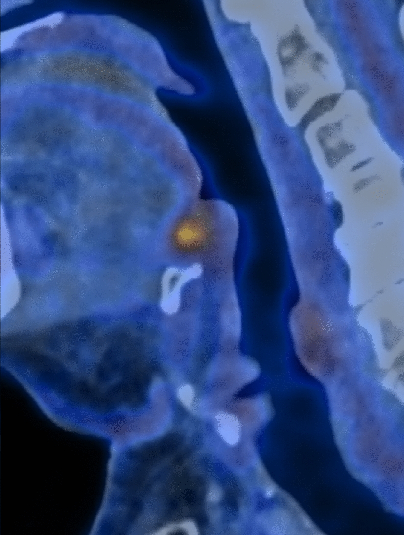
Outcome MeasuresPrimary OutcomeThe primary outcomes of this study were aimed to quantitatively assess the difference in dermal thickness, collagen level, Young's modulus of elasticity, viscoelasticity and retraction time of the surgical scar in the neck and adjacent normal tissue in patients of both groups using the ultrasound probe (USGP) and elasticity probe (EP) of DLC at 1st and 4th postoperative week.
Secondary OutcomeThe secondary outcome of this study was to qualitatively assess the patient and observer perspective of the surgical scar in the neck using POSAS on 1st and 4th postoperative week.
ProcedureDemographic data with respect to age, gender, site of primary tumor, histopathological description, clinical TNM staging, type of neck dissection and incision employed were collected. The patients included in the study were blinded and randomly divided into 2 groups. The study group Gr-I included patients for whom 2 ml of topical hemocoagulase (Botroclot®, Juggat Pharma) was applied at the site of the neck incision after completion of subcutaneous layer suturing. Each milliliter of Botroclot contains 0.2 CU of hemocoagulase, along with chlorhexidine gluconate solution IP 0.1% v/v as a preservative, and water for injection IP q.s. The solution was allowed to soak for 8–10 minutes before proceeding to skin layer closure. The control group Gr-II included patients for whom the closure of the neck incision proceeded without any intervention. In both the groups, to avoid bias, 3–0 vicryl was used for subcutaneous layer closure and 3–0 polypropylene was used for skin layer closure. Similarly, in both groups, simple buried suturing technique was used for subcutaneous layer closure and vertical mattress suturing technique was used for skin layer closure. All the patients had an uneventful postoperative recovery and were shifted to the Intensive Care Unit immediately after the surgery where they were monitored for 1–2 days before being shifted to the ward. Postoperative antibiotic therapy consisted of intravenous infusion of 1.5 g Magnus Forte and 80 mg Gentamicin, twice daily along with 500 mg Metronidazole, thrice daily. All the patients remained as in-patients for a minimum of 7 days after the surgery and conventional wound dressings with normal saline, betadine solution, betadine ointment and gauze were given every day. On discharge from the hospital, patients were instructed to maintain clean the surgical site and topical application of betadine ointment over the surgical site in the neck was advised once daily for both groups. The sutures were removed alternatively on postoperative day 7 and day 10.
The USGP measured dermal thickness and collagen levels, while EP assessed Young's modulus, viscoelasticity, and retraction time of the scar. Each measurement was taken three times on both scar and contralateral normal tissue for accuracy (Figs. 2 and 3). The jelly-coated USGP was held perpendicular to the skin, with thickness displayed on the monitor. EP, with a 10 mm suction aperture, was set to 'normal skin' mode at 3 mbar pressure for standardization. Each measurement involved three elevation/retraction cycles, with average values used for analysis. While the elasticity measurements were automatically averaged, dermal thickness and collagen levels required manual calculation.
Fig. 2
Quantitative assessment of scar characteristics using ultrasound probe of DermaLab Combo at scar and contralateral normal site
Fig. 3
Quantitative assessment of scar characteristics using elasticity probe of DermaLab Combo at scar site and contralateral normal site
Measurements were taken at two time points by a blinded assessor: 1 week and 4 weeks postoperatively which allowed for assessment of both early and later stages of scar healing. For each patient, every parameter in the primary outcome was measured at the site of neck incision and contralateral normal neck skin after which the difference between them was calculated thereby providing a quantitative measure of how the healing scar differed from normal skin. By using each patient's own contralateral normal site as a reference, this method effectively controls for individual variability, thereby reducing potential bias and the impact of confounding factors. A smaller difference between the scar and normal skin was considered indicative of a better outcome, whereas a larger difference was viewed as a poorer outcome (Fig. 4). At the same visit, POSAS was recorded by the blinded patient and the blinded assessor. Thus the precise, quantitative measurements from the DLC device were combined with the qualitative assessments from the POSAS.
Fig. 4
Flowchart depicting the methodology
Statistical AnalysisThe data were entered in Microsoft Excel Spreadsheet and analyzed using IBM SPSS (Version 20.0, Armonk, NY, USA: IBM SPSS for Windows). Normality of data was assessed using Kolmogorov–Smirnov test. The demographic data was analyzed using descriptive statistics while mean and standard deviation were used to summarize the quantitative data. Frequency and percentage were used to summarize qualitative and categorical data. Independent samples t-test was used for intergroup comparison of the primary outcomes while Mann–Whitney U test was used for intergroup comparison of the POSAS scores. Effect sizes were quantified using Cohen's d, and 95% confidence intervals were calculated to assess the precision of the estimated effects. Throughout the study, a p value of < 0.05 was considered as a statistically significant difference.
Comments (0)