A surrogate measure of LASr obtained through measurements of mitral annular displacement and LAV (MAPSE/LAV) provided similar diagnostic information as its strain-based counterpart. Since LA volumes are acquired in a standard echocardiographic examination, this measure could easily be obtained by adding MAPSE measurement to the examination. Given the prognostic value of MAPSE [14, 15, 25], in itself, the addition of this measurement thus adds both diagnostic and prognostic value to the echocardiographic examination. Both MAPSE/LAV and LASr were lower in patients with elevated left ventricular filling pressures as determined both by the ASE/EACVI algorithms, a quantitative echocardiographic PAWP estimation, and invasively measured PAWP. LASr has previously been shown to provide diagnostic information regarding left ventricular filling pressures [4, 8, 26, 27]. For example, when LASr was incorporated into the ASE/EACVI algorithm, both feasibility and accuracy in detection of elevated filling pressures were improved, and LASr could effectively be used as replacement when either E/e’ or tricuspid regurgitation velocity were unavailable [8]. Although, LAV constitutes an important part of LASr, LASr improves prediction of elevated PAWP beyond that obtained from LAV indexed-to-body surface area [28]. In this study, MAPSE/LAV and LASr showed acceptable discrimination of patients with normal or elevated PAWP (AUC ~ 0.75). However, MAPSE/LAV resulted in only a small increase in diagnostic accuracy compared to its constituents alone (MAPSE and LAV, ΔAUC + 0.04 and + 0.03, respectively).
Both MAPSE/LAV and LASr were only moderately correlated with PAWP and neither of these measures can be used as standalone noninvasive measures of PAWP. A stronger correlation with PAWP could be observed for MAPSE/LAV in patients with reduced LVEF, in comparison to those with preserved LVEF. Although a similar pattern has been reported for LASr previously [13], this was not evident for LASr in this study. MAPSE/LAV explicitly incorporates systolic function (MAPSE), and higher PAWP could be expected with worse systolic function. However, the sample size was small and the cause for the stronger correlation for MAPSE/LAV among patients with reduced LVEF cannot be readily explored.
Interobserver variability was substantial for both MAPSE/LAV and LASr, although higher for MAPSE/LAV. Possibly, this difference was caused by the semi-automatic procedure used for LA strain, in contrast to MAPSE/LAV, which likely lowers variability. However, coefficients of variation were similar for LAV and LASr, indicating differences in LA delineation between the two observers, and the addition of another measure (MAPSE) increased variability further. The high variability observed in this study contrasts previous reports of repeatability of LA strain measures, which have suggested low interobserver variability [29].
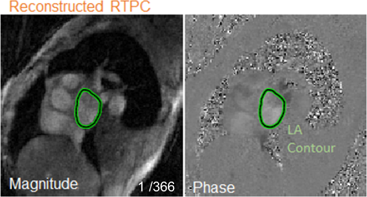
This can be explained by the use of single-beat measurements used in this study, suggesting that averaging beats over multiple cycles are needed to obtain adequate reproducibility. Also, we actively refrained from synchronizing measurement strategies between observers, to mirror clinical reality. For both MAPSE/LAV and LASr there was a systematic bias between observers, possibly caused by the different contour delineation of the LA or differences in placement of the annular detachments for both measures. The high variability of MAPSE/LAV, however, clearly limits clinical application. This finding needs further evaluation in future studies. Compared to real-time M-mode images, the temporal resolution of reconstructed M-mode images is limited but has been shown to provide accurate information for measurements of anatomical dimensions [30]. Still, variability could possibly be reduced by using real-time M-mode acquisitions instead of reconstructed M-mode images.
LASr is determined, to a large extent, by atrioventricular plane displacement and LA dimensions. This has been observed previously [12, 13]. Mălăescu showed excellent correlations between LV and LA strain curves across different cardiovascular pathologies. Similarly, Inoue, et al., showed LV global strain and LAV to be important determinants of LASr [13]. LV global strain describes the systolic longitudinal shortening of the LV in relation to the length of LV cavity. Thus, the main difference between MAPSE and LV global strain in relation to their association with LA strain is that LV global strain also incorporates information of LV dimensions, which is likely to be redundant. Despite this, we did not find a stronger relation between MAPSE and LASr compared to between LV global strain and LASr. Possibly because of a closer relationship of measurement techniques between the two strain modalities compared to the M-mode based measurement, or due to measurement variability.
Both MAPSE/LAV and LASr were lower in the cohort of patients with moderate aortic stenosis, compared to the cohort of patients who underwent clinical RHC. This could be explained by older patients in the aortic stenosis cohort, a higher proportion of male patients, or differences in the degree of LV disease [20, 31]. Also, reduced longitudinal function commonly occurs in patients with significant aortic stenosis [32], resulting in lower MAPSE/LAV and LASr.
Limitations
The cohort of patients with aortic stenosis did not include individuals with suspected heart failure, representing a limitation. However, the primary objective of analysing this cohort was to describe the correlation between MAPSE/LAV and LASr, and to determine reproducibility. Nonetheless, the potential clinical utility of MAPSE/LAV requires evaluation in settings that encompass patients with suspected or confirmed heart failure.
Since zero strain is set at end-diastole, i.e. minimum LAV, a stronger correlation with LASr could be achieved by dividing MAPSE with the minimum LAV instead of the maximum LAV. Using cardiovascular imaging, minimum LAV showed a stronger correlation with LASr than maximum LAV [33]. Pragmatically, we included the maximum LAV in the MAPSE/LAV calculation since this value is included in standard echocardiographic reports and protocols. Thus, only MAPSE would be required to be added to obtain a reasonable surrogate of LASr, if needed. Future studies could be performed to demonstrate a possibly improved diagnostic accuracy of MAPSE/minimum LAV instead of the measures included here.
There is a lack of robust non-invasive reference parameters for PAWP. In the non-invasive cohort, MAPSE/LAV and LASr was therefore described both in relation to the ASE/EACVI algorithm and to ePAWP. In a previous paper, with larger sample size than the current study, ePAWP provided better diagnostic accuracy and improved prognostic value compared to the ASE/EACVI algorithm [22]. Given the limited precision of ePAWP, however, the results from this study regarding the relation between MAPSE/LAV, and LASr, and PAWP must be interpreted with caution. Nonetheless, the pattern was similar both cohorts. Although such patients were part of the invasively examined study cohort, those patients constitute a mixed population with advanced disease unlikely to be representative of the typical population in which non-invasive determination of left ventricular filling pressures is sought. Nevertheless, our findings lend support to the hypothesis that comparable diagnostic information, as derived from strain-based LA measures, can be achieved without the use of advanced software.
Patients with atrial fibrillation were excluded from this study even though atrial fibrillation is common in patients with heart failure. The main aim of this study was to determine whether simple anatomical measures could provide similar diagnostic information as strain-based techniques, and atrial fibrillation could possibly make such comparisons more difficult, since this arrythmia would introduce further variability to both echocardiographic and invasive measures [34]. The findings of this study cannot be applied to patients with atrial fibrillation.
This study is further limited by small sample sizes. This is reflected, for example, in the wide confidence intervals for the AUC for both MAPSE/LAV and LASr, ranging from poor to excellent discrimination. These results, however, did not provide any evidence of improved diagnostic accuracy of LASr compared to the simple measures included in MAPSE/LAV.
Inter-reader variability was assessed after re-analysis of images from the same set of images. Although both readers could freely choose which 4- and 2-chamber views to use, it should be noted that variability can increase further if new acquisitions are performed [17]. Although inter-reader variability was higher in this study than previously reported, both LASr and MAPSE/LAV should be evaluated regarding their potential in detecting change in PAWP, since such information is affected by variability in repeated measurements. Such an analysis would require sequential invasive PAWP measurements, which was not available in this study.
MAPSE is affected by regional LV dysfunction, mitral annular calcification, conduction disorders, or small areas of fibrosis [35], which is a limitation both to the results of this study and to a general use of MAPSE/LAV. However, given the importance of the longitudinal excursion of the mitral annular plane to LASr, even LASr can be affected by these conditions, and future studies of both parameters in these specific conditions may be needed. Also, including lateral, or the septal, MAPSE measurement only in the calculation of MAPSE/LAV, did not show a stronger correlation with PAWP.
LA volumes were lower in the invasive cohort compared to the non-invasively examined cohort. This can be explained by differences in patient populations, e.g. the invasive cohort consisted of patients with a clinical referral for RHC including pulmonary hypertension not due to LV disease, while the non-invasive cohort consisted of patients with moderate aortic stenosis and thus, likely, a higher degree of LV and LA disease. Although this makes generalization of absolute values for both LASr and MAPSE/LAV difficult, it does not affect the head-to-head comparison of LASr to MAPSE/LAV and their respective association with LV filling pressures.
The above-mentioned limitations indicate a need for further studies in independent cohortsbefore clinical implementation. The results, however, can be considered as proof-of-concept of similar diagnostic value of MAPSE/LAV and LASr, although both methods are limited as stand-alone measures of LV filling pressures.


Comments (0)