
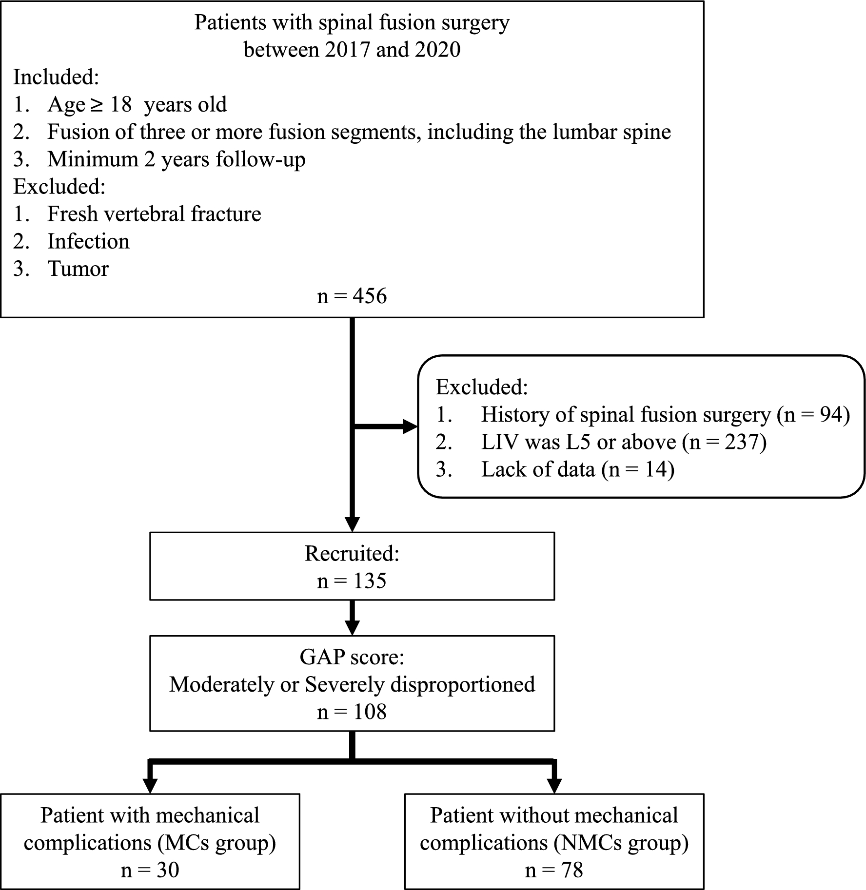
記住我
The most important finding of the study is that endplate weakening significantly reduced endplate resistance to subsidence of the endplates, which may promote cage migration or subsidence. The effect was even more pronounced when a uPLIF cage was used (70% (median)–36.6%) compared to TLIF cage (83.1% (median)–15%).
Lumbar interbody fusion techniques have evolved rapidly over the past few decades, leading to an expansion of indications [17]. Among these, PLIF and TLIF are the most commonly used techniques [18, 19]. They provide solid fusion and allow for neural decompression, provide an additional fusion interface with a higher fusion rate than bone graft alone [20], restore the intervertebral height and help restore alignment [21]. From a biomechanical point of view, the interbody fusion device is designed to support the anterior column and transmit compressive forces [22, 23]. However, failure to achieve bony fusion may result in cage migration or subsidence. Cage subsidence rates reported in the literature range from 0 to 65% [21, 24,25,26,27], with implant contact area, endplate condition, and bone mineral density appearing to play an important role [8, 28].
The contact area between the cage and the endplate depends on the morphology of the endplate (i.e., highly concave or irregularly shaped endplates reduce the contact area), and on the shape and size of the cage. The smaller the surface contact area, the higher the stress on the endplate [29,30,31]. Banana-shaped and anatomically contoured cages increase the contact surface compared to rectangular cages and therefore theoretically have a lower tendency to subside [11]. This may explain why the failure load of the uPLIF was slightly lower than that of the TLIF in the present study. Due to the concavity of the endplates, the PLIF cage often only came into contact with the bone at the edges, causing force peaks in these areas and resulting in fractures.
Violation of the endplates during cage bed preparation appears to have a relevant biomechanical effect: the exposure of cancellous bone reduced the failure force by 15% for the TLIF and 37% for the PLIF, respectively. To compare the distributions of forces measured for the intact and the weakened endplates, and to better assess the potential clinical implications of the experimental results, probability density functions were fitted to the failure force results for each cage configuration (uPLIF, bPLIF, and TLIF; Fig. 5).
Fig. 5
Illustration of the probability density functions of the intact and weakened endplate for three different cage configurations. For all three cage configurations, the cage loads in the upright position and during physical activity (such as lifting an object) are evident, as are the areas under the probability density function curves: the probability of endplate fracture is higher in the weakened state than in the intact state for all configurations
Preload and percentage of external load transfer through the cage were obtained from previously published biomechanical experiments involving axially compressed spinal segments with load cell instrumented vertebral cages [32]. Specifically, an average preload of up to 328N was measured (uPLIF 224N; bPLIF 328N; TLIF 317N), and up to 50% (uPLIF 40%; bPLIF 50%; TLIF 44%) of the external compressive load was transmitted through the cage to the ventral column of the spine, thus affecting the endplates. It was therefore possible to determine the likely loading of each cage configuration during upright standing (about 1000N acting on the spine) and during more vigorous daily activities (about 3000N on the spine) and to link it to the cage-specific failure forces of intact and weakened endplates [33, 34]. During upright standing, likely 624N, 828N, and 757N are going through the uPLIF, the bPLIF, and the TLIF, respectively. During more vigorous activities, the uPLIF, bPLIF and TLIF are subjected to 1424N, 1828N, and 1637N, respectively. Consequently, 12% (bPLIF) to 23% (TLIF) of intact endplates would fail during upright standing, whereas up to 39% (uPLIF) of the weakened endplates could fail under such loads (Fig. 5).
Considering the subsidence rates from clinical studies, the mean incidence of subsidence with bPLIF is 15.8% (10–65.1%) [21, 25, 35, 36], whereas the mean incidence of subsidence with TLIF is approximately 25.3% (0–51.2%) [8, 9, 21, 27, 37,38,39,40,41,42,43,44]. This clinically reported difference between TLIF and bPLIF subsidence is consistent with the experimentally determined distributions of endplate failure: both indicate a higher propability of failure with TLIF than with bPLIF (Fig. 5). In addition, the average clinically observed subsidence is within the range of endplates predicted to fail during standing (12–24% for bPLIF and 23–35% TLIF cages). This suggests that subsidence occurring in the early postoperative period may be induced by exposure to comparatively low spinal loads experienced during upright posture or non-impact activities.
Biomechanically, bone quality correlated positively with the absolute resistance force of the endplate: the higher the mean intracorporal HU values, the more axial compression force had to be applied until a fracture occurred in both the intact and weakened states. An increase of 10 HU in the mean intracorporal HU of an intact vertebral body resulted in an increase of 100N in the failure force (slope of the regression lines is approximately 10, Fig. 4). While this appears to be unchanged in a weakened vertebral body with the use of a TLIF cage, endplate weakening appears to be more detrimental with the use of a PLIF cage (Fig. 4). Reasons for this phenomenon could be the different positioning area for TLIF and PLIF, with a more “peripheral” placement of TLIF and the slightly larger contact area of TLIF.
This biomechanical understanding can guide clinical practice: in osteoporotic spines, where there is a higher risk of screw loosening and cage subsidence [9], bone density should be optimized preoperatively whenever possible [45], cages with large footprints should be preferred over small cages, and great care should be taken to preserve the endplates.
Some authors claim that neither fusion rates nor clinical outcomes are affected by cage subsidence [46], however, many of the advantages that come with a cage, as mentioned above, are lost with subsidence.
Overall, we believe that endplate preparation should be performed with caution, as cortical bone compromise is associated with significant loss of resistance to axial compression (Fig. 6). In addition, ventral force transmission is further compromised by the previous disc dissection required for cage insertion [47]. This results in increased stress at the screw-bone interface. Park et al. [9] demonstrated a correlation between cage positioning, endplate injury, single cage use, and cage migration. Cage migration did not result in subsidence in all cases. However, the rate of non-fusion and screw loosening was significantly higher in patients with cage subsidence. Understanding these biomechanical relationships can be used to improve clinical decision-making.
Fig. 6
Illustrative visualization of lumbar vertebral body segments without and with a cage, with cage subsidence and after successful bony fusion. Compared to the intervertebral disc, an inserted cage increases the stiffness of the construct and thus the force transmission to the underlying screws. With bony fusion, the stiffness is further increased. If cage subsidence is present, the cage cannot transmit interbody forces and the stiffness of the anterior column decreases
This in vitro biomechanical study has several limitations. Firstly, an isolated axial compression force was applied to the vertebral body. This is a simplified model that does not represent the complex movement of the spine. However, we know that the axial compression force is the main loading direction for all loading cases, therefore it is an appropriate study scenario. Secondly, the endplates were weakened with a shaver until cancellous bone was visible. It is possible that in some cases a little more substance was removed than in others. However, since the endplate in the cage area was completely removed during shaving, the effect of a little more or less depth should be negligible compared to the effect of a missing endplate. Thirdly, the degrees of freedom in our setup always allowed the cage to align perfectly with the endplate. In vivo, it is possible that a cage is loaded more unilaterally and collapses more quickly. However, in another study, it was found that under high loads, the cage is mostly loaded relatively evenly (anterior vs posterior load distribution) [32]. Fourthly, HU are widely used to assess bone quality, but the absolute values vary between scanners and they are notoriously difficult to compare between studies. However, within this study only relative comparisons between the samples were drawn, which are justifiable from our point of view, as the imaging parameters were the same. Lastly, we have tried to position the cages in the same area of the endplate, but small variations may have been unavoidable.
留言 (0)