Study design and eligibility criteria
Moderate and late preterm infants (32 + 0 to 36 + 6 weeks gestation) with moderate to severe RDS who were admitted to the NICU of AUMH between February, 2023, and September, 2023, participated in this randomized clinical trial. Neonates who needed intubation in resuscitation or had multiple major anomalies were excluded from the study.
This study was performed in accordance with the ethical standards of the institutional research committee and with the 1964 Helsinki Declaration and its later amendments. This trial was approved by the ethics committee of Alexandria University on 19/11/2020 with approval number 0201417, IRB no 00012098, and FWA no. 00018699.
Informed consent was obtained from patients’ guardians upon NICU admission.
Randomization and blinding
Randomization into the two groups was done with ratio 1:1 using random permuted blocks of 4 and 6 size prepared by a person not involved in the study. Allocation into the two groups was done using serially numbered opaque and sealed envelopes by neonatology residents. Patients allocated to one group have remained on the assigned intervention up to the final weaning with no permitted crossover. Blinding was not applicable due to nature of the interventions.
Intervention
At delivery room and during transfer to NICU, all enrolled infants were immediately supported with continuous positive airway pressure (pressure: 5–6 cm H2O) using the Neopuff infant resuscitator. Upon NICU admission, they were randomly assigned to undergo treatment with either NHFV (n = 50) or NCPAP (n = 50) as primary modes of NIV. The severity of RDS was determined based on clinico-radiological data and/or FIO2 requirement on admission FIO2 ≥ 40 and ≥ 50 for moderate and severe RDS, respectively.
NCPAP and NHFOV were provided by the SLE5000/6000 ventilators using single naso-pharyngeal tubes as interfaces that were inserted 4 cm beyond the naris. Size was chosen according to the nares diameter as the best fitting one to avoid nasal trauma. Naso-gastric tubes were inserted through the other nostril to reduce the leak.
Infants assigned to the NHFOV-group were subjected to initial settings of MAP of 6 cm H2O (equal to that of the NCPAP-group), a frequency of 10 Hz (range 8–12), an amplitude that was initially set at the double value of MAP or until the chest was seen to be “wiggling,” and an inspiratory time of 50% (1:1). Subsequent adjustments of MAPs have followed a physiology-based approach (open lung strategy), using oxygenation guided lung recruitment maneuver similar to that used in invasive HFOV [8]. Starting from the initially set MAP value, MAP was increased in a stepwise fashion by 2 cmH2O every 5 min to identify the value of the opening MAP at which FiO2 can be reduced to ≤ 30% with maintained SpO2 within the target range (90–94% or up to 96% if evidence of pulmonary hypertension). MAP was gradually lowered while maintaining the FiO2 fixed until oxygenation began to decline (closing MAP was attained). The final MAP was then set at 2 cmH2O above the closing MAP. Patients were kept on these final optimal MAPs provided that the targeted SPO2 was maintained using minimal FIO2; otherwise, these pressures were escalated again toward the opening MAPs. Values and times of the maximal MAPs were recorded for each patient throughout the period of NIV to identify the boundaries of the ventilatory strategy. The pressure amplitude was adjusted according to PaCO2 values.
Infants assigned to the NCPAP group were initially supported with a starting pressure of 6 cm H2O with subsequent increments up to a maximum level of 8 cm H2O for safety reasons [9].
NHFOV or NCPAP failure criteria (criteria for intubation) were [10] hypoxemia (persistent FiO2 > 0.55 with PaO2 < 50 mmHg from an arterial blood gas sample), severe respiratory acidosis (PaCO2 > 65 mmHg with pH < 7.20), severe apnea and bradycardia (defined as recurrent apnea with > 3 episodes per hour associated with heart rate < 100/min or a single episode of apnea that requires bag and mask ventilation), pulmonary hemorrhage, and cardiopulmonary arrest needing chest compression.
Weaning from NIV was accepted whenever patients exhibited minimal or no RDS, NHFOV or NCPAP pressure of 6 cm H2O, and FiO2 of ≤ 30% to achieve the target SpO2.
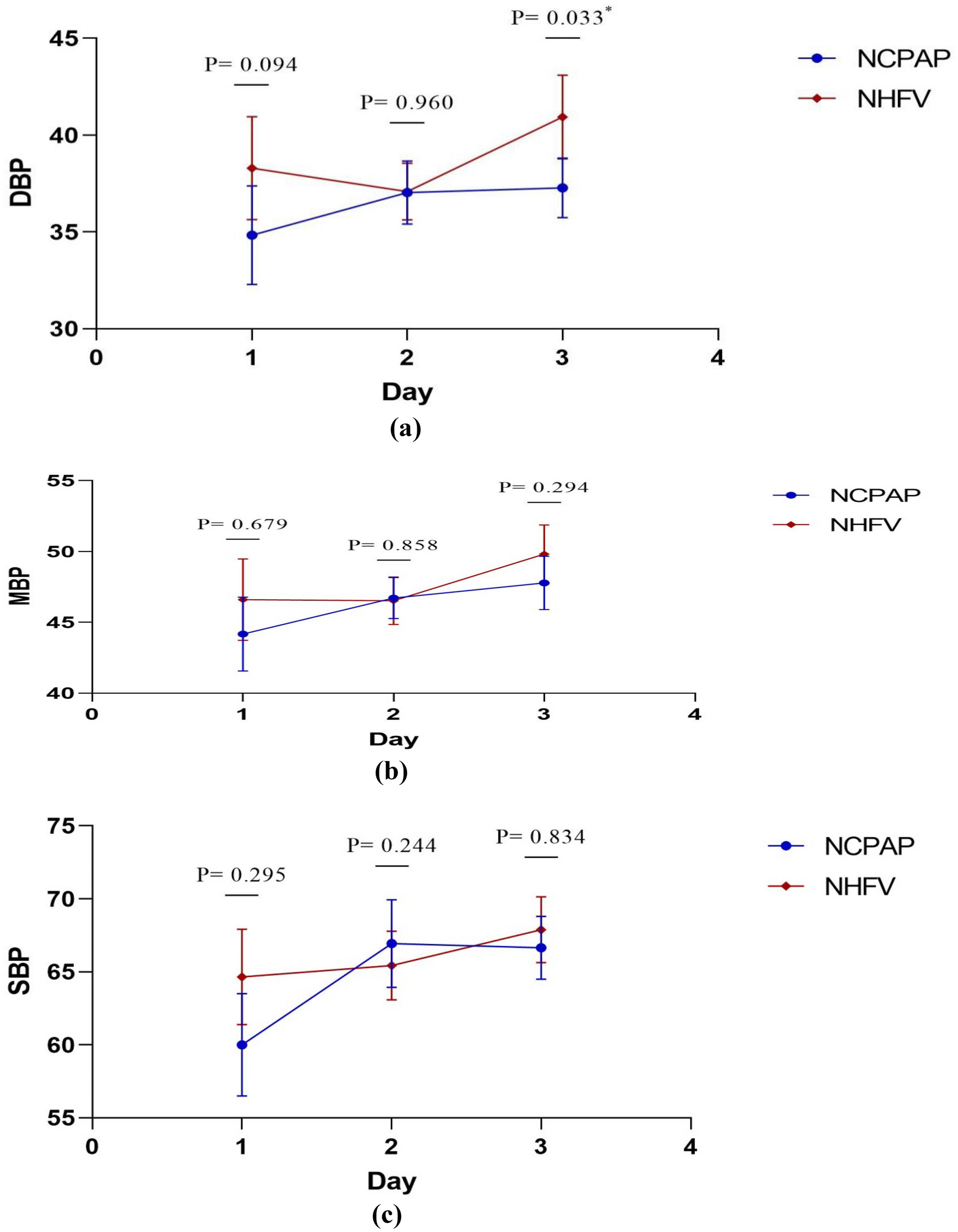
Functional echocardiography (FE) was performed using GE Vivid iq premium (probe: GE 12S-RS) with a frequency range of 5–11 MHz. Assessed parameters included LVO, EF, FS, RVO, TAPSE, and SVC blood flow [11, 12]. Echocardiography was performed within the first 24 h to all participants while they were on NIV modes (NCPAP or NHFOV) at the maximally recorded MAPs. Timing of the study in the NHFOV group was guided by the data obtained during the alveolar recruitment process, at times whenever the maximal MAPs were reached (MAPs within 2 cmH2O from the opening pressure). In patients who were maintained on their optimal MAPs for at least 4 h, the study was performed before any reductions in MAPs were done.
Cranial ultrasound using GE 8C-RS probe with a frequency range of 3.5–10 MHz was performed to detect IVH and measure anterior cerebral artery (ACA) doppler velocities in the first 72 h of life. The international guidelines of point of care ultrasound in neonates were followed while evaluating patients using the bedside ultrasound [13].
Primary outcomes
The primary outcome parameter was LVO in both study groups assessed while patients were on the maximally recorded MAPs.
Secondary outcome
Other FE-measured-hemodynamic parameters within the first 24 h during applying NIV, duration of NIV and need of IMV in the first 72 h, and short-term complications such as air leak syndromes, pulmonary hemorrhage, nasal trauma, IVH, PVL, or NEC were the secondary outcome parameters.
Statistical analysis
For sample size planning, we used SPSS program version 20. A minimal total sample size of (96) moderate and late preterm infants (48 per group) was needed to detect an assumed difference of 35 ml/kg/min in the mean LVO between both groups to study their hemodynamic effects with assumed group SD of (10, 30), respectively, using a two-sided independent t test. The statistical significance level was set at a type I error of 0.05 and the study statistical power of 80%.
Data analysis was made by using IBM SPSS software package version 20.0. The Kolmogorov–Smirnov test was used to verify the normality of distribution. Qualitative data were described using number and percent. Quantitative data were described using range (minimum and maximum), mean, and standard deviation, median, and interquartile range (IQR). Significance of the obtained results was judged at 5% level.
Student t-test Monte Carlo test, χ2: chi-square test, and Fisher exact test were used for comparison between the two groups regarding different variables. Kaplan–Meier survival plot was used representing time course of NCPAP and NHFOV failure.


留言 (0)