This study evaluated MRI morphology of LNs in ASCC patients with at least one PET-CT positive LN before treatment and found that both PET-CT-positive and negative LNs were predominantly characterized as homogeneous with nodal signal similar to the primary tumor. Most LNs, both PET-CT-positive and negative, showed round and regular morphology. As previous research on nodal disease in ASCC is scarce, these observations are novel and provide a foundation for future research on imaging criteria.
A substantial amount of PET-CT-negative LNs was additionally found on MRI (n = 78), which might indicate that not all LNMs in ASCC can be identified with PET-CT alone. The median size of PET-CT-negative LNs on MRI was statistically smaller than for PET-CT-positive nodes, despite sharing similar morphological characteristics. The smaller sizes of PET-CT-negative LNs could, at least partially, explain why metabolic uptake above the cut-off (DS ≥ 3) cannot be detected, indicating a potential threshold of size for LN detection on PET-CT. Hence, neither size nor hypermetabolism alone (defined as DS ≥ 3) seems to be reliable predictors of LNM in ASCC. According to the European Society of Gastrointestinal and Abdominal Radiology (ESGAR) consensus guidelines for nodal staging in rectal cancer, criteria for malignant nodes include LNs with a short-axis diameter of 5–8 mm and at least two suspicious morphological characteristics [20]. We therefore separated all PET-CT-positive LNs in size categories of 5–8 mm and ≥ 9 mm, respectively. The data presented in Table A1, Appendix, demonstrate that smaller PET-CT-positive LNs measuring 5–8 mm (n = 66) exhibit MRI morphological characteristics suggestive of pathology. As PET-CT could potentially miss smaller pathological LNs with insufficient 18F-FDG-uptake, multimodal imaging is of importance when assessing LNs in ASCC, where suspicious MRI morphological criteria could be applied for smaller LNs 5–8 mm similarly to the ESGAR guidelines for rectal cancer.
To our knowledge, this is the first study describing MRI morphological characteristics independent of PET metabolism in the setting of ASCC. The patient cohort was large considering the rarity of the disease and consisted of patients treated consecutively at a single institution with routine use of PET-CT. Nodal staging guidelines for primary rectal cancer may have been applied for nodal evaluation in ASCC, which in our opinion should not be done, considering the different anatomical and histological origins of rectal adenocarcinomas compared to ASCC. Morphological criteria such as round shape, irregular contour, loss of fatty hilum, and mixed/heterogeneous nodal signal intensity have all been proposed for detection of LNMs in rectal cancer before treatment [20,21,22]. In contrast to previous morphological findings for LNM in rectal cancer, the majority of all PET-CT-positive LNs in our ASCC patient cohort were judged to have a homogeneous nodal signal and a regular border, with similar characteristics seen in PET-CT-negative LNs identified on MRI. These results support that LNMs in the context of ASCC differ at least partly from LNMs seen in rectal adenocarcinomas, why the same criteria cannot be applied.
The study methodology poses some limitations. Firstly, and most importantly, nodal evaluations were of subjective nature without the possibility to compare to a reference standard, potentially introducing observer bias. Histopathology commonly serves as the gold standard reference in other diseases where surgery is the primary treatment. However, in ASCC, this reference standard is not obtainable since CRT is the standard treatment given and primary surgery is rare. In our cohort, none of the LNs were biopsied before CRT and locoregional nodal recurrence was rare after CRT, with confirmed histopathological metastasis in only two out of four patients who underwent either biopsy or surgical resection. For these reasons, the actual presence of metastases within the LNs detected with PET-CT and MRI remains unknown. Characteristics of PET-CT-positive and PET-CT-negative LNs were therefore presented descriptively, without the use of statistical analyses for comparison of LNs. However, only patients with at least one PET-positive LN were selected for inclusion in this study, which might increase the likelihood that additional PET-CT-negative LNs found on MRI are actual LNM containing micrometastases not reaching the cut-off value for hypermetabolism on PET-CT due to smaller size.
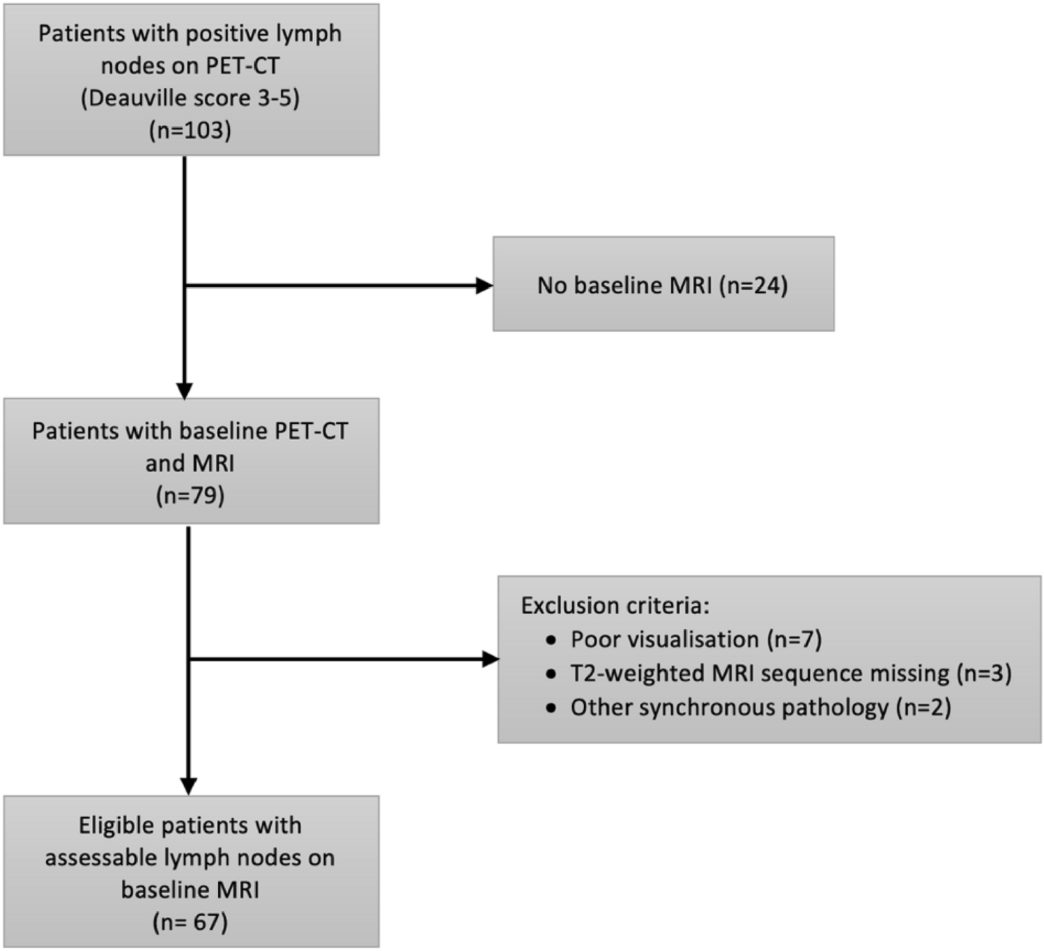
Secondly, different generations of PET-CT cameras and MRI scanners were used throughout the study period and patients were excluded if a baseline MRI scan was missing or if the image visualization was poor, which may have introduced selection bias. For this reason, patient characteristics of excluded patients were presented, and no major differences were noted.
Additionally, interobserver agreement for categorical morphological characteristics was poor to moderate, which is in accordance with previous results of interobserver agreement of nodal evaluation on MRI in ASCC [23]. This uncertainty underscores the importance of future studies to identify reliable nodal features. Possible contributors to the interobserver variability include the subjectivity involved in visual evaluation of nodal characteristics and varying experience of the reviewers. However, the overall distribution of morphological characteristics among PET-CT-positive LNs was relatively similar between the two reviewers. For nodal size, the random variation in size difference was relatively constant around the mean nodal size difference, and neither of the reviewers seemed to systematically under- or overestimate nodal size.
Another study limitation was that the anatomical location and symmetrical distribution of LNs were not considered when presenting nodal characteristics, as the amount of LNs assessed was deemed too low to find significant morphological differences in various pelvic locations or asymmetry between lymph nodes on different sides. The current patient cohort was used in a previous study, showing that PET-CT-positive LNs were most prevalent in the inguinal followed by the perirectal areas [18]. It is possible that inguinal LNMs differ morphologically from mesorectal LNMs. Even if subgroup analysis on location was not performed, we did observe that inguinal LNs tended to be necrotic to a greater extent compared to perirectal nodes. Necrosis was almost exclusively seen in PET-CT-positive LNs and is seemingly an important feature of LNMs in ASCC.
Importantly, we described MRI morphological characteristics of PET-CT-positive and negative regional LNs in ASCC patients (Tables 2 and 3), but their value for radiation therapy planning remains untested and needs to be examined in prospective clinical trials.

留言 (0)