There is no gold standard for the diagnosis of CD, which requires a combination of clinical manifestations, imaging, endoscopy and pathology. There are a variety of reference indicators for identifying whether it is in the active period, each of which has its own advantages and disadvantages. Dual-energy CT has 2 independent bulb-detector systems that can simultaneously obtain the attenuation coefficients of substances at different radiation energy levels, which not only enables the acquisition of conventional CT enterography findings, including increased bowel wall thickness, bowel wall hyperenhancement, comb sign and mesenteric fat infiltration, but also provides several parameters for diagnosis using the corresponding postprocessing workstations [12], including iodine content, virtual single-energy CT values, energy-spectrum curve slope K values, FF and DEI. DECTE scanning results in a shorter scanning time and reduced radiation dose relative to conventional CT scanning [13, 14]. In addition, direct measurement of iodine concentration using DECTE may be less dependent on scan parameters (manufacturer, energy, contrast material time) than conventional Hounsfield unit–based measurements [15]. This is important because patients often need to be imaged for years in different hospitals using different scanners.
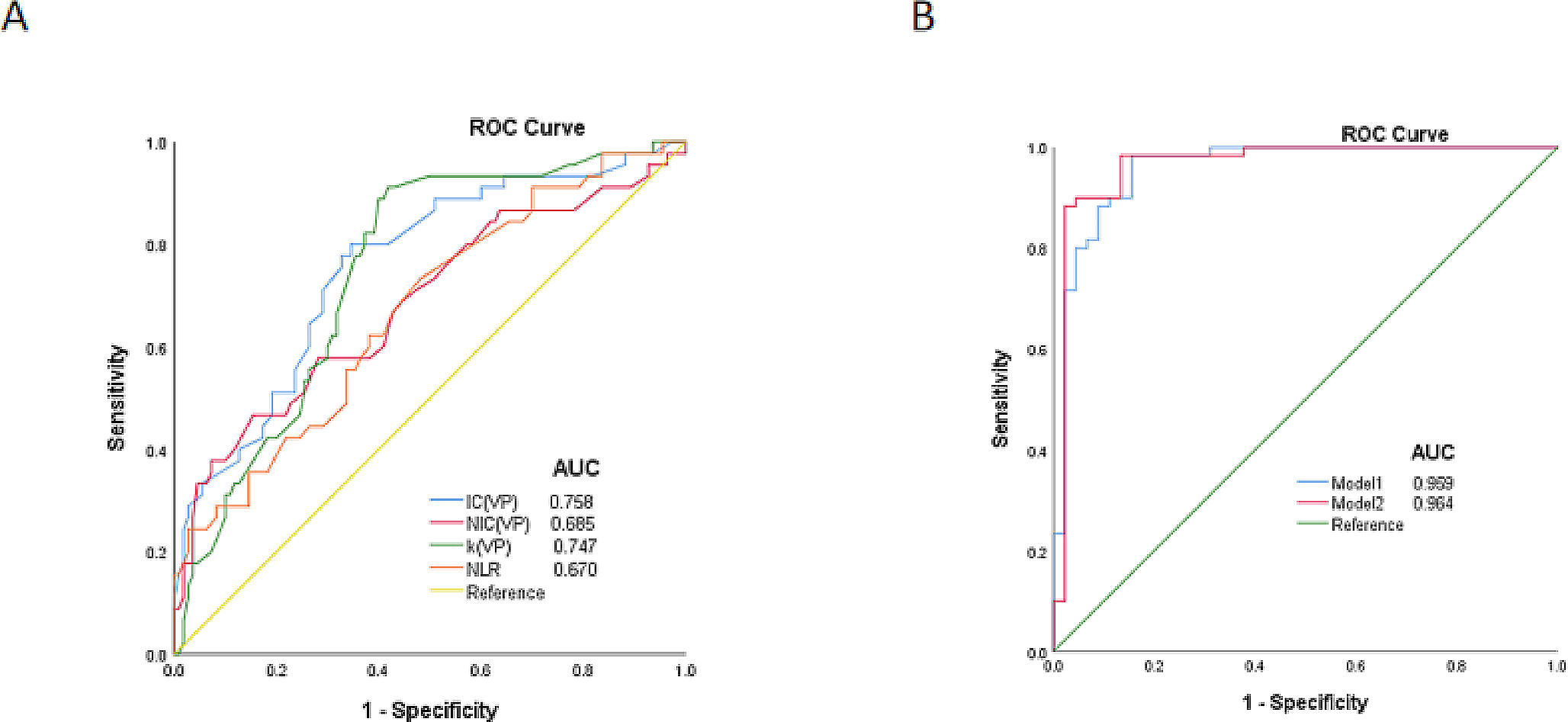
Previous studies [5, 16] have demonstrated that dual-energy iodine content is a good surrogate marker for perfusion in various body imaging application. NIC was used in this study to minimize the effects of a variety of confounders, such as individual differences and contrast flow rates. Previous research [17] has suggested a strong correlation between iodine content and the CDAI or endoscopy findings. Xiao et al. [18] assessed iodine contents in the arterial and venous phases using DECTE to differentiate between active and inactive Crohn’s disease. Using the CDAI as a reference standard, at arterial phase iodine concentrations greater than 2.55 mg/mL, the reported specificity and sensitivity were 100% and 61%, respectively. In this study, using pathology as the reference standard, the NIC of CD patients significantly differed between the active and inactive phases. This was because of the inflammatory cell infiltration in the mucosa and the increased blood supply; a more severe inflammatory reaction is associated with a more obvious the blood supply increase and thus a higher iodine content [18]. This study suggests that the AUC of NIC in the arterial phase is the largest at 0.908, and when the NIC is greater than 0.245, the sensitivity of NIC for diagnosing CD intestinal segments in the active phase is 0.833, and the specificity is 0.800.
In addition, the energy-spectrum curve slope K and DEI were also significantly different between active and inactive bowel segments, and the energy-spectrum curve slope K and DEI of active bowel segments were higher than those of inactive bowel segments. Infact, both the NIC and the energy-spectrum curve slope K can be considered direct or indirect expressions of the iodine content within the ROI and thus should not be significantly different from each other [19]. In accordance with the use to distinguish active from inactive CD intestinal segments, Xiao et al. [18] found that energy-spectrum curve slope K could effectively distinguish normal from active intestinal segments in CD patients. DEI represents the difference between the X-ray attenuation coefficients of a substance at two different energies. Different substances have different, fixed DEIs; for example, water has a DEI of 0, and fat has a DEI of − 0.0242, but the values are affected by the contrast agent, and when the contrast agent is injected, the DEI increases and is directly proportional to the concentration of the contrast agent.
This study also assessed the FF, but the results showed that they were not significantly different between the active and inactive phases. Relatively few studies have been conducted on dual-energy FF, and Villanueva Campos [20] found that FF was significantly higher in normal small intestines than in inflammatory bowel segments. Isabelle De Kock et al. [21] in contrast, showed that the FF was unable to differentiate between normal small bowel and active CD. However, their studies did not have a reliable reference standard such as endoscopy, histologic samples. Indeed, submucosal fat accumulation in the bowel wall, known as the "fat halo sign" on CT imaging, has been described in long-term CD patients and is associated with chronic intestinal inflammation, which is not typical of active disease [22].
In this study, we found that the AUC of NIC, energy-spectrum curve K and DEI was higher in arterial phase than in venous phase, and the difference between the AUC of NIC was statistically significant. The reason for this may be due to the fact that the fibrosis of the inactive bowel wall in CD is more severe and often shows progressive enhancement, whereas the small arteries that are significantly dilated and tortuous in the arterial phase of the active bowel wall show reduced enhancement in the venous phase, resulting in a higher difference in iodine content between active and inactive bowel segments in the arterial phase than in the venous phase, which leads to a reduction in the difference in X-ray attenuation between the two in venous phase [23].
It is well known that CD is a chronic, nonspecific inflammatory disease with recurrent episodes that result in patients having both active and inactive bowel segments. However, commonly used clinical activity indices or biochemical indicators of Crohn’s disease, such as the Harvey-Bradshaw index and CDAI or FC and CPR, default to the same status for all diseased bowel segments, which does not correspond to reality. This is why these indicators do not always correlate with endoscopic or radiological findings [4, 16, 24]. The analyzes conducted in this study were based on the use of the pathological criteria of a particular bowel segment as a reference and thus are more specific to the diseased bowel segment itself, which is more consistent with real-world conditions. In addition, CDAI is more dependent on the subjective impression of the physician, whereas endoscopic disease activity, such as SESCD and CDEIS, can only be assessed for mucosal status, while submucosal and muscular diseases cannot be shown.
Of course, there are some disadvantages in this study: (1) The sample size of this study is small, and more patients need to be included in future studies; (2) We performed manual ROI analyzes of the diseased bowel segments, which inevitably resulted in some errors, and we need to develop software for automated or semiautomated segmentation of the bowel in the future; (3) Consistency within the observation group was not assessed; and (4) There may be imprecise anatomical correlation between the assessed endoscopic specimens and imaging.

留言 (0)