
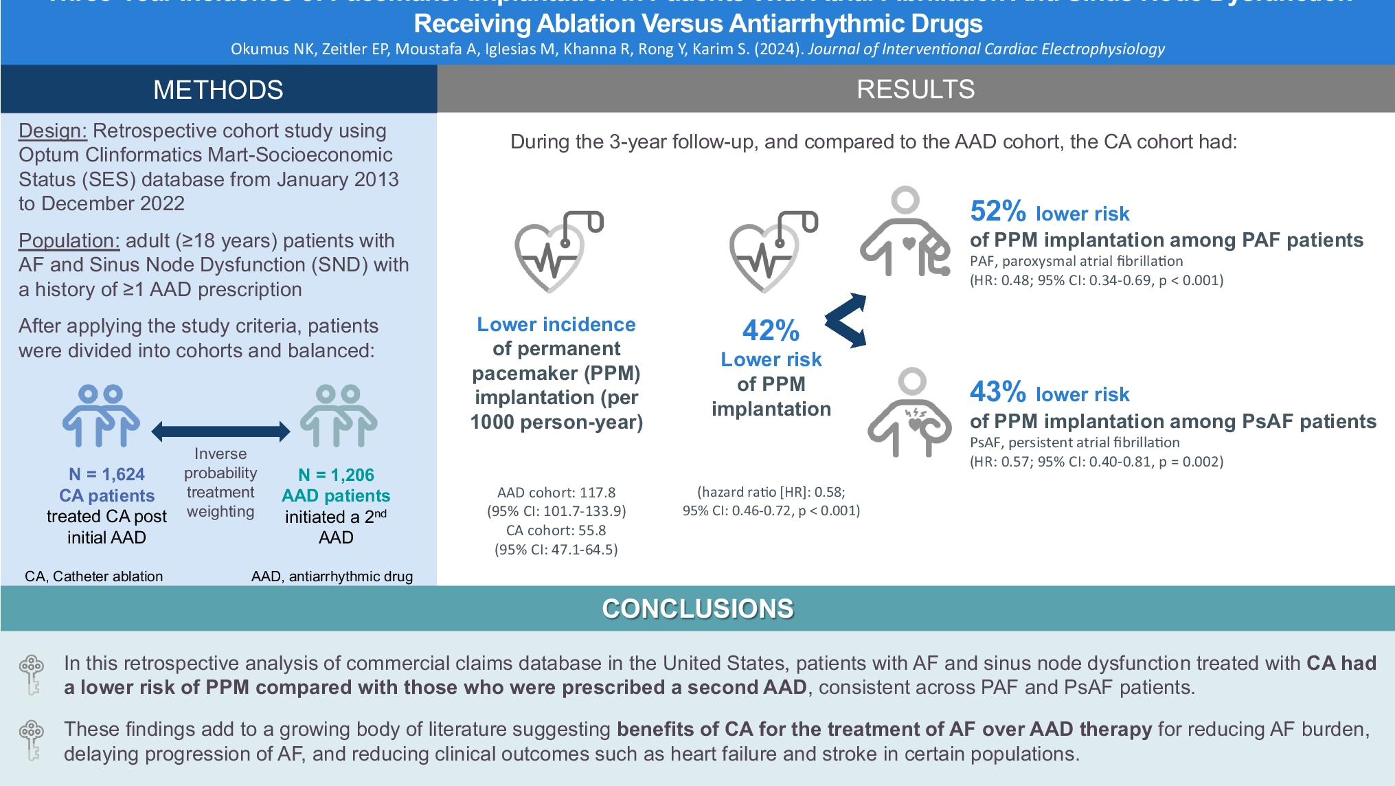
記住我

Patients with a left ventricular ejection fraction (LVEF) < 40% who either already had a cardiac resynchronization therapy device implanted (with leads positioned within the right atrium, His bundle, and right ventricle) or were attending for a clinically indicated invasive electrophysiology (EP) procedure (including VT stimulation/ablation procedures) were recruited with leads temporarily placed within the right atrium, His bundle and right ventricular (RV) apex.
2.1.2 Study 2: investigating novel pacing interventions during clinical VTWe investigated pacing interventions if VT was induced, with VT stimulation protocols, in the patients attending for an invasive EP procedure, including VT ablations.
We did not recruit patients who were in persistent atrial fibrillation or if patients were unable to give informed consent.
2.2 MeasurementsWe simultaneously recorded a continuous 3-lead ECG (Fukuda Denshi 7100, Fukuda Denshi, Japan) and beat-by-beat blood pressure measured either invasively or, if not clinically indicated, non-invasively with the use of a Finometer (Finapres Nova, Finapres Medical Systems, NL). We report the measured average systolic blood pressure over 15 s in the reference state immediately prior to a pacing transition and the following 15 s after the transition (the reference state was either sinus rhythm or simulated VT in Study 1 and clinical VT in Study 2). For patients undergoing invasive EP procedures, quadripolar EP catheters were positioned in the high right atrium, His bundle, and RV apex.
These experiments were conducted during simulated VT in Study 1 to enable accurate quantification of the impact of the different pacing interventions. While artificial, this serves to enhance the mechanistic understanding for Study 2 in which we tested these therapies during clinical VT.
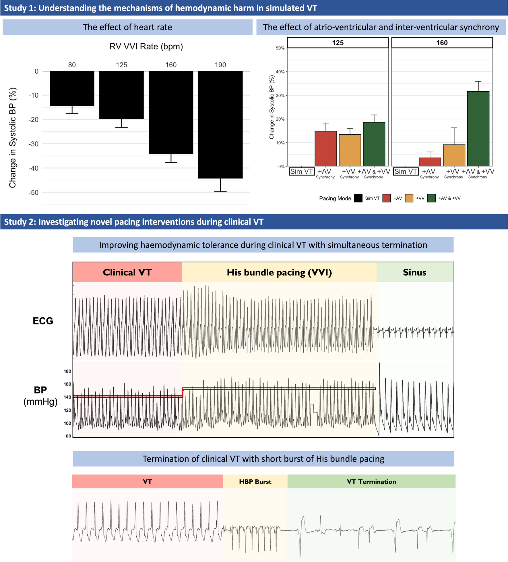
2.3 Pacing protocols2.3.1 Study 1: understanding the mechanisms of hemodynamic harm during simulated VTThe pacing protocol aimed to help delineate the relative hemodynamic effect of the following: (1) heart rate, (2) atrioventricular synchrony, (3) biventricular synchrony, and (4) combined atrioventricular and biventricular synchrony for patients with VT.
Thirty seconds of the patient’s intrinsic baseline rhythm were recorded before and after each episode of simulated VT.
Simulated VT was achieved by asynchronous ventricular pacing from the RV apex at various heart rates (80, 125, 160, and 190 bpm; Fig. 1). This resembled VT in that there was neither atrioventricular nor biventricular synchrony.
Fig. 1
Example trace of the impact of simulated VT rate (160 bpm) on hemodynamics
Example trace of the simultaneous ECG and beat-by-beat blood pressure recordings in a patient undergoing simulated VT generated from right ventricular apical pacing at a rate of 160 bpm with evidence of a profound change in hemodynamic status. BP, blood pressure; ECG, electrocardiogram; VT, ventricular tachycardia.
Participants underwent each of the pacing protocols three times, in a computer-generated random order.
To achieve restoration of atrioventricular synchrony alone, we performed synchronous dual chamber atrial and RV pacing at 125 bpm and 160 bpm (AV delay 140 ms).
Restoration of ventricular synchrony alone was performed by His bundle pacing at 125 bpm and 160 bpm in all patients. Standard His bundle capture criteria were utilized to confirm capture prior to the experimental protocol [8] and subsequently confirmed during the pacing maneuvers when the stimulus to ventricular electrogram interval was measured to confirm this reflected conduction down the His Purkinjee network. In all cases, non-selective capture was achieved.
Finally, restoration of atrioventricular and biventricular synchrony was achieved by synchronous dual chamber atrial and His bundle pacing at 125 bpm and 160 bpm (AV delay 100 ms).
For the restoration assessment protocols, asynchronous RV pacing at 125 or 160 bpm was performed for 20–30 s, and then the pacing mode was changed to determine the effect of the tested component on hemodynamic response.
2.3.2 Study 2: investigating novel pacing interventions during clinical VTIn Study 2, we aimed to assess the benefit derived through the correction of atrioventricular and biventricular synchrony during clinical VT. When VT did not occur spontaneously, induction approaches were utilized with a ventricular drive train and short coupled S2 and S3 beats as required.
Only patients who experienced spontaneous or induced VT after pace stimulation were included in this part of the study.
We allowed approximately 15 s of clinical VT before testing the pacing interventions described below. Where possible, three alternations were made between clinical VT and the tested pacing intervention.
The experimental pacing interventions we aimed to test included restoration of atrioventricular synchrony through the addition of atrial pacing at ~ 5 bpm above the clinical VT rate and attempted restoration of biventricular synchrony through the addition of His bundle pacing at ~ 5 bpm above the clinical VT rate. His bundle pacing confirmation approaches are detailed above.
It was not possible to try every pacing protocol in each patient. We prioritized His bundle pacing as this was both novel, and we felt that this had the greatest potential to deliver hemodynamic improvement. Following the unexpected termination of VT with His bundle pacing in some of the early study participants, the protocol was adapted to deliver a short burst of His bundle pacing (8–15 beats) at 91% of the tachycardia cycle length, aiming to be less aggressive than traditional anti-tachycardia pacing (ATP).
We measured the change in acute hemodynamic function relative to VT, for each of the performed interventions. During VT, we measured the stimulus timing in relation to the VT cycle length and the preceding ventricular beat to attempt to determine the ideal timing of stimuli to interact with the VT.
2.4 EthicsEthical approval was obtained by the local Research Ethics Committee (London – Central 16/LO/0339). All participants provided written informed consent.
2.5 Statistical analysis2.5.1 Study 1: understanding the mechanisms of hemodynamic harm during simulated VTTo assess the impact of the rate of VT on the hemodynamic harm for each patient, we calculated the percentage drop in systolic blood pressure, for each rate, as compared to the patient’s baseline rate. Group differences were assessed using ANOVA.
The importance of atrioventricular and biventricular synchrony during VT were assessed as the potential benefit that might be achieved through their restoration as compared to simulated VT at the same heart rate. Calculations were made at 125 and 160 bpm. The percentage increase in systolic blood pressure with the restoration of either atrioventricular or biventricular synchrony alone or in combination was compared to simulated VT (without atrioventricular or biventricular synchrony) and quantified.
2.5.2 Study 2: investigating novel pacing interventions during clinical VTTo assess the impact of the pacing intervention on hemodynamics during VT, the percentage change in systolic blood pressure was calculated between the VT state and the pacing intervention. Using the mean change for each patient with each potentially therapeutic pacing approach, the mean across all patients was then calculated. The t-test was used to determine if the change was significantly different from zero.
留言 (0)