Intravascular catheter insertion is common in hospitals and outpatient settings for various purposes, such as administering fluids, medication, blood, and nutrients. However, venous access through catheterization often induces blood clot formation. Implanted catheters rapidly acquire layers of fibronectin and fibrin upon insertion, accelerating the surface deposition and accumulation of foulants. It is estimated that nearly 18% of central venous catheter (CVC) insertion can lead to thrombotic complications. Consequently, these complications will downstream to more severe conditions such as pulmonary embolism, loss of venous access, infection, and post-thrombotic syndrome [1]. Furthermore, patients with long-term catheterization or underlying diseases such as cancer and hypercoagulability are at even greater risk for catheter-related thrombosis (CRT).
Systematic anticoagulants, such as heparin, have been used to stabilize and prevent blood clots from growing while waiting for the body's innate plasmin protein to dissolve the clot [2]. However, catheter-related thrombosis treatment often involves the administration of blood-thinning medication for approximately three months [2]. Once thrombosis is diagnosed, systematic therapy of heparin lacks long-term efficacy. It can cascade into unwanted side effects such as thrombocytopenia, osteopenia, and even severe bleeding, aside from insufficient evidence supporting that prophylactic anticoagulation leads to the prevention of thrombosis [1], [2], [3].
Other preventative actions can be taken to reduce the incident CRT, such as minimizing endothelial damage during catheter insertion and reducing blood flow stasis in the body. Regardless, some common risk factors, including the location of catheter insertion and the material and size of the CVC, cannot be easily altered in clinical practice. For these reasons, recent endeavors toward minimizing CRT have focused on developing surface modifications that prevent and reduce thrombus formation in the early stage of catheterization [4,5]. For example, heparin-bonded catheters and chlorhexidine/silver sulfadiazine-impregnated catheters have been developed, and they have shown success in the antifouling activity of intravascular catheters [6]. However, earlier-mentioned side effects of heparin are of concern and unavoidable, preventing the material from being used in the clinics.
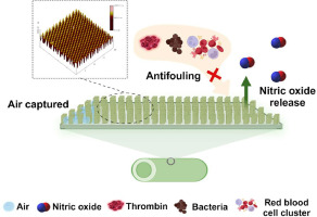
Recently, bioinspired surface modifications that mimic natural phenomena to reduce the adhesion of bacteria and foulants have been developed [7,8]. One of the well-known surface modification techniques is to recreate antifouling shark skin through micropatterned surfaces [7]. Shark skin-inspired micropatterns have been proven to repel the adhesion of pathogenic bacteria, even after blood protein exposure [9]. While these bioinspired antifouling materials generally possess micron-sized structured surfaces with dimensions ranging from 1 to 300 μm [10], submicron or nano-size textured patterns with dimensions less than the size of single platelet or bacterium may have more benefit for reducing fouling than larger patterns because the small dimension reduces the surface contact area for interactions between formed elements and surfaces [11]. Xu et al. have fabricated submicron textured polyurethane (PU) surfaces with pillared patterns of 400 nm and 500 nm diameter with 500 nm spacing and demonstrated effective antifouling activity against microbes [12]. Other submicron-textured surfaces also significantly reduced platelet adhesion under shear, showing that the surface can reduce thrombus formation on blood-contacting medical devices [13].
Another popular biomimetic approach is to create an endothelial layer-like interface through nitric oxide (NO) release from the polymer matrix. NO is a free radical gasotransmitter that attenuates platelet binding and activation to the adsorbed plasma protein layer present on foreign materials in the body. NO's antithrombotic properties have been widely reported [14], [15], [16]. However, due to the short half-life of NO, prolonged NO release from the surface can only be achieved by incorporating NO donors into materials. Among the extensively studied NO donors, S-nitroso-N-acetyl-DL-penicillamine (SNAP) is preferred due to its long-term stability and controlled release mediated by photolysis, thermal decomposition, and metal ion catalysis [17]. By incorporating SNAP into various polymers with different water uptakes, its NO release concentration and duration can be tuned to be application-based. For example, SNAP-doped polyurethane Elast-eon E2As polymer retained 82% of the initial SNAP after two months of storage at 37°C and released NO slowly at physiological flux levels for up to 3 weeks [18].
However, the approaches mentioned above have some inherent limitations (e.g., defects in surface texturing or limited NO release lifetime in donor-incorporated biomaterials) in controlling thrombosis on implanted biomedical devices, the combinatorial approach becomes more attractive since it has the potential to overcome the shortcomings of these individual approaches. The combination of surface texturing and NO-releasing mimics the inner layer of a blood vessel, which contains a rough surface with micro-grooves in the blood flow direction and nano-protuberances on the ridges, and where the endothelial cells produce NO simultaneously. Toward this, we fabricated NO-releasing submicron patterned PU surfaces where SNAP was either doped or impregnated in PU films [19,20]. Such biomimetic surfaces significantly increased the plasma coagulation time and exhibited reduced platelet adhesion and activation, thereby reducing the risk of blood coagulation and thrombosis [21]. It was the first study to combine physical surface modification via topographical texture and chemical modification via NO release to show the complementary bactericidal and anti-thrombosis effects on one surface. However, the above studies did not assess the hemocompatibility of the combination surface in vivo. Similarly, throughout the literature, the studies of in vivo responses to textured surfaces are also limited and are primarily focused on either antibacterial and blood-repellent metal surfaces, or increased fibroblast formation on textured silicone implants [22], [23], [24]. Among all the micro- or nano-patterned polymetric surfaces being developed, a knowledge gap exists on how these materials would perform under long-term assessment in the physiological environment.
To gain a deeper insight on whether the complementary efficacy of bioinspired topographical texture and NO release can translate into clinical settings, this study examined the hemocompatible characteristics of this dual functional surface in vivo. To our knowledge, no prior study has examined the in vivo hemocompatibility of textured NO-releasing surfaces. Herein, we report the development of textured NO-releasing CVCs and assess these catheters for their long-term hemocompatible characteristic via a 7 d rabbit catheter model. Surface characterization was carried out using atomic force microscopy (AFM), static contact angle, and water uptake assessments. The optimized NO-releasing profile was then evaluated through UV-vis spectrophotometer and chemiluminescence-based measurements. The biocompatibility of PU catheters was assessed using an in vitro cytotoxicity assay with BJ human fibroblast cells. In vitro blood compatibility of the catheter surface was examined in a blood flow loop system designed for 20 h. Finally, the in vivo antithrombotic efficacy of the surfaces was evaluated through a 7 d rabbit thrombogenicity model. The NO release and surface topography's combined effects on anti-thrombosis formation were evaluated against smooth control catheters.

留言 (0)