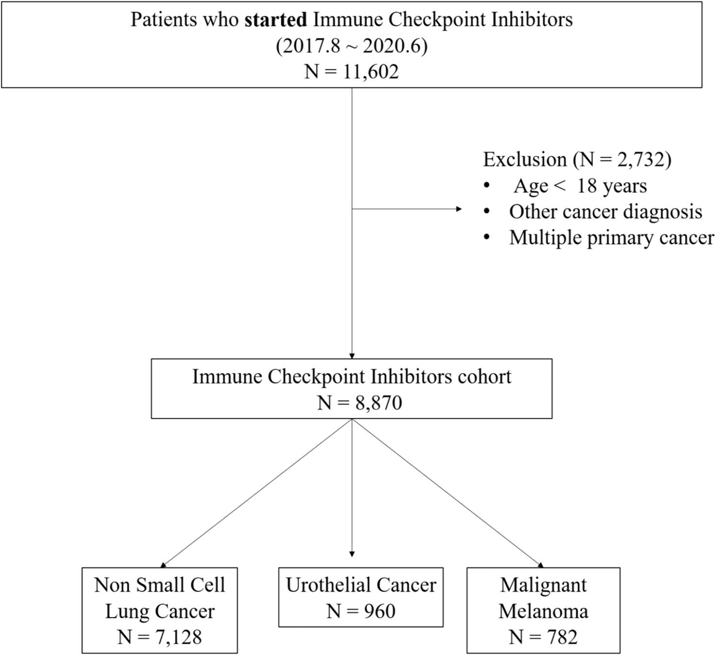
Cell lines, cell culture, drug and data source
DMEM medium and RPMI-1640 medium with 10% FBS and 1% penicillin–streptomycin were used to incubate ARPE-19 and CM cells (C918, Ocm-1, Omm2.3 and Mel270), respectively. All cells were grown in humidified environment at 37 °C and 5% CO2. ARPE-19 and Ocm-1 were bought from Beijing BeNa Culture Collection, C918 from Wuhan Procell Life Science & Technology Co., Ltd, Omm2.3 and Mel270 from Guangzhou Cellcook Biotech Co., Ltd. ART were purchased from Sigma. In previous study (Geng et al. 2021), we examined the toxic effects of ART and the IC50 concentration in CM cells. The significance of EFNA3 in CM was analyzed using University of Alabama at Birmingham Cancer data analysis portal (UALCAN; https://ualcan.path.uab.edu/index.html) and tumor immune estimation resources (TIMER v2.0; https://cistrome.shinyapps.io/timer/).
Quantitative real time polymerase chain reaction (qPCR)
According to instructions, RNA was extracted utilizing RNA-easy Isolation Reagent (Vazyme, Nanjing, China). Subsequently, RNA was reserve transcribed by HiScript III RT SuperMix for qPCR (+ gDNA wiper) (Vazyme, Nanjing, China) and amplified by qPCR. GAPDH was used to normalize the expression of target genes and the relative expression levels of gene were calculated according to the 2−ΔΔCT method. h-EFNA3-F: CGCACAGCCCCATCAAGT, h-EFNA3-R: AGACGAACACCTTCATCCTCAGA.
Western blot assay
Protein was isolated using RIPA with protease inhibitor and phosphatase inhibitors, separated using 10% SDA-PAGE, transferred to PVDF membranes, blocked with 5% nonfat milk at 37 °C for 1 h, and incubated with anti-GAPDH antibody (1:5000, Servicebio), anti-EFNA3 antibody (1:1000, HUABIO), anti-p-Stat3 antibody (1:2000, CST), anti-Stat3 antibody (1:2000, CST), anti-Akt antibody (1:1000, CST), anti-phosphor-Akt antibody (1:1000, CST) for overnight at 4 °C. Subsequently, PVDF was incubated with antibodies of secondary at room temperature for 1 h, detected with Sensitive ECL Luminescence Reagent (Meilunbio, Dalian, China).
siRNA and plasmid transfection
siRNA of EFNA3 were purchased from GenePharma company (Shanghai, China). EFNA3 siRNAs sequences: siRNA #1,5′-CGUGAACGACUAUCUGGAUAU-3′; siRNA #2,5′-GCAGGUGAACGUGAACGACUA-3′. The overexpression plasmids pcDNA-EFNA3 were purchased from Genechem company (Shanghai, China). siRNA and plasmid were transfected into CM cell using Lipo8000™ transfection reagent (Beyotime Biotechnology, Shanghai, China) according to instructions.
Assembly of EFNA3 lentivirus
First, suitable and transduction reagent were selected according to the requirements of manufacturer. Then cells were spread into 6-well plates and lentivirus was added into the plates when cells reached 20–30% confluence. Stable cells were screened using puromycin (1 μg/ml). EFNA3-downregulating lentivirus were bought from Genechem company (Shanghai, China).
Cell counting kit-8 assay (CCK-8)
Two thousand cells were added in 96-well plates and cultured for overnight. When removing the original medium, 10 μ CCK-8 reagent (MCE, Weehawken, USA) and 90 μ RPMI-1640 was added. After incubation of two hours, microplate reader was utilized to assessed the absorbance at 450 nm wavelength. CCK-8 assay was performed for cultures at 0 h, 24 h, 48 h, 72 h, 96 h.
Clonogenic assay
Each well was seeded 1000 cells in 6-well plates and cells were incubated with 10% FBS medium for 10–14 days. After 10–14 days, 4% paraformaldehyde was utilized to fix the colonies. Subsequently, crystal violet was employed to stained the colonies. Photoshop was used to count the number of colonies.
Transwell migration assay
Cells in medium without FBS were added into upper chamber and medium containing 10% (C918) and 30% (Ocm-1, Omm2.3) FBS added into lower chamber as a chemoattractant. After being incubated for 24 h (C918) and 48 h (Ocm-1, Omm2.3), cells on the lower surface were fixed, stained, and counted. The assay was repeated thrice.
Immunohistochemistry
Using 4% paraformaldehyde and paraffin to fixe and embed tumors, then cut into slices. After deparaffinization in dimethylbenzene, the slices were gradually dewatered in ethanol, treated with citrate buffer and immerged 3% hydrogen peroxide solution. Subsequently, slices were incubated using primary antibodies and secondary antibodies successively, and treated with diaminobenzidine, counterstained with hematoxylin.
Nude mouse xenograft study
The animal experiments were approved by the ethics committee of the Affiliated Hospital of Qingdao University (permit number: AHQU-MAK20230526). The mouse experiment was in accordance with the institutional guidelines of Animal Care and Use Committee. Fifteen female BALB/c nude mice aged 4 weeks were bought from Jinan Pengyue. C918 cells (downregulating EFNA3 and empty vector control) were digested with pancreatic enzymes and resuspended in PBS counting 1/2 polymerized Matrigel to a final cell count of 2 × 10⁷/mL. A volume of 100 μ of the cell suspension was injected in the right armpit of the mouse. After one week, mice were administered solvent (the mouse of vehicle and shEFNA3), ART (i.p., 70 mg/kg, qd) for 14 days. The size of tumor was measured using digital caliper. At the conclusion of the experiment, the tumor was excised and subsequently weighed.
Statistical analysis
All results were analyzed using t-teat (two groups) and one-way ANOVA (multiple groups) in GraphPad Prism 9.0 (GraphPad Software, USA). Data were presented as mean ± standard error of the mean (SEM). All experiments were performed at least in three times and p < 0.05 was regarded as significance (ns: no significance, *p < 0.05, **p < 0.01, ***p < 0.001, ****p < 0.0001).

留言 (0)