
Remember me


Hepatic fibrosis results from chronic liver injury and is attributed to multiple predisposing factors, including viral hepatitis, alcohol consumption, nonalcoholic steatohepatitis, autoimmune hepatitis, and cholestatic liver disease (1,2). A significant event during liver fibrosis is the activation of myofibroblasts, which originate from fibroblasts, including hepatic stellate cells (HSCs), portal fibroblasts, and fibrocytes (2). Proliferating HSCs lead to the accumulation of extracellular matrix through collagen secretion that forms fibrous scar tissue, which facilitates initial tissue repair; however, such formation can lead to cirrhosis if inappropriately regulated (2,3). Transforming growth factor-β1 (TGF-β1) is considered as a major profibrogenic cytokine within the liver (3). TGF-β1 indirectly stimulates the expression of type I collagen and α-smooth muscle actin (α-SMA) during stressed fibrogenesis through the activation of HSCs (4). However, long-term treatment of hepatitis B virus with nucleoside or nucleotide analogues has been reported to induce the histologic improvement and regression of liver fibrosis and cirrhosis (4–6), as well as the reduction of the risk of hepatocellular carcinoma (7). These findings verify that hepatic fibrosis can regress with the removal of pathophysiological causes and the elimination of activated myofibroblasts, resulting in the reabsorption of the scar tissue. Therefore, the regulation of myofibroblast activation, proliferation, and apoptosis is crucial in the management of liver fibrosis (6).
Exosomes are extracellular nanosized (40–100 nm) polymorphic vesicles secreted by all cell types and are widely localized in plasma, saliva, urine, and other body fluids (7). Their roles of delivering miRNA, lncRNA, DNA fragments, and proteins as stable cargos lead to a global interest in exploring their association with diseases. Recent accumulating studies revealed the new roles of exosomal miRNAs during fibrogenesis and as a therapy for fibrosis reversal, such as miR17-92 promoted HSCs activation (7,8), miR-21 participated in extracellular matrix remodeling (9), and miR214/199-5a clusters, reducing related protein cellular communication network factor 2 (CCN2) expression in activated HSCs (10). The above studies mainly focus on the role of exosomal miRNAs in the molecular mechanism of liver fibrosis. Notably, exosomal miR-155 was found to be closely associated with the progression of cirrhosis and clinical prognostic indicators of cirrhosis, suggesting that exosomal miR-155 can act as a noninvasive biomarker for the diagnosis and progression of hepatic fibrosis (11). However, this study is limited by lacking of relevant prospective liver biopsy standard for histologic assessment of hepatic necroinflammation and fibrosis and long-term longitudinal clinical data about miR-155, so it is difficult to apply to clinic in the short term. Moreover, exosomal miR-122 has been as a valuable noninvasive biomarker for liver fibrosis, particularly in patients with nonviral etiologies of chronic liver disease (12). However, few studies have been reported on the association between miRNAs derived from human serum exosomes and liver fibrosis, clinical validation, and related mechanism.
In this study, we aimed to screen and identify the association between serum exosome–derived miRNAs and fibrogenesis. This study was conducted on a cohort of patients with chronic hepatitis B (CHB) who had paired histologic data available and were enrolled in a multicenter randomized controlled clinical trial (5,6,13). In addition, the current study elucidated the underlying mechanism. It further revealed that serum exosome–derived miR-193a-5p and miR-381-3p promote fibrogenesis by regulating the AMPK/TGF-β/Smad2/3 signaling pathway.
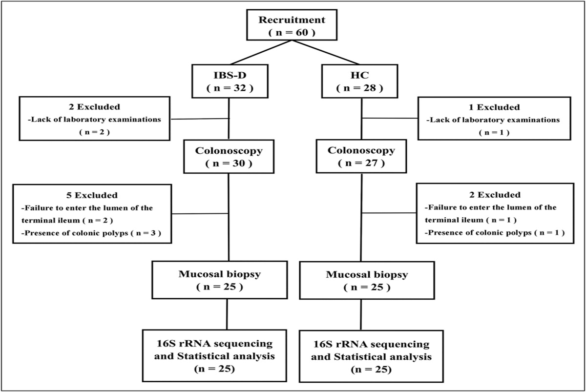
MATERIALS AND METHODS Collection of clinical samplesSix patients with CHB with paired histologic data were recruited from an extension study associated with a multicenter randomized controlled clinical trial (CHB with Significant Fibrosis Antiviral Long-Terms Treatment Against Hepatic Fibrosis, CHBALT-F; No. NCT01965418), and 6 healthy control subjects were recruited as the discovery cohort. The clinical characteristics at the baseline of these 6 patients, as well as the double-blind treatment at week 72, are listed in Supplementary Digital Content (see Supplementary Table 1, https://links.lww.com/CTG/B42). For the validation cohort, among 42 cases of advanced fibrosis or cirrhosis, 23 cases of hepatocellular carcinoma (HCC) and 23 healthy donors (HDs) were recruited. The clinical characteristics of enrolled patients with advanced fibrosis and cirrhosis, patients with HCC, and HDs in this validation cohort are listed in Supplementary Digital Content (see Supplementary Table 2, https://links.lww.com/CTG/B43). Written informed consent was provided by each participant. This study was conducted in accordance with the local regulatory requirements, good clinical practice, and the Declaration of Helsinki. The study protocol was approved by the ethics committee (Ethical approval number: 2018101D).
Exosome isolation and identificationPeripheral blood samples from individuals were collected in ethylene diamine tetraacetic acid tubes. After the serum was centrifuged at 3,000g for 15 minutes at 4 °C, it was aspirated and stored at −80 °C. The exosomes were isolated by size exclusion chromatography methods as described previously with minor modifications (14,15). In brief, 1 mL of 0.8-μm filtered plasma was 1.5-fold diluted with phosphate buffer saline (PBS) and further purified using Exosupur columns (Echobiotech, China). The samples were then eluted with further 0.01 M PBS, and a total of 2 mL eluate fractions were collected according to the manufacturer's instructions. Fractions were concentrated to 200 μL by 100 kDa molecular weight cutoff Amicon Ultra spin filters (Merck, Germany). Vesicle suspensions with concentrations between 1 × 10⁷/mL and 1 × 10⁹/mL were examined using the ZetaView PMX 110 (Particle Metrix, Meerbusch, Germany) equipped with a 405-nm laser to determine the size and quantity of particles isolated. A video of 60 seconds duration was taken with a frame rate of 30 frames/s, and particle movement was analyzed using nanoparticle tracking analysis (NTA) software (ZetaView 8.02.28). Ten-microliters exosome solution was placed on a copper mesh and incubated at room temperature for 1 minute. After washing with sterile distilled water, the exosome was contrasted by uranyl acetate solution for 1 minute. The sample was then dried for 2 minutes under incandescent light. The copper mesh was observed and photographed under a transmission electron microscope (H-7650; Hitachi, Tokyo, Japan).
Quantification and differential expression analysis of miRNAThe Bowtie software was used to align clean reads with the Silva, GtRNAdb, Rfam, and Repbase databases. Repeats and ncRNAs, such as ribosomal RNA, transfer RNA, small nuclear RNA, and small nucleolar RNA, were then excluded.
The remaining reads were used to detect known miRNA and new miRNA by comparing known miRNAs from miRBase and the human genome, respectively. The expression matrix containing quantified unique molecular identifier (UMI) counts of miRNAs was normalized to transcript per million. The relative log expression values were then calculated using the edgeR package.
RNA extraction and quantitative RT-PCRThe total RNA of plasma was extracted in accordance with the miRNeasy Serum/Plasma Advanced Kit protocol (Qiagen, Cat. No. 217204; Hilden, Germany). Meanwhile, the total RNAs of tissues and cells were extracted using the RNAiso for Small RNA method (Takara, 9753A; Kyoto, Japan). The quality of the total RNAs was assayed using RNA NanoDrop measurements.
miRNA expression levels were determined by real-time quantitative polymerase chain reaction (RT-qPCR). The Bulge-Loop miRNA qRT-PCR Starter Kit (RIBOBIO, C10211-3; Guangzhou, China) was used for miRNA quantification. The specific miRNA reverse transcription and PCR primers were obtained from the Bulge-Loop Primer kit to which RNase-free H2O was added. A 10-μL reaction system was configured as instructed. A qPCR reaction system was prepared and then run using the set program. U6 was used as an internal control for the analysis.
Cell line and cell cultureLieming Xu-2 (LX-2) cells from Shanghai Fuheng Biotechnology were cultured in Dulbecco's modified Eagle's medium (DMEM) (Boster, Wuhan, China) supplemented with 10% fetal bovine serum (Vivacell, Shanghai, China) and 1% penicillin-streptomycin. The cells were stored in incubators with 5% CO2 at 37 °C.
Transfection in vitroLX-2 cells were seeded in 12-well plates until 80% confluence was reached. miRNA mimics, inhibitors, and their corresponding negative controls (miR-NC and inhibitor-NC) were synthesized by Suzhou GenePharma (Suzhou, China) and then transfected into LX-2 cells by using jetPRIME (Polyplus-transfection, Strasbourg, France). Transfection efficiency was verified by RT-qPCR. The target sequences used in the study are listed in Supplementary Digital Content (see Supplementary Table 2, https://links.lww.com/CTG/B43).
Western blot analysisRadioimmunoprecipitation assay lysis buffer was used for protein extraction. Protein samples were heated to 105 °C and then subjected to electrophoresis. The proteins were then transferred to polyvinylidene fluoride membranes and blocked with 5% milk (nonfat milk in tris-buffered saline tween [TBST]). The membranes were then incubated with the primary antibodies required in this study at 4 °C overnight. The antibodies used in the study are listed in Supplementary Digital Content (see Supplementary Table 3, https://links.lww.com/CTG/B44). On the following day, the membranes were incubated with secondary antibodies for 1 hour at room temperature after being washed with TBST 3 times. Protein expression levels were assessed using commercially enhanced chemiluminescence kits (Huaxing Bio, HX1868; China) with enhanced chemiluminescence assay membranes in accordance with the instruction manual.
5-Ethynyl-2'-deoxyuridine proliferation assayCell proliferation was determined by incorporating 5-ethynyl-2'-deoxyuridine (EdU) by using the EdU Cell Proliferation Assay Kit (Beyotime, BeyoClick EdU Cell Proliferation Kit with Alexa Fluor 488). LX-2 cells were incubated with EdU for 2 hours, fixed in 4% paraformaldehyde, and incubated with a click reaction solution for 30 minutes. Cell nuclei were stained with Hoechst 33342. The proportion of cells incorporating EdU was determined using a high-content screening instrument (Thermo).
In vitro wound-healing assayCells were seeded in 12-well plates to near confluence. Wounds were created using a micropipette tip. Wound-healing assays were performed in complete medium and photographed at the following time points: 0, 12, and 24 hours. The widths of the wounds were measured in different positions by using ImageJ at each time point to quantify cell migration. The migration distance was calculated as the difference in width between consecutive time points. The migration distance for each sample was initially normalized to the initial wound width and then compared with the other samples.
Apoptosis assayThe Annexin V-fluorescein isothiocyanate (FITC)/propidium Iodide (PI) apoptosis detection kit (Procell, Wuhan, China) was used for apoptosis assay. Cells were resuspended 48 hours post-transfection in 1 × Annexin V Binding Buffer at concentration of 1 × 10⁵ cells/mL. The cells were thoroughly mixed with 5 μL of Annexin V-FITC, incubated at room temperature for 15 minutes, and washed with PBS. After 5 μL (50 μg/mL) of PI was added, the samples were subjected to flow cytometry by using FACSCanto II (BD Biosciences, Franklin Lakes, NJ) and then analyzed using NovoExpress 1.5.0.
Statistical analysisData analysis was performed using GraphPad Prism 9.5.1 (a learning biological drawing software application known for its capabilities in biological data visualization and analysis). Experimental data were presented as mean ± SDs. For comparison of 2 groups, the unpaired Student t test was used, and one-way analysis of variance (ANOVA) with Dunnett post hoc test was used to compare multiple groups. Statistical significance was denoted as follows: *P < 0.05, **P < 0.01, and ***P < 0.001. No significance was indicated using NS.
RESULTS Characterization of exosomes derived from the serum of patients with liver fibrosisThe successful isolation of the plasma exosomes was confirmed by transmission electron microscopy, nanoparticle tracking analysis, and Western blot (WB) analysis. Transmission electron microscopy revealed small vesicles with diameters ranging from approximately 50 to 150 nm and a lipid bilayer (Figure 1a,b). Exosomes were identified by WB analysis, which revealed specific bands corresponding to CD9, CD63, and Tsg101 (Figure 1c). These bands were observed in the exosome pellet but not in the exosome-depleted supernatant. On the basis of the aforementioned evidence, we verified the presence of exosomes in the serum samples. This confirmation complies with the requirements of the minimal information for studies of EVs (16), that is, “3 positive protein markers and one negative marker.”
 Figure 1.:
 Characterization of exosomes derived from serum samples of patients with liver fibrosis. TEM was used to observe the morphology of exosomes (scale bar = 0.5 um (a) and 200 nm (b)); Western (WB) analysis was performed to detect exosomal surface markers CD9, Tsg101, CD63, and calnexin, as well as to compare their levels with those in the cellular lysate (c). Nanoparticle tracking analysis was performed to analyze the exosomes (d). TEM, transmission electron microscopy.
Identification of serum exosome–derived miRNAs associated with fibrogenesis by transcriptomic analysisUsing comprehensive bioinformatic analyses, we performed screening for signature serum exosome–derived miRNAs associated with fibrogenesis in CHB. The signature serum exosome–derived miRNAs were obtained: 147 in the significant liver fibrosis group (Figure 2a,e), 206 in the advanced liver fibrosis group (Figure 2b,f), and 263 in the cirrhosis group (Figure 2c,g). In the 3 sets of comparison, 123 potential common differentially expressed miRNAs closely correlated with liver fibrosis were obtained using the Veen analysis.
 Figure 2.:
 Identification of serum exosome–derived miRNAs associated with fibrogenesis by transcriptomic analysis. Heatmap showing differentially expressed miRNAs of the healthy control group (n = 6) vs the liver fibrosis group (n = 6) (a), the advanced liver fibrosis group (S = 4, 5, 6, n = 3) (b), and the cirrhosis group (S = 5, 6, n = 5) (c). Volcano plots of differentially expressed miRNAs are shown. Each point represents one detectable miRNA (e, f, g). In the regression group, heatmap showing differentially expressed miRNAs between the healthy control group (n = 6) and the pretreatment group (n = 3) (d), as well as between the pretreatment group (n = 3) and the posttreatment group (n = 3) (i). Volcano plots reflecting the 2 results are shown (h, j). Veen analysis to screen miRNAs closely related to liver fibrosis and that may affect the reversal of liver fibrosis after treatment (k). miR-193a-5p and miR-381-3p filtered out with their expression levels were detected by qPCR and displayed separately in scatter plots (l, m). Scatter plots showing changes in the expression levels of these 2 miRNAs before and after treatment (n, o). miRNA, microRNA.
To further explore the association of dysregulated miRNAs with histologic outcomes, we analyzed the differential expression of miRNAs in the regression group. Compared with the control group, a total of 198 miRNAs were dysregulated during the regression of liver fibrosis in CHB (Figure 2d,h). Relative to those in the baseline, there were 17 miRNAs dysregulated during the regression of liver fibrosis after treatment for 72 weeks (Figure 2i,j). Considering the 2 aforementioned, we found 6 common miRNAs expressed differentially in CHB that were closely related to liver fibrosis reversal. Ultimately, miR-193a-5p and miR-381-3p were confirmed to be closely related to the outcome of liver fibrosis (Figure 2k). The values of the log2 fold change and P values for these comparisons are listed in Table 1. The quantitative analysis of these 2 miRNAs in each group is presented in Figure 2l–o.
Table 1. - Differences in miRNA levels detected in sequencing analysis miRNA log2FC P valuea Regulated HD vs fibrosis miR-193a-5p 1.60809062 7.62E-05 Up miR-381-3p 2.42614368 3.18E-06 Up HD vs advanced fibrosis miR-193a-5p 1.03666851 0.00687305 Up miR-381-3p 2.56642842 3.70E-06 Up HD vs cirrhosis miR-193a-5p 1.09692583 0.00159236 Up miR-381-3p 2.12983747 5.99E-05 Up HD vs pretreatment in the regression group miR-193a-5p 0.81918381 0.03738767 Up miR-381-3p 2.04984473 0.00062881 Up Pretreatment vs posttreatment in the regression group miR-193a-5p 1.89330461 0.00169791 Up miR-381-3p −1.26979 0.03942891 DownmiRNA, microRNA; log2FC, log2 fold change; HD, healthy donor.
aP value significance threshold ≤0.05.
To confirm the potential miRNAs associated with histologic outcomes, we performed qRT-PCR assays on 2 differentially expressed exosome miRNAs (miR-193a-5p and miR-381-3p). After these miRNAs were filtered, the serum exosome–derived miR-193a-5p and miR-381-3p were significantly upregulated in CHB-related liver fibrosis relative to those in the control group (Figure 3a,b). Moreover, serum exosome–derived miR-193a-5p and miR-381-3p efficiently predicted significant liver fibrosis in CHB cases. This finding was indicated by the area under the receiver operating characteristic curves of 0.8706 (95% confidence interval [CI]: 0.7837–0.9574) with a sensitivity of 83.33% and a specificity of 82.61% for miR-193a-5p, and 0.8799 (95% CI: 0.7918–0.9680) with a sensitivity of 85.71% and a specificity of 86.96% miR-381-3p (Figure 3f,g).
 Figure 3.:
 Validation of the relevance of exosome-derived miR-193a-5p and miR-381-3p to histologic outcomes. Scatter plots of serum exosome–derived miR-193a-5p and miR-381-3p significantly upregulated liver fibrosis in the CHB group relative to the HD group (a, b). AUROCs of miR-193a-5p (0.8706) and miR-381-3p (0.8799) (f, g). Hematoxylin and eosin (HE), Masson staining, and reticular fiber staining to confirm the occurrence of fibrosis and hepatocellular carcinoma in the tissue samples recruited in another cohort are given (e). The expressions levels of miR-193a-5p and miR-381-3p in the liver cirrhosis group were significantly higher than those in the HD group and lower in the HCC group; however, the difference was not significant unlike that in the fibrotic group (c, d). Data are shown by mean ± SE of mean (SEM). *P < 0.05, **P < 0.01, ***P < 0.001, ****P < 0.0001. AUROC, area under the receiver operating characteristic curves; CHB, chronic hepatitis B; HCC, hepatocellular carcinoma; HD, healthy donor.
To further explore whether this difference can be detected in the target organ (i.e., liver tissue), we recruited another cohort consisting of 23 patients with CHB-related HCC with adjacent cirrhosis tissue and tumor tissue, in addition to 10 patients with no history of chronic liver diseases, who comprised the control group (Figure 3e). Relative to those in the HD group, the expression levels of miR-193a-5p and miR-381-3p in the liver cirrhosis group were significantly upregulated, which was consistent with serum findings. Moreover, histologic images revealed the disappearance of collagen fibers and the replacement of abnormally activated HSCs by tumor cells. These 2 miRNAs were poorly expressed in the HCC group, but the difference was not significant, contrary to that in the fibrotic group (Figure 3c,d). Upregulated miR-193a-5p and miR-381-3p in serum exosomes could be secreted by these abnormally activated HSCs.
Overexpression of miR-193a-5p and miR-381-3p promotes fibrogenesis in LX-2 cellsTo assess the effects of miR-193a-5p and miR-381-3p on liver fibrosis, a model of HSC activation was established using TGF-β1 in LX-2 cells. After the LX-2 cells were treated with 20 ng/mL TGF-β1, the expression of α-SMA, a profibrotic protein marker, became significantly increased (Figure 4a,b). In addition, the expression levels of miR-193a-5p and miR-381-3p were significantly increased after treatment with 20 ng/mL TGF-β1 for 48 hours, as determined by qPCR. This finding also confirmed their high expression in vitro cell models (Figure 4c,d). Subsequently, mimics of miR-193a-5p and miR-381-3p were transfected into LX-2 cells (Figure 4e,f). Overexpression of serum exosome–derived miR-193a-5p and miR-381-3p significantly promoted the proliferation of HSCs (Figure 4g), as well as wound healing and migration (Figure 4h), relative to those in the NC mimic group. Flow cytometry showed that miR-193a-5p and miR-381-3p mimics could significantly inhibit the apoptosis of LX-2 cells (Figure 4i). Meanwhile, qPCR results showed that the expression levels of profibrotic markers, including collagen-I, α-SMA, and TIMP1, were markedly increased (Figure 4j,k). This outcome was consistent with the results of the WB analysis (Figure 4l).
 Figure 4.:
 Overexpression of miR-193a-5p and miR-381-3p promotes fibrogenesis in LX-2 cells. mRNA (a) and protein levels (b) of α-SMA, as determined by qPCR and Western blot (WB) analysis in activated LX-2 cells treated with 20 ng/mL TGF-β for 24 and 48 hours. Expression levels of miR-193a-5p and miR-381-3p in activated LX-2 cells, as determined by qPCR (c, d). Mimics of miR-193a-5p and miR-381-3p transfected into LX-2 cells and transfection efficiency, as confirmed by qPCR (e, f). In vitro EdU cell proliferation assay (g) and wound-healing (h) images. Apoptosis evaluated by flow cytometry 48 hours after transfection with mimics of miR-193a-5p and miR-381-3p (i). mRNA levels of COL1A1, α-SMA, and TIMP1 in LX-2 cells activated by 20 ng/mL TGF-β for 48 hours, as determined by qPCR (j, k). Protein levels of α-SMA, measured by WB analysis (l). Data presented as means ± SE of mean (SEM). *P < 0.05, **P < 0.01, ***P < 0.001, ****P < 0.0001. COL1A1, collagen, type I, alpha 1; EdU, 5-ethynyl-2'-deoxyuridine; miRNA, microRNA; TGF-β, transforming growth factor beta; TIMP1, tissue inhibitors of metalloproteinase 1; α-SMA, α-smooth muscle actin.
Inhibition of miR-193a-5p and miR-381-3p ameliorates fibrogenesis through the AMPK/TGF-β/Smad2/3 signaling pathway in vitroTo further explore the mechanism underlying the upregulated miR-193a-5p and miR-381-3p modulating the fibrogenesis, analyses of the Kyoto Encyclopedia of Genes and Genomes pathway (Figure 5a) and Gene Ontology (GO) (Figure 5b) were conducted. The TGF-β signaling pathway was among the 10 most highly affected signaling pathways. The enriched GO (molecular function) bubble plot showed the top 10 terms, including TGF-β receptor activity, Smad binding, and TGF-β receptor binding. Moreover, target genes analysis indicated the potential regulatory function of miR-193a-5p and miR-381-3p on the expression of C18orf25. This gene, which regulates skeletal muscle function (17), was identified as a prospective target for both miRNAs (Figure 5c,d). Subsequently, qPCR was conducted to verify that miR-381-3p downregulated C18orf25 at the mRNA level. However, contrasting results were observed with miR-193a-5p, suggesting that miR-193a-5p played a regulatory role through other target genes (Figure 5e,f). Ultimately, the results were supported by WB analysis. When the inhibitors of miR-193a-5p and miR-381-3p were transfected into LX-2 cells (Figure 5g,h), AMPK phosphorylation was increased, whereas α-SMA, p-Smad2, and p-Smad3 were significantly reduced (Figure 5i). The combined results demonstrated that these 2 miRNAs regulated HSC activation in vitro by regulating the AMPK and TGF-β signaling pathways (Figure 6).
 Figure 5.:
 Inhibition of miR-193a-5p and miR-381-3p ameliorates fibrogenesis through the AMPK/TGF-β/Smad2/3 signaling pathway in vitro. KEGG analysis of significant pathway of differentially expressed miRNAs in the “regression” group (a) and Gene Ontology (GO) analysis of significantly differentially expressed miRNAs molecular function (b) were conducted. The common target genes of miR-193a-5p and miR-381-3p were selected by the Veen diagram (c). The binding position and sequences between these 2 miRNAs and C18orf25 were acquired through a target scan. Org (d). miRNA mimics were transfected into LX-2 cells to verify the regulatory effect of miRNAs on C18orf25 by qPCR (e, f). The respective inhibitors of miR-193a-5p and miR-381-3p were transfected into LX-2 cells, and the efficiency was confirmed by qPCR (g, h). Protein levels of α-SMA, p-AMPK, AMPK, p-Smad2, Smad2, p-Smad3, and Smad3 were determined by WB analysis (i). Data presented as mean ± SE of mean (SEM). *P < 0.05, **P < 0.01, ***P < 0.001, ****P < 0.0001. AMPK, Adenosine 5'-monophosphate (AMP)-activated protein kinase; KEGG, Kyoto Encyclopedia of Genes and Genome; MiRNA, microRNA; TGF-β, transforming growth factor beta; α-SMA, α-smooth muscle actin.
 Figure 6.:
 Proposed model for the effect of plasma exosome–derived miR-193a-5p and miR-381-3p on liver fibrosis. Serum exosome–derived miR-193a-5p and miR-381-3p were found significantly upregulated in patients with CHB with advanced fibrosis. Inhibiting the expression of miR-193a-5p and miR-381-3p promoted AMPK phosphorylation and inhibited the activation of the TGF-β/Smad2/3 signaling pathway, ultimately effectively reversing fibrosis. AMPK, adenosine 5'-monophosphate (AMP)–activated protein kinase; CHB, chronic hepatitis B; TGF-β, transforming growth factor beta.
DISCUSSIONLiver fibrosis is associated with adverse outcomes in chronic liver diseases, including cirrhosis, liver failure, portal hypertension, and HCC. This study provides compelling evidence that the serum exosome–derived miR-193a-5p and miR-381-3p are closely linked to liver fibrosis and can reflect the reversal process of liver fibrosis. Notably, we found that serum exosome–derived miRNA-193a-5p and miR-381-3p regulate the AMPK/TGF-β/Smad2/3 signaling pathway and promote fibrogenesis.
To the best of our knowledge, this study is the first to confirm that serum exosome–derived miR-193a-5p and miR-381-3p are significantly associated with liver fibrosis in patients with CHB with paired histologic data. This confirmation is from an extension study of a multicenter randomized controlled clinical trial (No. NCT01965418). Previous studies have demonstrated the relevance of miR-193a-5p to nonalcoholic fatty liver disease, HCC, and other diseases (18–21). Another literature showed that miR-193a-5p from tumor-associated macrophage–derived exosomes downregulates the TIMP2 gene to facilitate the development of renal cell carcinoma (20). Meanwhile, miR-381-3p has been reported to be associated with breast cancer and other tumor-related diseases (22) and has been identified as a fibrosis-related miRNA in human primary HSCs by high-throughput sequencing (23). In addition to these studies, few studies have also reported on the association of serum exosome–derived miR-193a-5p and miR-381-3p with liver fibrosis. The current study elucidates the role of serum exosome–derived miR-193a-5p and miR-381-3 in fibrogenesis and the mechanism by which they regulate the reversal of fibrosis. Our findings suggest that miR-193a-5p and miR-381-3p can be used as novel diagnostic markers and serve as potential therapeutic targets for liver fibrosis.
Owing to their stability, miRNAs play a crucial role in biological functions and disease pathologies, facilitated by the protective lipid bilayer membrane in exosomes. Therefore, exosome-derived miRNAs have been widely used in mechanistic studies and have emerged as a potential novel, noninvasive biomarker for the diagnosis of hepatic fibrosis and for monitoring its progression (7). Numerous studies have emphasized that certain miRNAs provide distinct advantages as biomarkers for the early diagnosis, prognosis, and evaluation of liver fibrosis, compared with conventional biomarkers (11,24,25).
In the current study, we identified 2 potential exosome-derived miRNAs (i.e., miR-193a-5p and miR-381-3p) highly associated with liver fibrosis and cirrhosis. These miRNAs were also considerably upregulated in fibrogenesis and exhibited significant diagnostic potential for advanced liver fibrosis. The expression levels of miR-193a-5p and miR-381-3p could potentially predict the histologic and clinical outcomes of patients with hepatitis B and advanced liver fibrosis during treatment. Patients with higher expression levels are likely to have a lower rate of liver fibrosis reversal after treatment for 72 weeks than patients with lower expression. The close link between miR-193a-5p and miR-381-3p and the progression of cirrhosis and clinical prognostic indicators of cirrhosis emphasize the rationale for using serum exosomes for liquid biopsy in the noninvasive detection and predictive staging of liver diseases.
Several studies have verified the application of exosome-derived miRNAs as diagnostic markers for diseases. EV-delivered miR-185-5p and EV-miR-193a-5p have shown potential as biomarkers for diagnosing colorectal cancer (26,27). When combined, hsa-miR-4271, hsa-miR-1275, and hsa-miR6891-5p may form a new biomarker to improve the diagnosis and prognosis of intrahepatic cholestasis of pregnancy (28). However, studies have rarely been reported on exosome-derived miRNAs as diagnostic biomarkers for liver fibrosis, particularly in the context of histologic alterations and clinical outcomes during treatment among patients with CHB and advanced liver fibrosis.
This study demonstrated that serum exosome–derived miR-193a-5p and miR-381-3p can be novel diagnostic biomarkers with area under curves (AUCs) of 0.8706 and 0.8799, specificity levels of 82.61% and 86.96%, and sensitivity levels of 83.33% and 85.71%, respectively. Compared with other established nonconventional serum markers, such as aspartate aminotransferase to platelet ratio index with an AUC of 0.77 for the diagnosis of significant fibrosis, 0.80 for severe fibrosis, and 0.83 for cirrhosis, and Fibrosis 4 score with an AUC of 0.73 (29), the exosome-derived miR-193a-5p and miR-381-3p can be used as noninvasive markers to avoid complications caused by liver biopsy. It can potentially reflect these dynamic changes, which can help identify patients at risk for progressive fibrosis to allow for earlier intervention.
This study demonstrated that the distinct expression of exosome-derived miRNAs in the regression of fibrosis in patients with CHB after treatment is closely related to the molecular functions of the TGF-β signaling pathway and its downstream molecules by Kyoto Encyclopedia of Genes and Genomes and GO enrichment analyses, which is one common target gene, C18orf25 (i.e., as an AMPK substrate) (17). We further demonstrated that inhibiting the expression levels of miR-193a-5p and miR-381-3p promoted AMPK phosphorylation and suppressed the activation of the TGF-β/Smad2/3 signaling pathway, ultimately reversing fibrosis. Accumulating evidence has shown that AMPK inhibits the TGF-β signaling pathway. The activation of AMP-activated protein kinase (AMPK) prevents TGF-β1-induced epithelial-mesenchymal transition and myofibroblast activation (30,31). Together, our findings suggest that serum exosome–derived miR-193a-5p and miR-381-3p regulate the AMPK/TGF-β/Smad2/3 signaling pathway and promote fibrogenesis. This finding emphasizes the potential use of serum exosome–derived miR-193a-5p and miR-381-3p as a promising cell-free agent for fibrosis reversal.
The findings of this study have important clinical and research implications. Regardless, this study has several limitations. First, relevant long-term longitudinal and clinical data about serum exosome–derived miR-193a-5p and miR-381-3p are lacking, impeding their short-term clinical application. Second, further studies are needed to elucidate the functional roles of the molecules containing serum exosome–derived miR-193a-5p and miR-381-3p, such as DNA, mRNA, noncoding RNA, and specific growth factors.
In conclusion, we first demonstrated that serum exosome–derived miR-193a-5p and miR-381-3p were significantly upregulated in patients with CHB with advanced fibrosis. Notably, inhibiting the expression of miR-193a-5p and miR-381-3p promoted the AMPK phosphorylation and inhibited the activation of the TGF-β/Smad2/3 signaling pathway, ultimately reversing fibrosis. Our findings provide experimental data and supporting theories for clinical applications.
CONFLICTS OF INTERESTGuarantor of the article: Yongping Yang, MD.
Specific author contributions: Y.p.Y., S.h.W., Y.C.: conception and design. Y.p.Y., Y.C., Z.f.B., X.y.Z., S.h.W.: study supervision. Y.p.Y.: financial support. Y.p.Y., S.h.W., Y.C.: manuscript writing. S.h.W., L.L.: statistical analysis. Y.p.Y., Z.f.B., S.h.W., Y.C.: technical support (operator). Y.p.Y., G.l.L., Z.f.B., Y.C., X.m.M., H.W., L.j.A.: provision of study materials or patients. S.h.W., Y.C., Y.L., Z.S., Q.w.H., R.X.: collection and assembly of data. S.h.W., L.L.: data analysis and interpretation. Y.p.Y.: critical revision of manuscript. All authors had access to the study data and reviewed and approved the final manuscript.
Financial support: This study was funded by the State Key Projects Specialized in Infectious Disease, Chinese Ministry of science and technology (2013ZX10005002; 2018ZX10725506).
Potential competing interests: None to report.
Study Highlights
WHAT IS KNOWN ✓ Understanding the molecular mechanism of liver fibrosis promotes the prospect of developing therapies for the disease reversal. ✓ Exosome-based therapies in the regression of liver fibrosis have become a major concern. ✓ Few studies have been reported on exosome-derived microRNAs as diagnostic biomarkers of liver fibrosis, particularly histologic alterations and clinical outcomes in patients with chronic hepatitis B and advanced liver fibrosis during treatment. WHAT IS NEW HERE ✓ Serum exosome–derived miR-193a-5p and miR-381-3p are associated with fibrogenesis. ✓ Serum exosome–derived miR-193a-5p and miR-381-3p are closely related to histologic changes and clinical outcomes. ✓ Inhibition of miR-193a-5p and miR-381-3p ameliorates fibrogenesis through the adenosine 5'-monophosphate (AMP)–activated protein kinase/transforming growth factor beta/Smad2/3 signaling pathway in vitro. ✓ Our finding provides experimental data and supporting theories for clinical applications. ACKNOWLEDGEMENTSWe thank all the individuals who participated in this study.
REFERENCES 1. Parola M, Pinzani M. Liver fibrosis: Pathophysiology, pathogenetic targets and clinical issues. Mol Aspects Med 2019;65:37–55. 2. Kisseleva T, Brenner D. Molecular and cellular mechanisms of liver fibrosis and its regression. Nat Rev Gastroenterol Hepatol 2021;18(3):151–66. 3. Roehlen N, Crouchet E, Baumert TF. Liver fibrosis: Mechanistic concepts and therapeutic perspectives. Cells 2020;9(4):875. 4. Marcellin P, Gane E, Buti M, et al. Regression of cirrhosis during treatment with tenofovir disoproxil fumarate for chronic hepatitis B: A 5-year open-label follow-up study. Lancet 2013;381(9865):468–75. 5. Rong G, Chen Y, Yu Z, et al. Synergistic effect of biejia-ruangan on fibrosis regression in patients with chronic hepatitis B treated with entecavir: A multicenter, randomized, double-blind, placebo-controlled trial. J Infect Dis 2022;225(6):1091–9. 6. Ji D, Chen Y, Bi J, et al. Entecavir plus Biejia-Ruangan compound reduces the risk of hepatocellular carcinoma in Chinese patients with chronic hepatitis B. J Hepatol 2022;77(6):1515–24. 7. Chen L, Brenner DA, Kisseleva T. Combatting fibrosis: Exosome-based therapies in the regression of liver fibrosis. Hepatol Commun 2019;3(2):180–92. 8. Povero D, Panera N, Eguchi A, et al. Lipid-induced hepatocyte-derived extracellular vesicles regulate hepatic stellate cell via microRNAs targeting PPAR-γ. Cell Mol Gastroenterol Hepatol 2015;1(6):646–63.e4. 9. Wang T, Feng Y, Sun H, et al. miR-21 regulates skin wound healing by targeting multiple aspects of the healing process. Am J Pathol 2012;181(6):1911–20. 10. Chen L, Charrier A, Zhou Y, et al. Epigenetic regulation of connective tissue growth factor by MicroRNA-214 delivery in exosomes from mouse or human hepatic stellate cells. Hepatology 2014;59(3):1118–29. 11. Niu LJ, Zhang YM, Huang T, et al. Exosomal microRNA-155 as a biomarker for hepatic fibrosis diagnosis and progression. Ann Transl Med 2021;9(2):137. 12. Chang Y, Han JA, Kang SM, et al. Clinical impact of serum exosomal microRNA in liver fibrosis. PLoS One 2021;16(9):e0255672. 13. Qu J, Yu Z, Li Q, et al. Blocking and reversing hepatic fibrosis in patients with chronic hepatitis B treated by traditional Chinese medicine (tablets of biejia ruangan or RGT): Study protocol for a randomized controlled trial. Trials 2014;15:438. 14. Böing AN, van der Pol E, Grootemaat AE, et al. Single-step isolation of extracellular vesicles by size-exclusion chromatography. J Extracell Vesicles 2014;3(1):3. 15. Théry C, Amigorena S, Raposo G, et al. Isolation and characterization of exosomes from cell culture supernatants and biological fluids. Curr Protoc Cell Biol 2006. Chapter 3:Unit 3.22. 16. Mateescu B, Kowal EJ, van Balkom BW, et al. Obstacles and opportunities in the functional analysis of extracellular vesicle RNA–an ISEV position paper. J Extracell Vesicles 2017;6(1):1286095. 17. Blazev R, Carl CS, Ng YK, et al. Phosphoproteomics of three exercise modalities identifies canonical signaling and C18ORF25 as an AMPK substrate regulating skeletal muscle function. Cell Metab 2022;34(10):1561–77.e9. 18. Johnson K, Leary PJ, Govaere O, et al. Increased serum miR-193a-5p during non-alcoholic fatty liver disease progression: Diagnostic and mechanistic relevance. JHEP Rep 2022;4(2):100409. 19. Wang JT, Wang ZH. Role of miR-193a-5p in the proliferation and apoptosis of hepatocellular carcinoma. Eur Rev Med Pharmacol Sci 2018;22(21):7233–9. 20. L
Comments (0)